

Loading, please wait...
Loading, please wait...

Ankle sprains represent the most frequent musculoskeletal injuries encountered in sports medicine and general orthopedic practice. Although most patients recover after conservative treatment, approximately thirty percent progress to chronic lateral ankle instability. Consequently, recurrent instability predisposes active individuals to persistent synovitis and early degenerative changes. Modern arthroscopic Broström repair provides a reliable anatomic solution that addresses ligamentous laxity and concurrent joint pathology.
Chronic lateral ankle instability severely compromises joint kinematics and impairs athletic function. Historically, surgeons relied on open reconstruction to restore the anterior talofibular ligament. While open repairs yield satisfactory mechanical stabilization, they require extensive incisions through sensitive capsular tissues. Furthermore, open exposure increases postoperative wound complications and delays functional rehabilitation. Arthroscopic techniques have emerged as a formidable alternative over the past decade. Minimally invasive approaches allow direct intra-articular inspection of the entire talocrural joint. Consequently, clinicians can identify and debride concurrent chondral lesions, loose bodies, and impingement tissue during the same operative sitting. Moreover, preserving cutaneous soft tissue envelopes reduces scar tethering and postoperative pain. In addition, patients undergoing arthroscopic stabilization experience faster recovery milestones and earlier weightbearing progression. As a result, sports physicians increasingly adopt minimally invasive repair strategies for symptomatic athletes. However, executing an anatomical reconstruction through minimal incisions demands exceptional technical accuracy. Surgeons must consistently reconstruct the exact fibular footprint to restore natural biomechanics. Therefore, modern sports medicine continues to refine arthroscopic instrumentation to make anatomical restoration both reliable and reproducible for practicing clinicians.
Despite substantial advancements, standard techniques for arthroscopic Broström repair present several technical hurdles. First, conventional arthroscopy typically requires an accessory anterolateral working portal to achieve proper drill angles. Adding this extra portal noticeably increases overall surgical trauma. In addition, an accessory portal places the superficial peroneal nerve branches at direct risk of iatrogenic laceration or entrapment. Neurovascular injury remains one of the most frustrating complications in foot and ankle surgery. Furthermore, standard four-millimeter arthroscopes often struggle to visualize the tight anterior talofibular ligament footprint on the distal fibula. Limited visualization can lead to inaccurate anchor positioning or poor bone preparation. Consequently, misaligned suture anchors fail to recreate native ligament kinematics, leading to residual laxity or early failure. Moreover, managing multiple portals requires coordinated assistance to hold traction, retract soft tissue, and manipulate fluid flow. Therefore, conventional setups increase operating room complexity and surgical fatigue. Surgeons need a more streamlined strategy that preserves anatomical precision without adding surgical incisions. Overcoming these mechanical barriers requires innovative optical technology and simplified instrumentation.
The targeted anchor placement technique revolutionizes lateral ligament repair by utilizing an ultra-compact 1.9-millimeter needlescope. Operating teams establish standard anteromedial and anterolateral portals for preliminary diagnostic arthroscopy. Subsequently, the surgeon advances the slender needlescope directly through the anterolateral portal toward the fibular attachment. This miniature scope navigates tight recesses effortlessly without causing cartilage scuffing. Furthermore, the needlescope cannula doubles as a rigid working conduit. Surgeons introduce decortication burrs and drill guides directly through this single sheath. As a result, the technique completely eliminates the requirement for an accessory anterolateral viewing portal. The clinician places knotless suture anchors directly onto the footprint under crystal-clear optical control. Knotless anchors achieve immediate bone purchase and provide stable fixation without bulky subcutaneous knots. In addition, this integrated approach allows a single surgeon to control visual orientation and anchor deployment simultaneously. Therefore, the operating team eliminates redundant assistants and shortens turnover time. Ultimately, targeted anchor placement ensures precise anatomical restoration while reducing surgical footprint and technical frustration.
Iatrogenic neurological damage represents a severe threat during lateral ankle reconstruction. Specifically, the intermediate dorsal cutaneous branch of the superficial peroneal nerve crosses near the distal fibula. Traditional multi-portal approaches frequently jeopardize these delicate neural structures during portal dilation and suture passage. Consequently, injured patients suffer from painful neuromas, paresthesias, or chronic hyperalgesia across the dorsum of the foot. Such complications often prove more debilitating than the original ankle instability. The needlescope targeted anchor placement technique directly mitigates these hazards. By restricting operative access to standard portals, clinicians keep instruments well clear of hazardous superficial nerve trajectories. Furthermore, direct visualization along the fibular neck allows the surgeon to identify surrounding capsular boundaries clearly. The knotless anchor design also avoids subcutaneous suture knots that might entrap terminal nerve branches. As a result, postoperative neuropathic complaints diminish dramatically. In addition, patients experience less soft tissue swelling and minimal cutaneous scarring. Thus, prioritizing neurological safety through portal reduction transforms the safety profile of arthroscopic lateral stabilization.
Successful lateral ankle stabilization depends equally on precise surgical execution and structured postoperative rehabilitation. Surgeons begin the procedure with a comprehensive diagnostic sweep to evaluate chondral surfaces and syndesmotic integrity. After addressing intra-articular lesions, the operator targets the anterior talofibular ligament footprint using the needlescope. Following secure knotless anchor placement, the surgeon tensions the repair in slight eversion and neutral dorsiflexion. Consequently, the construct provides robust mechanical restraint against inversion stresses immediately. Postoperatively, the clinical team places the patient into a protective removable boot for two weeks. Furthermore, early protected range-of-motion exercises commence during the second week to encourage collagen remodeling. In addition, physical therapists introduce controlled neuromuscular retraining and balance exercises by the fourth week. Patients subsequently transition into supportive lace-up braces and initiate functional agility drills around week eight. Because the needlescope technique minimizes surgical dissection, soft tissue healing progresses rapidly with negligible inflammatory effusion. Ultimately, athletes regain proprioception efficiently and safely return to competitive sports within three to four months.
Traditional procedures often require an accessory anterolateral working portal that passes extremely close to branching divisions of the superficial peroneal nerve. Consequently, iatrogenic nerve entrapment or direct laceration can occur. By introducing a 1.9-mm needlescope through established standard portals, surgeons directly visualize the fibular footprint without creating additional soft tissue tracts. Therefore, this streamlined technique avoids dangerous neurological territories entirely, substantially decreasing nerve injury risks while maintaining optimal anatomical visualization.
Surgeons generally indicate arthroscopic reconstruction for chronic lateral ankle instability refractory to extensive conservative rehabilitation. Furthermore, this approach proves ideal when patients present with concomitant intra-articular pathology, such as osteochondral talar lesions, syndesmotic impingement, or recalcitrant synovial inflammation. Conversely, clinicians should carefully avoid this minimally invasive method in cases exhibiting poor remaining ligament tissue quality, severe generalized hypermobility, or gross varus hindfoot malalignment requiring concurrent corrective osteotomies.
Knotless suture anchors eliminate prominent intra-articular and subcutaneous knots that frequently cause painful mechanical impingement and soft tissue irritation over the distal fibula. In addition, knotless systems provide consistent, reproducible tensioning across native ligament tissues without manual knot slippage. As a result, the construct delivers robust mechanical fixation immediately. This allows earlier guided mobilization protocols, promotes physiological ligament healing, and lowers the long-term revision rates associated with symptomatic hardware prominence.
Disclaimer: This content is for informational and educational purposes only... Refer to the latest local and national guidelines for clinical practice.
References

Read summarized clinical updates, watch expert medical content, and earn CME certifications right from your smartphone.
Open your smartphone camera to scan and install.





This technique guide details a targeted anchor placement method for arthroscopic Broström repair using a 1.9-mm needlescope. By avoiding an accessory anterolateral portal, surgeons preserve the superficial peroneal nerve while maintaining anatomical restoration and reducing surgical morbidity.
Today
Acute uncompensable heat stress impairs the gut barrier, increasing small intestinal permeability. A landmark physiological trial reveals that elevated baseline oestradiol correlates with augmented intestinal barrier disruption in females, offering vital insights into heat illness and systemic endotoxemia.
Today
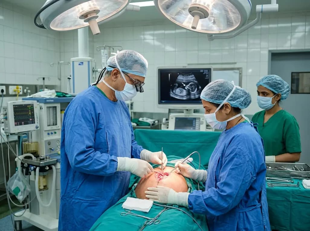
In a historic medical milestone, surgeons at AIIMS New Delhi successfully performed India's first open prenatal repair of a severe neural tube defect at 25 weeks of gestation. This pioneering in-utero procedure shields delicate neural structures, mitigating hydrocephalus and lifelong motor deficits before birth.
Today
A randomized crossover trial reveals that daily fresh watermelon consumption significantly reduces baseline TMAO levels and favorably modulates lipid profiles, lowering triglycerides and LDL cholesterol in adults with overweight and obesity.
Today
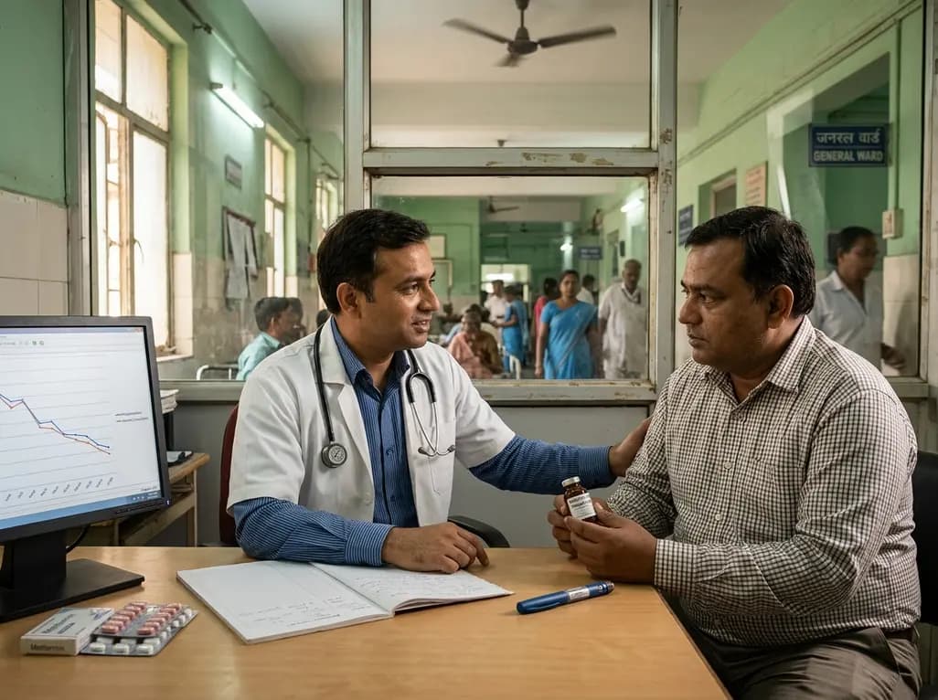
A large-scale retrospective cohort study demonstrates that GLP-1 receptor agonists significantly reduce all-cause hospitalizations, alcohol-related admissions, major cardiovascular events, and mortality in patients with overweight or obesity and alcohol use disorder, providing profound clinical insights.
Today
