

Loading, please wait...
Loading, please wait...
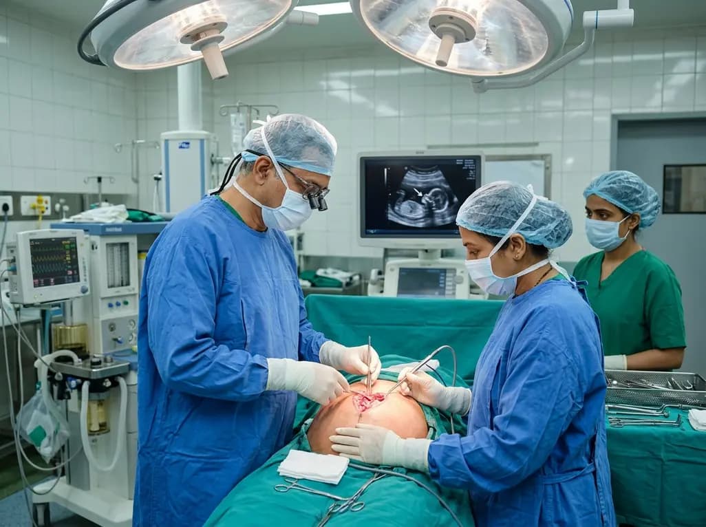
Fetal myelomeningocele repair represents one of the most remarkable frontiers in modern fetal medicine. Recently, clinicians at the All India Institute of Medical Sciences in New Delhi achieved an unprecedented breakthrough. The multidisciplinary team performed India's first open intrauterine repair of a neural tube defect at twenty-five weeks. Consequently, this surgical milestone introduces transformative in-utero neurosurgical possibilities to India. Previously, surgeons could only address this debilitating spinal anomaly postnatally. However, this innovative prenatal intervention directly targets early neural injury, offering vulnerable infants substantially improved long-term neurological prognoses.
Myelomeningocele remains the most severe and debilitating phenotypic expression of spina bifida. The condition arises when the embryonic neural tube fails to close completely during the fourth week of embryonic life. Consequently, the malformed spinal cord and protective meninges protrude through the defective posterior vertebral arches. Therefore, the delicate neural tissue suffers continuous, progressive damage throughout gestation. Neuroscientists explain this progressive deterioration through the established two-hit hypothesis. First, the primary embryonic failure creates incomplete neural tube closure. Second, toxic amniotic fluid exposure and mechanical trauma inflict secondary destructive injuries upon the exposed neural placode. In addition, persistent cerebrospinal fluid leakage through the spinal opening alters intracranial hydrostatic dynamics. This pressure disruption frequently triggers hindbrain herniation, also known as the Arnold-Chiari type II malformation. Furthermore, impaired ventricular drainage causes progressive hydrocephalus in affected infants. Hence, prenatal closure acts as a biological shield against relentless chemical degradation. By eliminating persistent intrauterine trauma, timely intervention preserves critical lower-limb motor tracts and pelvic visceral nerve function. Ultimately, early in-utero reconstruction halts neural tissue degradation before irreversible destruction takes place.
For decades, pediatric surgeons deferred neural tube reconstruction until after delivery. However, the landmark Management of Myelomeningocele Study transformed global pediatric guidelines. This prospective randomized clinical trial compared open prenatal intervention directly against standard postnatal closure. Notably, the trial demonstrated profound, undeniable therapeutic advantages for prenatal surgery candidates. Specifically, fetal repair significantly reversed hindbrain herniation across a large majority of treated cases. Moreover, the trial revealed that intrauterine surgery drastically diminished the subsequent need for ventriculoperitoneal shunting. At twelve months, forty percent of fetal surgery patients required shunts, compared to eighty-two percent of postnatal controls. Furthermore, researchers observed substantial improvements in ambulatory outcomes at thirty months of follow-up. Children receiving prenatal repair exhibited improved composite motor function scores and greater independent walking ability. In addition, extended evaluations confirmed sustained functional independence into school-age years. Therefore, randomized clinical trial evidence unequivocally demonstrates that prenatal repair alters the natural history of this congenital disorder. Nevertheless, clinicians must thoughtfully weigh these profound neurodevelopmental benefits against potential maternal and obstetrical morbidities. Accordingly, meticulous multidisciplinary protocols ensure optimal patient selection to maximize infant developmental potential while preserving maternal safety.
The pioneering procedure at AIIMS Neurosciences Centre represents extraordinary interdisciplinary coordination and technical precision. Prior to surgery, clinicians dedicated five weeks to exhaustive fetal imaging, patient counselling, and multi-team operative rehearsals. Subsequently, a specialized team of over thirty medical professionals executed the demanding six-hour operation. The maternal-fetal medicine and obstetric teams successfully performed a meticulous laparotomy and hysterotomy. Meanwhile, obstetric anesthesiologists maintained maternal hemodynamic stability while preventing dangerous uterine contractions. Working through the small uterine incision, neurosurgeons utilized high-resolution microscopy and continuous ultrasonographic guidance. The surgical team carefully dissected the neural placode away from surrounding tissues. Then, they restored the protective anatomical envelopes to halt cerebrospinal fluid leakage. Plastic surgery specialists assisted with delicate tissue mobilization, ensuring watertight, tension-free dural and cutaneous closure. Crucially, the public health framework enabled full accessibility for this family. The surgery occurred entirely free of cost under the Janani Shishu Suraksha Karyakram initiative. Consequently, this remarkable feat proves that highly sophisticated fetal therapies can succeed within the public healthcare infrastructure of India. Additionally, both the expectant mother and the developing fetus recovered smoothly following the intervention, demonstrating excellent postoperative recovery.
Despite its transformative therapeutic promise, fetal surgery introduces non-negligible maternal and fetal dangers. Consequently, surgical candidates must undergo rigorous evaluation before specialists recommend an operative intervention. Open maternal-fetal hysterotomy inherently increases the risk of preterm premature rupture of membranes and premature labor. Furthermore, maternal risks include intraoperative hemorrhage, chorioamnionitis, and complications from the uterine hysterotomy scar. Because the procedure involves uterine incision, future deliveries strictly require planned Cesarean section to prevent uterine rupture. Therefore, multidisciplinary fetal boards must evaluate strict inclusion and exclusion criteria before offering surgery. Eligible candidates typically present with isolated myelomeningocele between nineteen and twenty-six weeks of gestation. Additionally, fetuses must demonstrate an intact motor level above specific sacral thresholds without major kyphoscoliotic deformities. Conversely, fetal chromosomal anomalies, maternal medical contraindications, and pre-existing preterm labor strictly preclude surgical candidacy. Clinicians must conduct non-directive counselling so parents comprehend these delicate trade-offs. Ultimately, fetal surgery provides functional mitigation rather than a definitive cure for this complex condition. Moreover, close post-procedure surveillance remains mandatory to detect subclinical uterine activity and ensure uninterrupted fetal growth throughout the remaining trimesters.
The triumph at AIIMS Delhi marks a historic beginning for pediatric neurosurgery and perinatology across South Asia. Neural tube defects impose a substantial disease burden across India, where incidence rates remain clinically significant. Therefore, expanding tertiary fetal care infrastructure represents an urgent national healthcare priority. Advanced maternal-fetal centers must implement comprehensive ultrasound screening programs to facilitate early anomaly detection. When sonographers identify spinal dysraphism early, timely referral enables comprehensive multidisciplinary counseling and evaluation. Furthermore, institutional leaders plan to establish a structured, protocol-driven national fetal-care network. Such programmatic advancements will bridge the divide between specialized tertiary capability and community healthcare delivery. In addition, successful fetal surgery depends on robust neonatology, pediatric urology, and neuro-rehabilitation services after delivery. The comprehensive continuum of care supports bladder management, musculoskeletal development, and cognitive progress throughout childhood. As clinical training expands, more academic institutions will likely adopt these advanced surgical techniques. Ultimately, this paradigm shift offers renewed hope, turning previously untreatable intrauterine pathologies into viable opportunities for early recovery. Consequently, Indian tertiary hospitals can systematically lower childhood disability rates through preemptive intrauterine surgical intervention.
Q1: What clinical criteria determine eligibility for in-utero myelomeningocele repair?
Candidate eligibility requires an isolated myelomeningocele lesion with upper boundary between T1 and S1 confirmed by high-resolution neurosonography and magnetic resonance imaging. Furthermore, the gestational age must strictly fall between nineteen and twenty-six weeks. The mother must possess a normal singleton pregnancy without chromosomal aneuploidy, previous classical hysterotomy scars, or short cervical length. In addition, maternal health must permit general anesthesia without significant cardiovascular or systemic maternal contraindications.
Q2: Why does prenatal repair yield superior neurological outcomes compared to postnatal surgery?
Prenatal intervention mitigates progressive neurological injury by shielding exposed spinal neural tissue from caustic amniotic fluid and mechanical intrauterine shearing forces. Furthermore, halting cerebrospinal fluid leakage reverses hindbrain herniation and dramatically diminishes the incidence of Arnold-Chiari type II malformations. Consequently, this cranial decompression reduces the subsequent need for ventriculoperitoneal shunt placement by more than fifty percent. Moreover, children achieve significantly improved lower-limb motor function and higher rates of independent ambulation.
Q3: What are the primary maternal and obstetrical risks associated with open fetal surgery?
Open maternal-fetal surgery requires a full-thickness hysterotomy, which substantially elevates the risk of preterm labor and preterm premature rupture of membranes. Additionally, mothers face potential risks of intraoperative hemorrhage, placental abruption, chorioamnionitis, and complications associated with uterine incision healing. Because the uterine integrity is permanently altered, future pregnancies strictly necessitate planned Cesarean deliveries to avoid catastrophic uterine rupture during labor. Therefore, clinical teams must maintain vigilant multidisciplinary surveillance throughout pregnancy.
Disclaimer: This content is for informational and educational purposes only. It does not constitute medical advice or replace professional judgment. Refer to the latest local and national guidelines for clinical practice.
References

Read summarized clinical updates, watch expert medical content, and earn CME certifications right from your smartphone.
Open your smartphone camera to scan and install.





In a historic medical milestone, surgeons at AIIMS New Delhi successfully performed India's first open prenatal repair of a severe neural tube defect at 25 weeks of gestation. This pioneering in-utero procedure shields delicate neural structures, mitigating hydrocephalus and lifelong motor deficits before birth.
Today

A randomized crossover trial reveals that daily fresh watermelon consumption significantly reduces baseline TMAO levels and favorably modulates lipid profiles, lowering triglycerides and LDL cholesterol in adults with overweight and obesity.
Today
Catheter ablation from the noncoronary cusp provides a safe and effective approach for eliminating para-Hisian accessory pathways in Type B WPW syndrome when standard right-sided ablation fails or risks complete AV block despite misleading surface ECG patterns.
Today
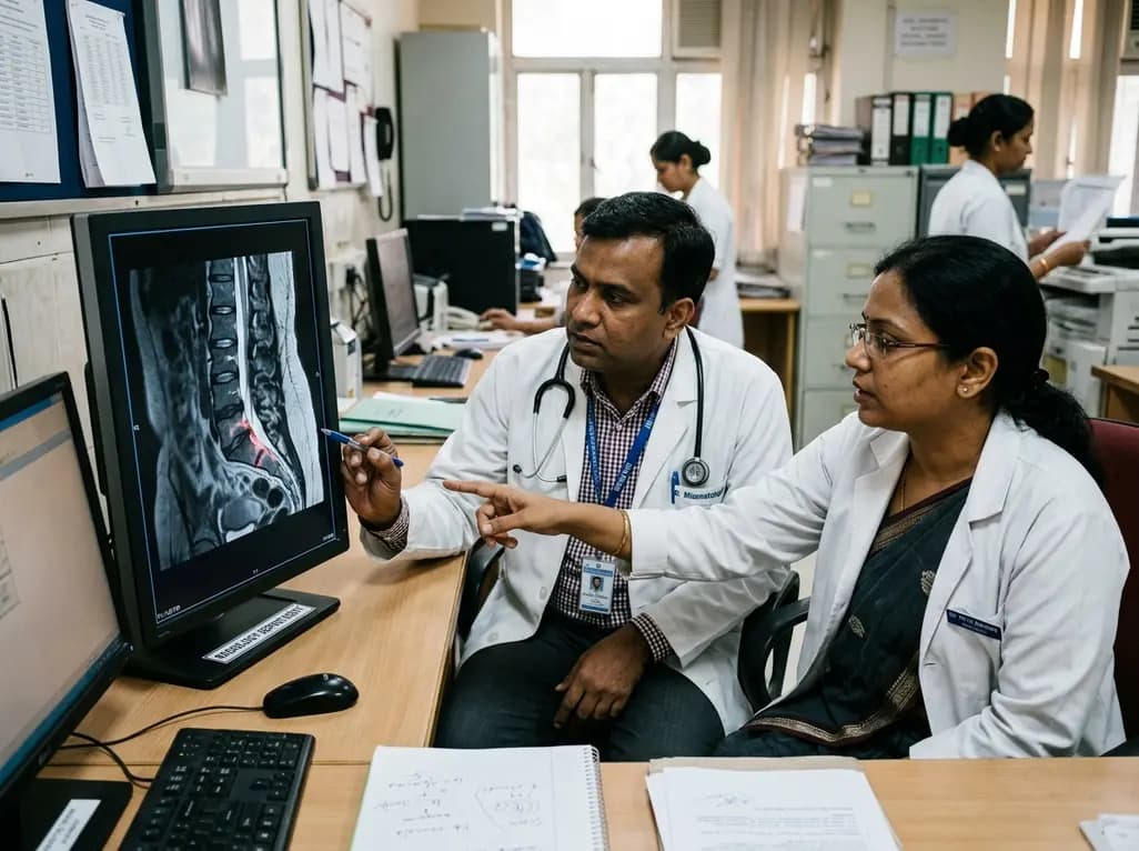
A qualitative study highlights the clinical role of MRI in axial spondyloarthritis, exposing disparities in access, reporting gaps in private healthcare, and the urgent need for standardized imaging protocols and multidisciplinary collaboration between rheumatologists and radiologists.
Today
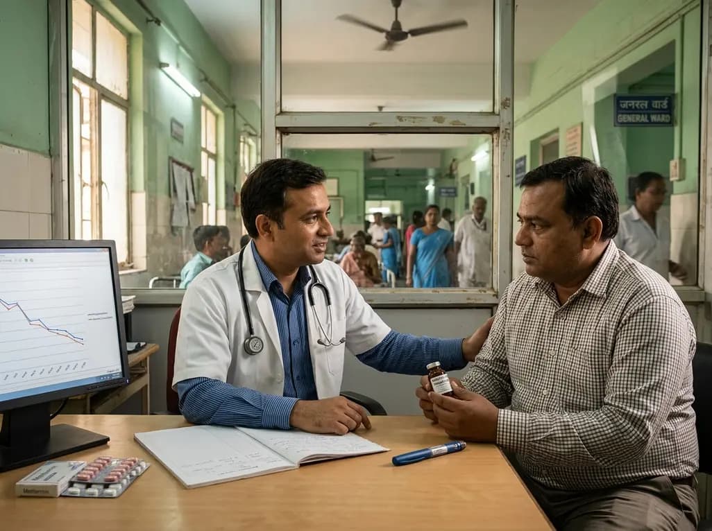
A large-scale retrospective cohort study demonstrates that GLP-1 receptor agonists significantly reduce all-cause hospitalizations, alcohol-related admissions, major cardiovascular events, and mortality in patients with overweight or obesity and alcohol use disorder, providing profound clinical insights.
Today
