

Loading, please wait...
Loading, please wait...
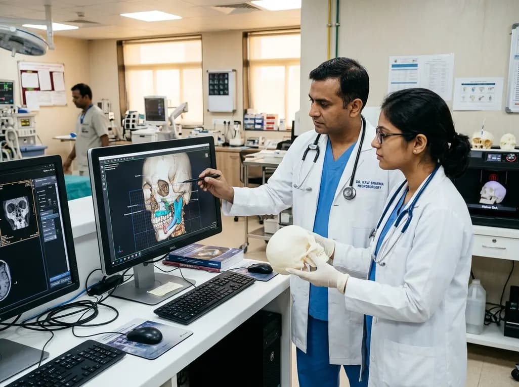
Modern reconstructive surgery demands high precision when restoring complex bony anatomy and soft tissue symmetry. Over the last decade, virtual surgical planning has emerged as an indispensable cornerstone of patient-specific care in head and neck surgery. Craniofacial defects resulting from high-velocity trauma, extensive oncological resections, or severe congenital anomalies present complex geometric challenges. Consequently, surgical teams are transitioning away from freehand manipulation toward sophisticated digital simulation. Recent bibliometric analyses illustrate a dramatic rise in computational modeling within maxillofacial and craniofacial surgery. By integrating advanced imaging with intelligent design tools, clinicians can preoperatively simulate osteotomies, model bone grafts, and anticipate aesthetic contours. This digital shift optimizes surgical precision while dramatically improving functional outcomes for diverse reconstructive candidates.
Historically, reconstructive surgeons relied heavily on two-dimensional radiographs and intraoperative trial-and-error to restore facial symmetry. However, modern digital protocols have fundamentally revolutionized preoperative preparation. Through high-resolution computed tomography, virtual surgical planning enables clinicians to manipulate intricate skeletons in three dimensions before making any incision. Specialized software allows teams to perform virtual osteotomies, reposition displaced fragments, and determine accurate contour alignment. Moreover, surgeons can design patient-specific cutting guides and prefabricated titanium plates with submillimeter accuracy. Consequently, operating room time decreases significantly because teams avoid tedious manual contouring during surgery. Furthermore, donor-site harvesting becomes far more precise when using tailored cutting templates. For example, surgeons reconstruct complex mandibular defects using vascularized fibular free flaps with predictable bony union and dental rehabilitation. In addition, reduced anesthesia duration decreases perioperative complications and accelerates patient recovery. Thus, digital planning bridges the gap between diagnostic imaging and operative execution, elevating the standard of surgical reconstructive care.
Earlier iterations of digital reconstructive surgery depended primarily on static navigation and mechanical guide fabrication. Nevertheless, recent computational advancements have propelled the field into dynamic machine learning workflows. Automated image segmentation now accelerates skeletal modeling by extracting bone contours within seconds. Previously, manual segmentation demanded hours of painstaking slice-by-slice contouring by radiologists or surgical engineers. Today, deep learning architectures automatically delineate intricate structures, such as the mandibular canal, delicate orbital walls, and vascular pedicles. Furthermore, convolutional networks identify anatomical landmarks to calculate symmetry planes autonomously. These intelligent systems also assist in predicting soft-tissue draping following significant skeletal movements. As a result, surgeons can evaluate aesthetic changes and simulate postoperative profiles with remarkable fidelity. In addition, predictive algorithms help multidisciplinary teams choose optimal reconstructive vectors and graft dimensions for complex defects. Consequently, surgical teams save valuable planning hours while enhancing procedural consistency. This technological synergy highlights how artificial intelligence is transforming static surgical blueprints into interactive, patient-tailored simulations.
The intellectual landscape of digital craniofacial reconstruction has expanded dramatically across multiple scientific domains. A comprehensive bibliometric evaluation reveals that annual scientific publications accelerated markedly after 2010. Interestingly, research output bridges clinical disciplines like plastic surgery, oral and maxillofacial surgery, and dentistry with computational fields. Computer vision and engineering sciences frequently supply the underlying algorithmic models that power modern surgical software. Prominent collaborative networks have emerged across North America, Europe, and Asia, with substantial contributions from institutions in the United States and China. Moreover, expanding participation from centers across India and other emerging healthcare markets is driving global innovation. However, this multidisciplinary overlap also introduces substantial variations in terminology across different scientific journals. For instance, computer scientists frequently describe computational facial modeling using concepts distinct from clinical reconstructive terms. Therefore, establishing uniform scientific vocabularies and standardized reporting benchmarks remains essential for effective interdisciplinary collaboration. Fostering structured communication between surgical practitioners and data scientists will ensure that computational innovations address authentic operative needs.
Despite promising technological advancements, moving computational models from research laboratories into daily clinical practice presents significant challenges. Many proposed machine learning algorithms train on modest, highly homogeneous patient cohorts. Consequently, models often lack robust external validation and may underperform when encountering rare anatomical deformities or atypical trauma patterns. Furthermore, closed-source proprietary software frequently limits transparency, creating a black-box environment for operating surgeons. Medical teams must maintain clear accountability and cannot rely blindly on automated recommendations. Therefore, researchers must emphasize rigorous multicenter clinical trials to validate algorithmic accuracy, safety, and reproducibility. In addition, regulatory bodies require robust evidence regarding the biomechanical integrity of patient-specific implants and guides. Ethical considerations regarding patient data privacy, imaging storage, and algorithmic bias also demand careful management. Ultimately, safe clinical adoption requires transparent algorithms that operate under direct human oversight. Clinicians must always retain final decision-making authority, ensuring that technological tools assist rather than replace seasoned surgical judgment.
For surgeons operating within India, the integration of advanced digital planning holds exceptional clinical significance. The domestic healthcare burden includes a high prevalence of high-velocity road traffic collisions and complex oral cancer resections. Furthermore, pediatric teams encounter a substantial volume of untreated congenital craniofacial anomalies requiring intricate reconstruction. Historically, the high costs of imported software and external proprietary engineering services restricted digital planning to select tertiary institutions. However, the emergence of open-source artificial intelligence frameworks and localized three-dimensional printing is shifting this paradigm. Indian academic medical centers are increasingly adopting in-house digital workflows to optimize surgical precision cost-effectively. As a result, reconstructive teams can design customized surgical splints, guides, and graft plates within hospital-based modeling laboratories. Furthermore, developing indigenous training programs and sharing open datasets across Indian centers will build essential local expertise. By embracing these accessible digital technologies, Indian reconstructive surgeons can enhance procedural efficiency, reduce operative complications, and democratize access to world-class patient outcomes across diverse patient populations.
Virtual surgical planning allows surgeons to simulate complex osteotomies and reconstructive steps on high-resolution 3D models before entering the operating room. By designing patient-specific cutting guides and plates, surgical teams achieve greater skeletal accuracy and symmetry. Furthermore, pre-bending fixation plates and predetermining graft dimensions dramatically shortens operative time and ischemia duration. Consequently, patients experience lower complication rates, improved functional dental rehabilitation, and better overall aesthetic outcomes following major trauma or oncological resections.
Artificial intelligence enhances digital surgical planning primarily by automating time-consuming tasks like anatomical image segmentation and landmark identification. Deep learning models rapidly isolate bone structures, soft-tissue boundaries, and critical neurovascular paths from computed tomography scans. Additionally, predictive algorithms assist in simulating post-surgical soft-tissue drape, helping clinicians anticipate aesthetic changes. By accelerating digital workflow preparation, machine learning reduces planning overhead, enabling reconstructive teams to formulate customized operative strategies with greater precision and consistency.
Primary barriers to in-house planning include high initial capital investment for specialized design software and biocompatible three-dimensional printers. Furthermore, hospitals often face a steep learning curve requiring dedicated training for surgeons and biomedical engineers. Regulatory compliance, quality control standards, and sterilization verification also present logistical hurdles for point-of-care manufacturing. However, as localized printing hardware becomes more affordable and open-source software matures, tertiary centers are increasingly overcoming these hurdles to deliver tailored patient care.
Disclaimer: This content is for informational and educational purposes only and should not be considered as medical advice or clinical guidance. Healthcare professionals must rely on their clinical judgment, institutional protocols, and relevant diagnostic evidence when evaluating and managing individual patients. Refer to the latest local and national guidelines for clinical practice.
References

Read summarized clinical updates, watch expert medical content, and earn CME certifications right from your smartphone.
Open your smartphone camera to scan and install.





A global bibliometric analysis highlights the rapid integration of artificial intelligence and virtual surgical planning into craniofacial reconstruction. The convergence of computational imaging and surgical engineering enhances operative precision, patient-specific implants, and functional restoration.
Today
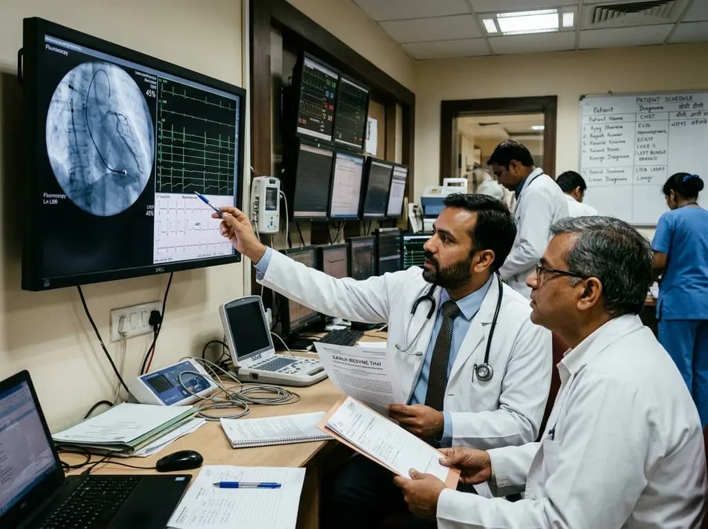
The randomized EARLY-RESYNC trial reveals that early left bundle branch pacing combined with guideline-directed medical therapy significantly improves LVEF and reduces LV end-systolic volume compared to medical therapy alone in patients with complete LBBB and mildly reduced ejection fraction (36%-50%).
Today
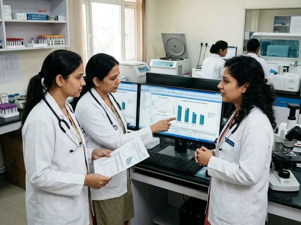
A comparative evaluation of INNO-LiPA, Cobas 4800, and Amplicor highlights significant assay-dependent variation in high-risk HPV detection, emphasizing the clinical distinction between analytical sensitivity and screening specificity.
Today
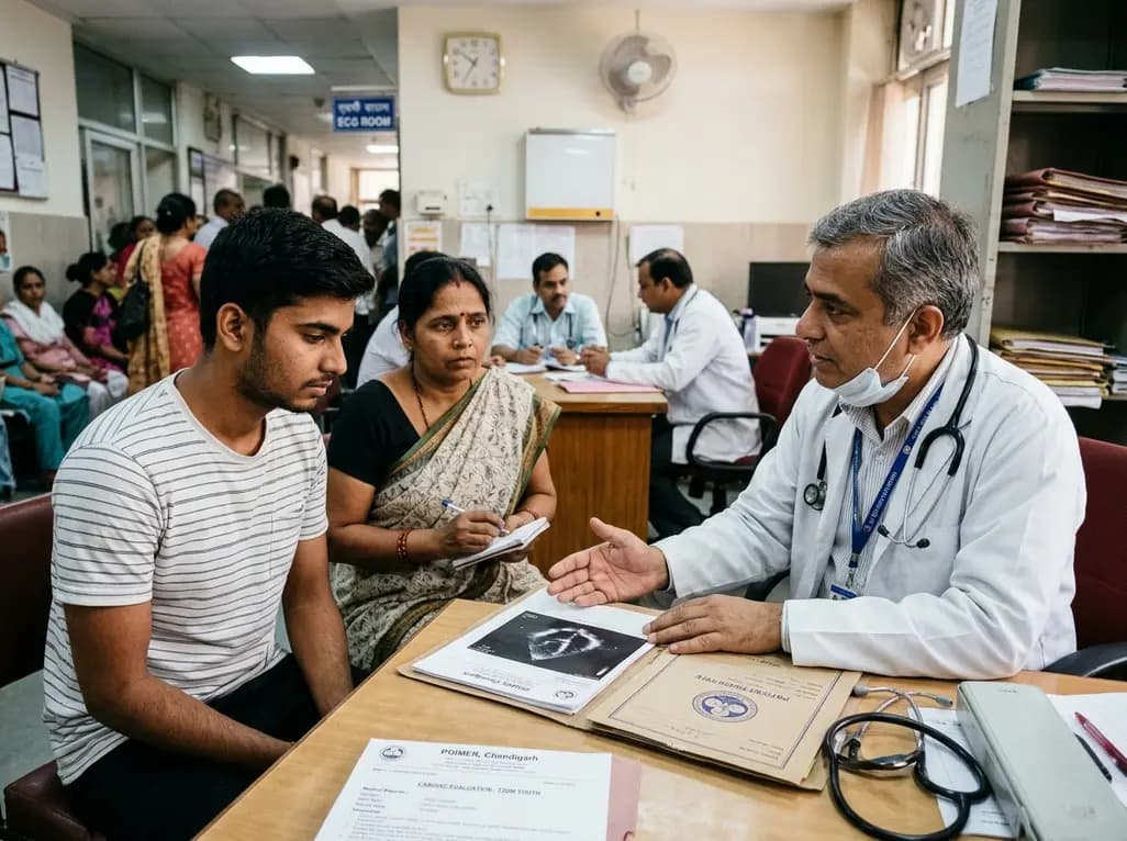
Recent data from PGIMER reveals that young adults diagnosed with type 2 diabetes face an aggressive disease trajectory. They encounter severe cardiovascular and microvascular damage within five years, occurring at higher rates than in type 1 diabetes despite lower mean blood glucose levels.
Today
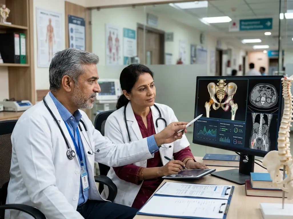
Discover the six core principles of responsible AI in orthopaedics, emphasizing algorithmic fairness, transparency, clinical reliability, and specialist oversight to safeguard patient trust and outcomes.
Today
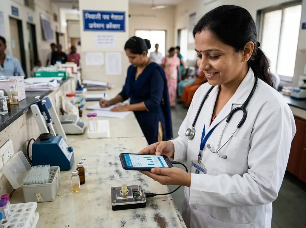
A novel smartphone-assisted biosensor utilizing molecularly imprinted polymers and catalytic nanozymes offers antibody-free, enzyme-free urinary microalbumin testing, enabling rapid, low-cost point-of-care screening for early chronic kidney disease.
Today
