.webp&w=384&q=75)

Loading, please wait...
Loading, please wait...
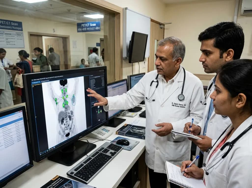
Modern clinical oncology relies heavily on 18F-fluorodeoxyglucose positron emission tomography combined with computed tomography for staging lymphoma. However, manual contouring of multiple pathological hypermetabolic lymph nodes remains time-consuming and introduces substantial inter-observer variability. Recently, deep learning algorithms have emerged to automate this tedious task. Implementing fully automated AI lesion segmentation in whole-body FDG PET/CT provides reproducible quantitative metrics, which fundamentally accelerates clinical decision-making. Consequently, automated frameworks enable nuclear medicine specialists to evaluate extensive tumor volumes without compromising diagnostic precision.
Evaluating disease burden across Hodgkin and non-Hodgkin lymphomas demands precise volumetric assessment. Clinicians frequently extract total metabolic tumor volume and total lesion glycolysis to predict patient survival and assess treatment response. Nevertheless, manual boundary delineation across dozens of nodal stations requires substantial physician time. Furthermore, distinct readers frequently draw diverging boundaries around identical tumors, which generates inconsistent risk stratification scores. This variability particularly complicates multi-center clinical trials where standardized measurement criteria remain indispensable. In addition, physiological tracer uptake in the brain, myocardium, liver, and urinary tract often mimics malignant lesions. Readers must therefore continuously cross-reference CT anatomical landmarks with functional PET signal intensity to eliminate false positives. Consequently, high daily scan volumes in busy cancer centers exacerbate specialist fatigue. Under such demanding conditions, radiologists can occasionally miss subtle pathological lesions displaying faint fluorodeoxyglucose avidity. Moreover, delayed report turnaround times can impede swift clinical interventions for aggressive disease. Thus, medical centers urgently require robust computational assistance to streamline whole-body evaluations and preserve diagnostic consistency.
To overcome manual contouring challenges, researchers developed an automated pipeline that leverages a hybrid deep learning model. Specifically, the framework couples the powerful nnUNet segmentation engine with a lightweight ResNet18 convolutional backbone. This hybrid 2D-3D design processes spatial context efficiently while capturing high-resolution volumetric details across comprehensive whole-body acquisitions. Moreover, the architecture seamlessly ingests paired PET and CT volumes, merging structural anatomy with functional radiotracer concentration. The engineering team incorporated sophisticated post-processing algorithms directly into the workflow to eliminate anatomical confounders. Consequently, the software filters out standard physiological excretion within the bladder, kidneys, and gastrointestinal structures. In addition, rule-based spatial thresholding suppresses false-positive signals stemming from non-malignant inflammatory nodes. The algorithm simultaneously refines tumor margins, minimizing quantitative errors that frequently plague conventional volumetric calculations. Furthermore, automated preprocessing prepares standardized voxel representations, ensuring smooth performance across diverse acquisition geometries. Therefore, this streamlined computational pipeline delivers reliable volumetric segmentations within minutes, providing oncologists with immediate quantitative readouts.
Rigorous clinical validation confirmed the exceptional diagnostic fidelity of the automated segmentation pipeline. Investigators tested the deep network against manual delineations performed by experienced physicians across dozens of lymphoma patients. Overall, the automated model achieved an outstanding Dice similarity coefficient of 89.2 percent across hundreds of distinct lesions. Furthermore, the tool demonstrated a balanced clinical profile, attaining an 82.9 percent sensitivity alongside a 96.5 percent positive predictive value. This remarkably high precision confirms that the system effectively prevents spurious non-cancerous over-segmentation. In addition, correlation analyses revealed near-perfect alignment between artificial intelligence calculations and manual benchmarks across all quantitative metabolic parameters. Specifically, correlation coefficients consistently exceeded 0.94 for standardized uptake values, total metabolic tumor volume, and total lesion glycolysis. Bland-Altman statistical analyses similarly displayed minimal quantitative bias without systematic skewing across diverse tumor burdens. Moreover, intraclass correlation coefficients remained above 0.94 across both Hodgkin and non-Hodgkin subtypes. Accordingly, the algorithmic output reliably matches the rigorous standards established by seasoned nuclear medicine specialists.
Although the deep learning tool demonstrated remarkable overall accuracy, fine image properties distinctly impacted its segmentation consistency. Notably, researchers identified a moderate negative correlation between the Dice similarity coefficient and liver signal-to-noise ratio. This statistical relationship indicates that background noise and hepatic metabolic variability can alter boundary precision around neighboring lymph nodes. However, the system maintained robust global lesion identification across varied digital scanner acquisitions. Modern digital PET systems, such as advanced high-sensitivity silicon photomultiplier scanners, provide superior timing resolution and count sensitivity. Consequently, these hardware improvements optimize signal clarity, which directly facilitates algorithmic feature extraction. Moreover, patient-related factors, including body habitus and tracer uptake timing, subtly influence PET signal heterogeneity. Clinicians must therefore ensure strict adherence to standardized scanning protocols to maximize algorithmic performance. In addition, ongoing quality control procedures guarantee that background physiological fluctuations do not distort automated tumor metrics. Thus, harmonizing acquisition parameters remains crucial for maintaining peak neural network efficacy across distinct imaging platforms.
Integrating deep learning tools into routine hospital practice substantially transforms clinical oncology pathways. Currently, lymphoma staging under the Lugano classification necessitates meticulous visual examination of multiple nodal stations and extranodal sites. Artificial intelligence automates this burdensome measurement phase, allowing radiologists to focus on holistic case interpretation. Furthermore, rapid volumetric extraction enables precise calculation of total metabolic tumor volume during baseline scans. Multiple prospective clinical trials show that baseline tumor volume serves as an independent prognostic factor across diverse lymphoma cohorts. Therefore, automated volumetric tools allow oncologists to identify high-risk individuals requiring intensified systemic chemotherapy regimens promptly. Similarly, precise automated segmentation assists interim therapy evaluation by calculating standardized metabolic changes with minimal user bias. In addition, deploying validated algorithmic assistants frees valuable physician hours in high-volume tertiary cancer centers. Consequently, close collaboration between computational algorithms and experienced clinicians establishes an unprecedented benchmark for personalized cancer care. Ultimately, scalable AI deployment will standardize metabolic quantification across academic medical centers and community oncology practices alike.
The hybrid architecture combines whole-body CT anatomical imaging with functional PET metabolic maps to identify malignancy accurately. In addition, the system incorporates rule-based post-processing algorithms to recognize typical physiological accumulation patterns. The software successfully filters out normal radiotracer excretion within the urinary bladder, kidneys, and bowel loops. Consequently, the model isolates pathological lymph nodes while rejecting non-malignant hypermetabolic structures, yielding an outstanding positive predictive value of ninety-six percent.
Liver parenchyma serves as the standard clinical reference organ for PET background metabolic activity. When hepatic background noise increases, image contrast degrades, which complicates subtle border delineation between pathological lymph nodes and adjacent normal tissues. Notably, statistical analyses show that liver signal-to-noise ratio correlates moderately with boundary segmentation accuracy. Therefore, maintaining strict scanner calibration and standardizing radiotracer uptake intervals remain vital for maximizing automated algorithmic precision in routine clinical imaging.
Total metabolic tumor volume provides powerful prognostic information regarding disease progression and overall patient survival. However, manual calculation across multiple nodal stations takes excessive time and invites substantial inter-observer disagreement. Automated segmentation software extracts this critical metric within seconds, delivering reproducible, standardized tumor burden evaluations. Consequently, oncologists can promptly identify high-risk lymphoma patients at baseline, enabling timely therapy intensification and enhancing personalized treatment planning without increasing clinical workload.
Disclaimer: This content is for informational and educational purposes only... Refer to the latest local and national guidelines for clinical practice.
References

Read summarized clinical updates, watch expert medical content, and earn CME certifications right from your smartphone.
Open your smartphone camera to scan and install.





A deep learning AI tool using nnUNet and ResNet18 achieves 89.2% Dice similarity and 96.5% PPV in segmenting lymphoma on whole-body FDG PET/CT. Automated volumetric quantification closely mirrors expert evaluations, offering high diagnostic precision and efficiency for nuclear medicine and oncology.
Today

A recent qualitative synthesis highlights how physical labour, dignity preservation, and weight stigma intersect when caring for larger-bodied patients in acute care. Overcoming these challenges requires ergonomic equipment, adequate staffing, flexible workflows, and size-inclusive institutional policies.
Today

Novel preclinical research demonstrates that antisense oligonucleotides targeting essential genes such as DARS1, DYNC1I2, and EIF2S3 selectively induce apoptosis in ovarian and lung cancer cells. Conditional biomarker activation provides a viable strategy to spare normal tissue while overcoming therapy resistance.
Today
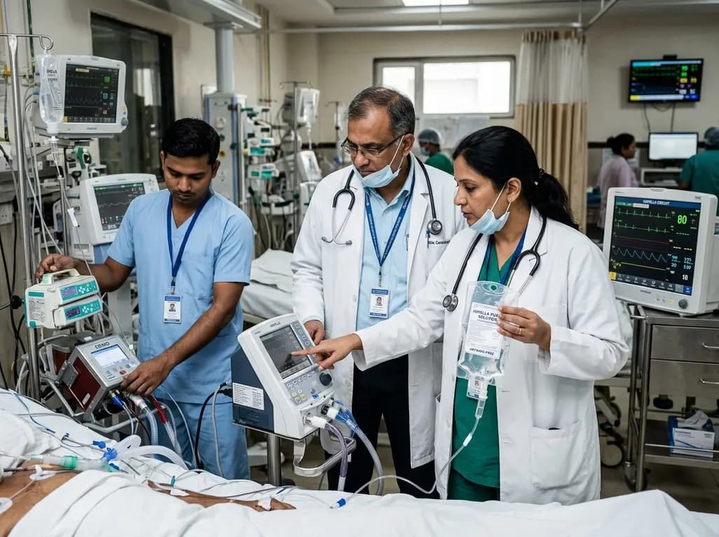
This clinical analysis examines heparin-free versus heparin-containing Impella purge solutions in patients receiving concomitant ECMO support (ECPELLA). Evidence suggests anticoagulant-free purges maintain device patency while reducing bleeding risks, offering a viable strategy alongside systemic anticoagulation.
Today

A 10-year Markov state-transition model shows minimally invasive tubular decompression dominates open laminectomy for pure lumbar spinal stenosis, yielding 5.95 vs 5.91 QALYs and saving $4,167 per patient through reduced complications and lower index costs.
Today

A preclinical study demonstrates that naringin-loaded phytosomes significantly enhance oral bioavailability and provide neuroprotective efficacy in diabetic neuropathy by dampening inflammation, reducing oxidative stress, and relieving neuropathic pain.
Today
