.webp&w=384&q=75)

Loading, please wait...
Loading, please wait...

Spine surgeons frequently encounter compromised bone mineral density in aging surgical cohorts. Addressing undetected osteoporosis in spine surgery remains a paramount priority because poor bone strength precipitates devastating hardware loosening, cage subsidence, and revision procedures. Traditional dual-energy x-ray absorptiometry examinations often fail to capture regional trabecular weaknesses due to degenerative osteophytes and vascular calcifications. Consequently, clinicians require rapid, opportunistic screening methodologies to detect skeletal vulnerability prior to instrumented arthrodesis. Surgeons routinely obtain computed tomography scans for preoperative anatomical planning, providing a valuable reservoir of bone attenuation data. Trabecular attenuation measured in Hounsfield units reflects regional bone mineral content with exceptional precision. However, manual quantification of attenuation across multiple cross-sectional images consumes valuable clinician time and introduces operator-dependent measurement error. Modern deep learning architectures now offer an elegant solution to these logistical bottlenecks. By implementing automated neural networks, surgical teams can extract quantitative densitometry from routine scans without ordering supplementary imaging or increasing radiation exposure. Therefore, automated evaluation represents an important evolutionary step toward individualized surgical risk stratification and optimized bone health management.
Compromised bone density drastically alters biomechanical load distribution across spinal instrumentation constructs. Consequently, patients with unaddressed bone loss face elevated rates of catastrophic pedicle screw pullout and proximal junctional failure. When surgeons place hardware into osteoporotic bone, the interface between metal and trabeculae experiences immense mechanical strain. Therefore, failure to identify fragile bone before operation frequently leads to delayed union, pseudoarthrosis, and debilitating chronic pain. Furthermore, sudden vertebral compression fractures may occur adjacent to rigid fusion constructs, necessitating complex revision surgeries. In contemporary practice, preoperative optimization with teriparatide, abaloparatide, or antiresorptive agents significantly enhances bone mineral density and accelerates fusion rates. Moreover, spine specialists can adjust surgical techniques when they anticipate low bone density in advance. For example, surgeons may employ cement augmentation, bicortical screw purchase, or expandable pedicle screws to secure stable fixation. However, clinical teams frequently miss the window for medical optimization when patients lack prior fragility screening. In addition, routine surgical referrals often bypass standard endocrinological evaluations due to urgent neural decompression requirements. Thus, incorporating reliable opportunistic screening directly into routine surgical pathways fundamentally transforms preoperative preparation and protects patient outcomes.
Dual-energy x-ray absorptiometry represents the historic diagnostic standard for systemic osteoporosis, yet it presents severe shortcomings in spinal assessment. Degenerative spine disease frequently introduces hypertrophic osteophytes, facet arthrosis, and aortic calcifications. Consequently, planar DEXA projection artificially overestimates areal bone mineral density in elderly cohorts, creating a false impression of skeletal integrity. In contrast, computed tomography isolates trabecular bone compartments within the vertebral body without interference from cortical spurs or calcified vessels. Hounsfield units directly quantify x-ray beam attenuation, which correlates robustly with volumetric trabecular density and biomechanical compressive strength. Multiple biomechanical investigations demonstrate that lower lumbar Hounsfield unit values directly correlate with screw loosening and cage subsidence. Specifically, L1 vertebral attenuation below approximately one hundred and ten Hounsfield units strongly correlates with true osteoporosis. Therefore, measuring attenuation on preoperative CT provides surgical teams with superior site-specific bone quality assessments. However, manual measurements require a trained clinician to draw elliptical regions of interest on axial slices. This manual workflow demands extra time, creates inter-observer variability, and limits routine clinical adoption. Automated artificial intelligence protocols resolve these persistent barriers by instantly evaluating bone quality during routine radiographic workups.
To eliminate manual calculation bottlenecks, researchers recently engineered a specialized artificial intelligence pipeline utilizing the nnU-Net framework. The investigators acquired two comprehensive computed tomography scan datasets comprising 501 clinical images to develop this diagnostic tool. Subsequently, they partitioned the imaging library into robust training, validation, and testing subsets to train the deep learning model. The nnU-Net framework excels in biomedical image analysis because it self-configures preprocessing, network architecture, and postprocessing parameters dynamically. Specifically, the neural network rapidly segments the L1 vertebral body from surrounding bony and soft tissue structures. Following precise volumetric segmentation, an automated script measures attenuation in Hounsfield units within the central trabecular space. Importantly, this algorithm avoids degenerative cortical endplates, venous plexuses, and posterior elements to ensure pristine trabecular sampling. The automated model executed vertebral segmentation with extraordinary precision, achieving a mean Dice similarity coefficient of 0.91. Consequently, the convolutional architecture successfully identified boundaries and isolated anatomical contours across diverse anatomical variations. This high anatomical fidelity proves that deep learning models can perform complex volumetric segmentation autonomously without manual operator correction or supervisory adjustment during image processing.
The investigators validated the performance of the artificial intelligence tool against manual calculations performed by expert raters on 56 computed tomography scans. Notably, statistical analysis revealed a Pearson correlation coefficient of 0.96 between algorithm-derived attenuation and manual human measurements. This remarkable correlation indicates an exceptionally strong linear relationship across wide attenuation spectrums. Furthermore, the intraclass correlation coefficient reached 0.95 for the first rater and 0.94 for the second rater, demonstrating outstanding interrater reliability. In addition, Bland-Altman plots revealed minimal systematic discrepancy, displaying a mean difference of only 7.0 Hounsfield units between artificial intelligence and human measurements. The ninety-five percent limits of agreement ranged from negative 21.1 to positive 35.2 Hounsfield units across the validation cohort. Crucially, a paired t-test confirmed no statistically significant difference between human and algorithmic measurements, producing a non-significant p-value of 0.21. Therefore, the automated framework mirrors expert radiologist evaluations with rigorous accuracy and consistency. These quantitative findings demonstrate that deep learning automation provides reproducible densitometric assessments that match standard manual measurements. Consequently, surgical teams can place substantial confidence in automated Hounsfield unit estimations for immediate clinical decision-making.
Integrating artificial intelligence into picture archiving and communication systems offers substantial pragmatic benefits for tertiary spine centers. First, automated evaluation converts every routine thoracolumbar computed tomography scan into an opportunistic bone health diagnostic report. Because surgeons routinely order preoperative scans for navigation, patients avoid supplemental appointments and additional radiation exposures. Furthermore, the automated software operates quietly in the background without imposing clerical burdens on overworked radiologists or orthopedists. When the model detects abnormally low attenuation values, the system flags the patient for immediate clinical bone optimization. Consequently, surgical teams can coordinate bone-forming pharmacotherapy or modify instrumentation strategies several weeks prior to elective spine procedures. In addition, automated screening alerts primary care physicians and endocrinologists to address systemic osteoporosis, preventing future peripheral fragility fractures. However, widespread clinical translation requires ongoing multicenter validation across diverse patient populations, various scanner manufacturers, and heterogeneous slice thicknesses. Future software updates must also address contrasting intravenous agents and severe scoliosis deformities. Nevertheless, this computational advancement bridges the critical diagnostic gap between spinal diagnostics and bone health management, delivering safer, personalized spine surgery.
DEXA scans frequently produce artificially elevated bone density scores because spinal degenerative changes, aortic calcifications, and osteophytes obscure planar measurements. In contrast, computed tomography scans evaluate isolated trabecular compartments directly. Measuring Hounsfield units provides site-specific bone density estimations that accurately predict pedicle screw fixation strength and implant subsidence risk.
The model utilizes the nnU-Net deep learning framework to segment the L1 vertebra autonomously from routine thoracolumbar computed tomography scans. Afterwards, a specialized computational algorithm calculates mean attenuation values exclusively within trabecular bone regions. This automated process avoids cortical edges, vascular channels, and degenerative deformities without requiring manual clinician input.
Opportunistic screening does not replace comprehensive medical management, but it identifies undiagnosed bone fragility early. When the artificial intelligence identifies low attenuation values, clinicians should initiate metabolic workups and bone-optimizing pharmacotherapy. This workflow ensures that surgeons reinforce instrumentation techniques and protect patients from impending postoperative mechanical failure.
Disclaimer: This content is for informational and educational purposes only and does not constitute formal medical advice. Healthcare professionals should evaluate individual clinical presentations independently. Refer to the latest local and national guidelines for clinical practice.
References

Read summarized clinical updates, watch expert medical content, and earn CME certifications right from your smartphone.
Open your smartphone camera to scan and install.





An innovative AI model leverages nnU-Net to measure L1 Hounsfield units on preoperative CT scans, enabling automated opportunistic screening for osteoporosis in spine surgery. The model demonstrates high correlation with expert manual analysis, facilitating timely preoperative bone optimization.
Today
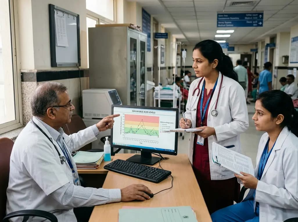
A comprehensive analysis of serum bile acid variability establishes robust reference ranges and reveals how sex, age, BMI, and smoking alter circulating profiles.
Today

A mixed-methods study evaluates a digital perinatal navigator app for high-risk pregnancies, demonstrating high usability, reduced distress, and improved awareness of vital healthcare services.
Today
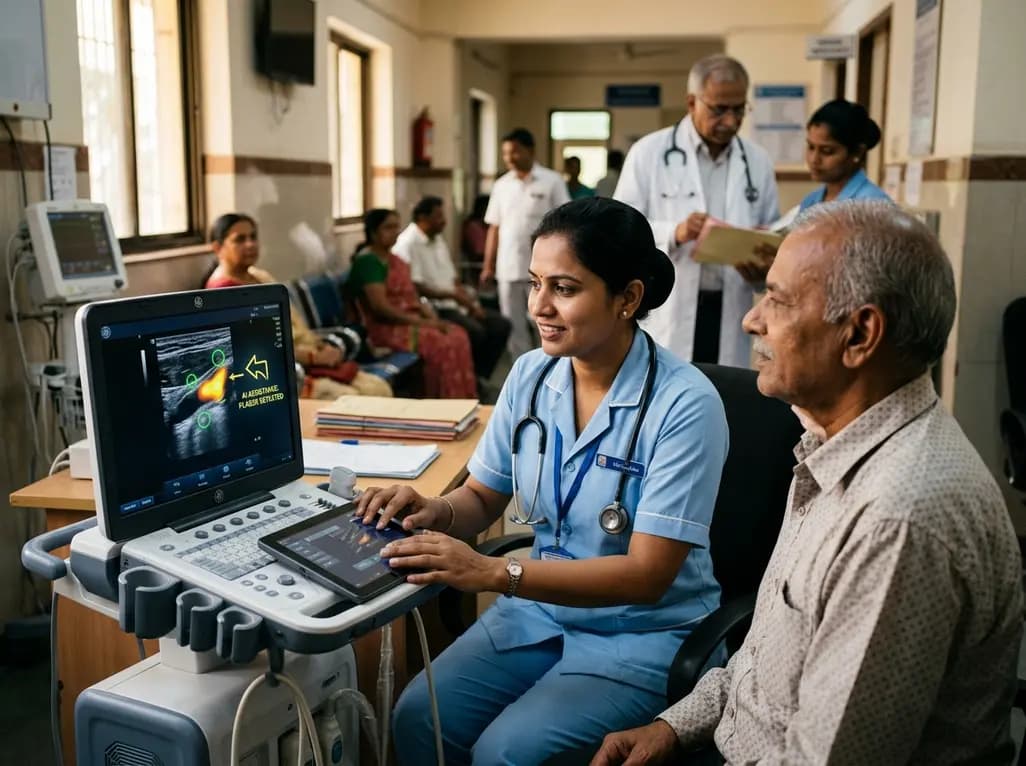
A user-centered study demonstrates that AI-assisted carotid ultrasound enables nonexpert primary care staff to detect subclinical atherosclerosis. With real-time guidance and workflow optimization, this technology supports early cardiovascular risk communication and task-shifting in routine clinical practice.
Today
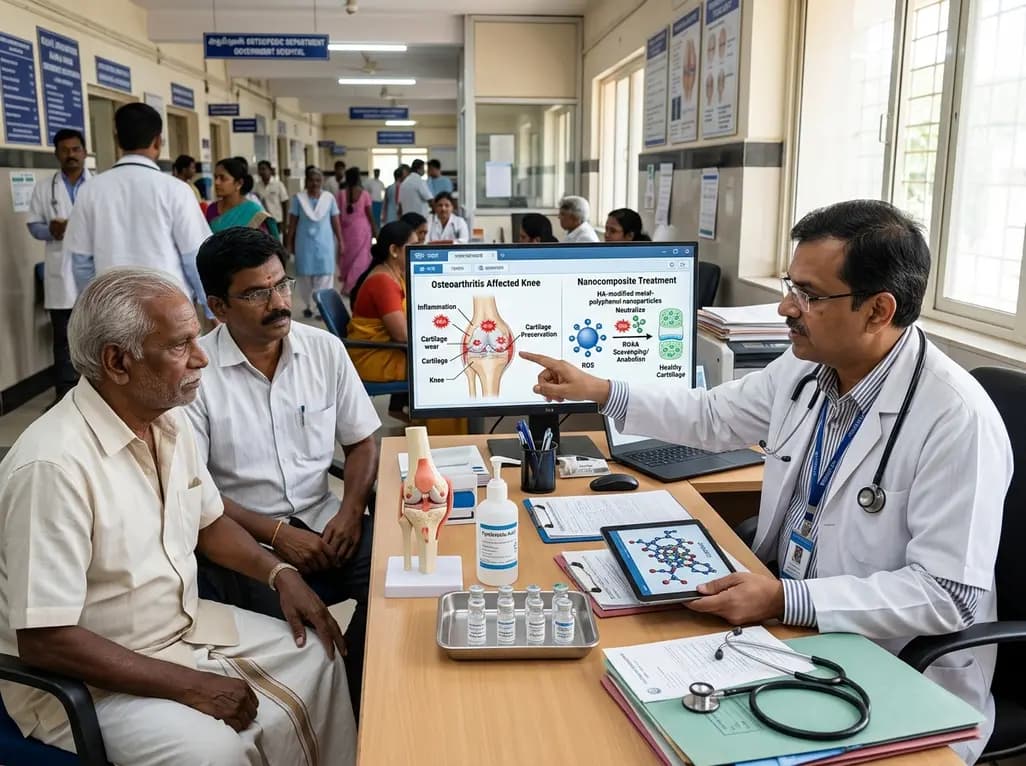
A hyaluronic acid-modified metal-polyphenol nanocomposite successfully eliminates reactive oxygen species in chondrocytes, halts cartilage breakdown, and promotes tissue anabolism, presenting a novel disease-modifying strategy for early osteoarthritis.
Today
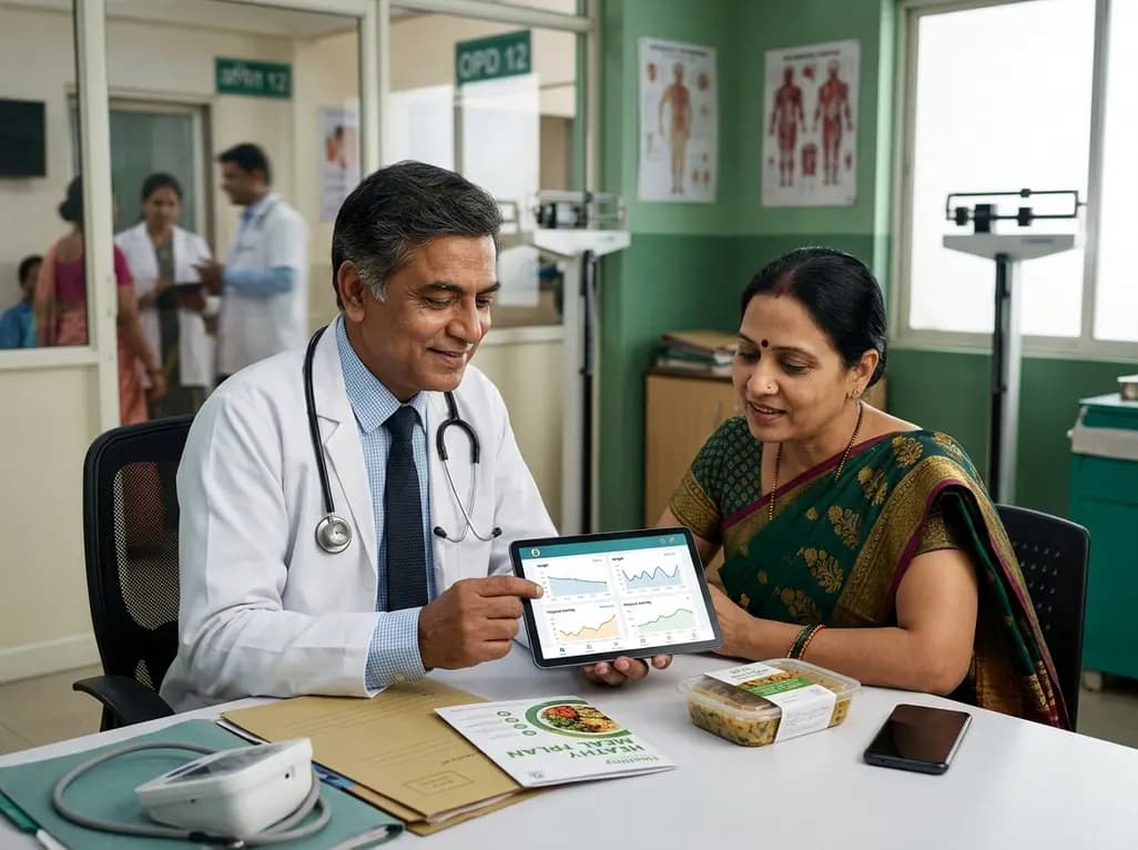
A pilot randomized trial demonstrates that digital wellness applications and medically tailored meals significantly attenuate rapid weight regain following GLP-1 receptor agonist discontinuation, providing valuable transitional support for long-term obesity management.
Today
