.webp&w=384&q=75)

Loading, please wait...
Loading, please wait...
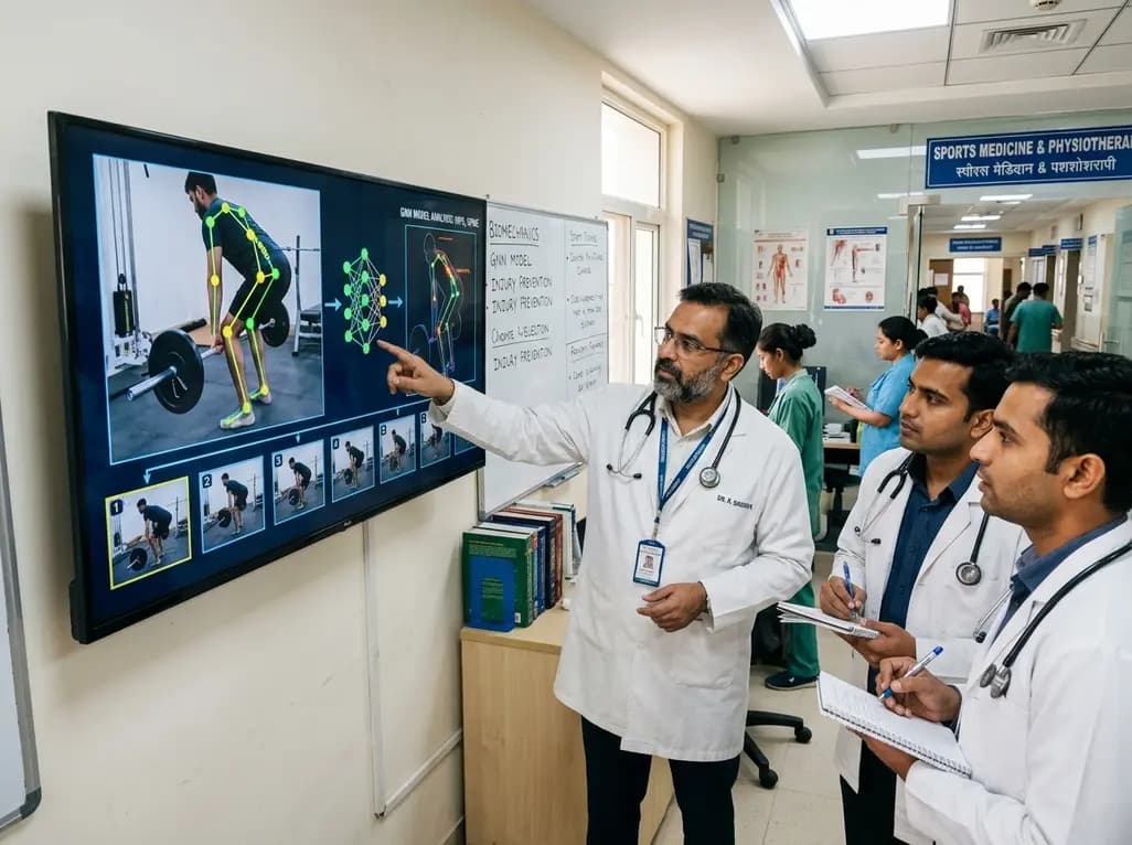
Precision deadlift form assessment represents an essential component of sports medicine and orthopedic injury prevention programs. Strength training athletes frequently execute the deadlift to enhance posterior chain power and systemic musculoskeletal endurance. However, incorrect biomechanical execution imposes extreme shearing forces and compressive loads across the lumbar intervertebral discs. When lifters lose spinal neutral positioning, excessive flexion dramatically magnifies stress on passive ligamentous structures and the lumbosacral junction. Consequently, subtle technical errors during heavy barbell lifts can rapidly trigger disc herniations, severe muscle strains, or debilitating facet joint irritation.
Automated video analysis offers an objective methodology to evaluate these dynamic movement patterns without relying solely on subjective observation. Unfortunately, traditional monocular RGB video capture presents profound technical hurdles for accurate analysis. Standard video streams produce massive volumes of redundant data, obscuring the brief, critical moments where technical breakdown occurs. Furthermore, conventional algorithms often struggle to distinguish minor biomechanical deviations from natural human movement variability. Therefore, developing precise, real-time diagnostic tools is necessary to safeguard athletes from catastrophic spinal trauma while maximizing athletic potential. Additionally, sports physicians require scalable technologies that detect harmful kinematics across diverse clinical and athletic environments.
To overcome high computational demands, investigators developed an innovative framework utilizing a modified Search-Map-Search algorithm for frame selection. Typical high-frame-rate video feeds contain hundreds of static or repetitive frames that overwhelm commodity hardware processors. In contrast, this novel approach systematically extracts only six representative frames per repetition. As a result, the algorithm reduces redundant video frames by an astounding 89.8% without discarding crucial kinematic waypoints. These preserved frames capture the precise lift phases, including initial barbell lift-off, knee clearance, and final hip lockout.
Moreover, reducing video input volume drastically diminishes computational processing latency. Clinical motion laboratories frequently rely on bulky multi-camera arrays and lengthy offline data processing. Conversely, this sparse methodology processes kinematic information rapidly on standard graphics hardware. The graph classification stage finishes processing in merely 0.4 seconds, whereas the complete end-to-end pipeline operates in roughly 0.8 seconds. Hence, practitioners can deliver immediate real-time kinematic feedback to lifters before dangerous movement habits solidify. Consequently, this engineering advancement bridges the longstanding divide between complex laboratory biomechanics and accessible everyday clinical practice. Furthermore, streamlining data processing enables sports medicine clinicians to evaluate high-volume patient cohorts without expensive computing infrastructure.
In addition to sparse sampling, the system leverages sophisticated graph-based deep learning models to evaluate skeletal joint relationships. Traditional convolutional neural networks treat human movement as simple grids of pixels, frequently ignoring anatomical constraints. Conversely, Graph Convolutional Networks model the human skeleton as interconnected graphs, where nodes represent joints and edges represent skeletal segments. Consequently, this structural representation allows the artificial intelligence model to track spatial and temporal dynamics with remarkable precision.
Specifically, the proposed Temporal Graph Convolutional Network achieved an impressive 89.5% accuracy in distinguishing correct lifting technique from faulty execution. The model exhibited high precision and recall, demonstrating reliable identification of subtle postural faults. Furthermore, researchers evaluated a compact Spatio-Temporal Graph Convolutional Network variant containing only 4,514 parameters. Remarkably, this ultra-lightweight architecture maintained an 85.3% diagnostic accuracy despite its minimal computational footprint. Therefore, these compact models operate seamlessly on consumer-grade mobile devices and standard monocular RGB cameras. By mapping biomechanical coordinates across time, graph architectures provide clinicians with robust algorithmic tools that mirror human kinematic expertise. Additionally, certified sports specialists rigorously annotated the underlying training dataset, ensuring that the model reflects verified clinical movement standards.
From an orthopedic standpoint, automated kinetic analysis offers profound benefits for mitigating spinal trauma among resistance training athletes. During the deadlift, the lumbar spine endures substantial compressive stresses reaching thousands of Newtons alongside dangerous shear loads. If an athlete rounds their lower back under load, mechanical forces shift away from active muscles onto passive spinal ligaments. Consequently, repetitive lumbar flexion under high loads accelerates intervertebral disc degeneration and predisposes athletes to acute posterior disc herniations.
Furthermore, improper hip hinge mechanics frequently induce compensatory pelvic tilt, aggravating sacroiliac joint dysfunction and secondary spinal instability. By identifying these biomechanical flaws during the earliest repetitions of a training set, automated monitoring prevents cumulative microtrauma. Sports medicine physicians can utilize these diagnostic insights to identify movement compensations caused by hip mobility deficits or weak core stabilizers. As a result, clinicians can prescribe targeted neuromuscular rehabilitation drills before tissue failure occurs. Ultimately, continuous kinematic screening transforms athletic injury prevention from a reactive treatment model into an objective, proactive paradigm. Moreover, providing objective kinematic validation empowers physical therapists to guide post-injury rehabilitation programs with unprecedented anatomical precision.
The integration of commodity RGB hardware and graph neural networks represents a transformative breakthrough for outpatient physical therapy and athletic coaching. Historically, rigorous biomechanical assessment required expensive optoelectronic motion capture laboratories, limiting accessibility for Indian athletes and community clinics. However, this lightweight framework operates successfully using standard smartphone video or single-lens webcams. Therefore, physiotherapists and sports physicians can easily deploy automated movement screening during routine outpatient consultations.
Additionally, remote telerehabilitation platforms can incorporate these algorithms to monitor patients performing strength exercises at home. Patients receive instant audiovisual alerts whenever their posture deteriorates, substantially reducing the risk of unsupervised exercise injury. Furthermore, coaches and clinicians gain longitudinal movement data, allowing them to track functional motor improvements across comprehensive rehabilitation cycles. Because the model executes within 0.8 seconds, athletes receive real-time biofeedback without disruptive training delays. Consequently, accessible computer vision democratizes advanced biomechanical assessment, elevating sports medicine care across both elite sports academies and grassroots fitness facilities. Moreover, orthopedists can utilize these quantitative movement metrics to establish objective return-to-play criteria following spinal or lower limb trauma. In conclusion, combining sparse frame extraction with graph learning establishes a new standard for functional movement screening and athlete safety.
Automated movement analysis identifies subtle technical errors, such as premature lumbar flexion and improper hip hinge mechanics, before severe spinal trauma develops. By continuously tracking joint coordinates, the algorithm detects dangerous shear loading across lumbosacral discs. Consequently, lifters receive immediate corrective feedback, preventing repetitive spinal microtrauma, muscle strain, and disc herniation. This objective evaluation enables athletes and clinicians to preserve neutral spinal alignment throughout heavy resistance training sessions.
Standard video capture produces substantial redundant data that burdens computing processors without adding diagnostic value. In contrast, sparse frame selection extracts only six critical kinematic waypoints per repetition, reducing frame volume by 89.8%. This targeted approach preserves essential biomechanical information, including barbell lift-off and lockout phases. Furthermore, minimizing data volume enables rapid processing on standard mobile hardware, ensuring real-time feedback for athletes and clinical practitioners.
While multi-camera optoelectronic systems remain the reference standard in biomechanics laboratories, lightweight graph convolutional networks bridge the accessibility gap. Advanced artificial intelligence accurately estimates three-dimensional skeletal coordinates from single-lens two-dimensional video. Consequently, commodity RGB cameras provide clinical-grade kinematic assessment at minimal expense. This technological progression allows grassroots athletic centers, physiotherapy clinics, and telemedicine providers to implement reliable, objective movement screening without investing in expensive laboratory motion infrastructure.
Disclaimer: This content is for informational and educational purposes only... Refer to the latest local and national guidelines for clinical practice.
References

Read summarized clinical updates, watch expert medical content, and earn CME certifications right from your smartphone.
Open your smartphone camera to scan and install.





A groundbreaking study in Scientific Reports presents a lightweight graph neural network that performs accurate deadlift form assessment using only six video frames, reducing computational load while preserving key biomechanical data for musculoskeletal injury prevention.
Today

A breakthrough study reveals that propionyl-CoA catabolism acts as a vital metabolic gatekeeper for fatty acid oxidation in pancreatic ductal adenocarcinoma. By preventing toxic metabolite accumulation and protecting mitochondrial enzymes, this pathway exposes critical therapeutic targets for oncology.
Today
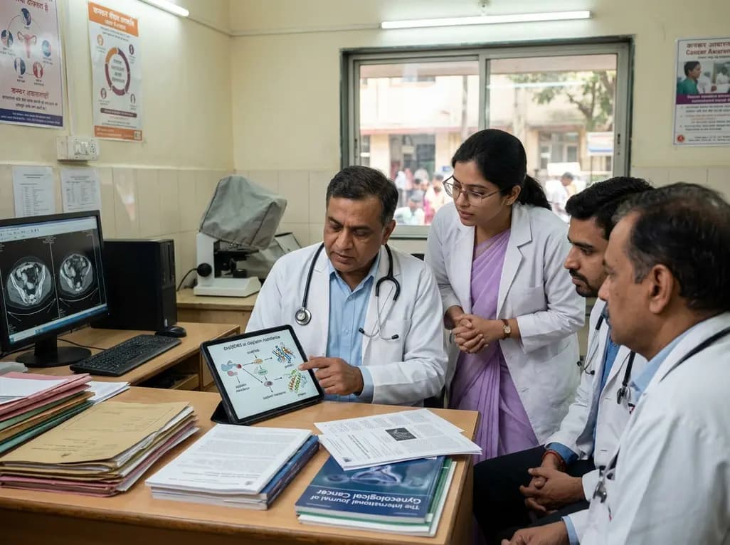
A landmark study reveals that circMCM3 promotes cisplatin resistance in cervical squamous cell carcinoma by recruiting USP49 to stabilize PTBP1 and CCND1. Targeting this newly identified molecular axis presents a promising therapeutic avenue to restore chemotherapy sensitivity and improve gynaecological outcomes.
Today

A 24-week trial shows that patients starting amlodipine/benazepril maintain stable blood pressure across all seasons, whereas those on benazepril/hydrochlorothiazide require significantly more up-titration when initiated in winter (18.9% vs. 5.0%), highlighting amlodipine's superior seasonal hemodynamic stability.
Today

A rare case of a pediatric pathological femoral fracture caused by occult telangiectatic osteosarcoma demonstrates the risks of misdiagnosis after trauma, where intramedullary nailing prompted iatrogenic tumor dissemination and required modular total endoprosthetic reconstruction.
Today
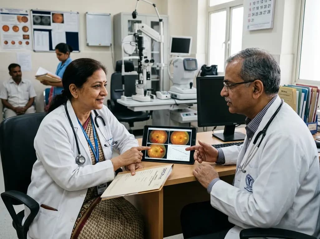
A comprehensive analysis of DRCRnet studies reveals that 15.1% of participants discontinue diabetic retinopathy clinical trials. Identifying baseline demographic and clinical predictors helps ophthalmologists and endocrinologists mitigate real-world treatment dropouts and protect long-term visual outcomes.
Today
