.webp&w=384&q=75)

Loading, please wait...
Loading, please wait...
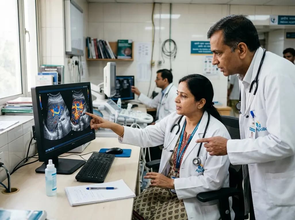
Microvascular perfusion evaluation remains essential across modern diagnostics, cardiovascular risk stratification, and oncology. Contrast-enhanced ultrasound has emerged as a frontline imaging modality because it delivers real-time, non-invasive microvascular visualization without ionizing radiation or nephrotoxic agents. However, conventional manual analysis faces significant hurdles due to operator dependency and massive dynamic cine-loop data volumes. Integrating artificial intelligence addresses these persistent diagnostic bottlenecks. Today, AI-assisted CEUS transforms dynamic perfusion interpretation into an objective, standardized, and highly reproducible clinical workflow.
Contrast-enhanced ultrasound relies on gas-filled microbubble contrast agents that resonate under low mechanical index acoustic fields. Consequently, this acoustic response yields exquisite microvascular signal enhancement while suppressing background tissue signals. Clinicians routinely employ this technology to evaluate tissue perfusion dynamics across multiple organs. For instance, CEUS allows real-time observation of arterial wash-in, peak enhancement, and capillary clearance kinetics. These dynamic characteristics provide indispensable physiological insight into tumor neoangiogenesis, plaque vulnerability, and parenchymal perfusion deficits.
Despite these distinct advantages, traditional image review encounters substantial operational bottlenecks. A standard examination generates thousands of dynamic video frames across diverse perfusion phases. Therefore, manual frame-by-frame scrutiny consumes substantial clinical time and demands exceptional sonographer expertise. Furthermore, qualitative visual inspection inevitably introduces high inter-observer variability, which impairs diagnostic reproducibility. Clinicians often struggle to extract stable quantitative parameters, such as time-to-peak intensity and mean transit time, amidst respiratory motion. As a result, subtle perfusion abnormalities might remain undetected during routine visual evaluations.
The structured workflow of AI-assisted CEUS integrates several sophisticated computational stages to convert raw sonographic cine-loops into actionable clinical metrics. First, automated data pre-processing mitigates acoustic artifacts and standardizes brightness variations across different scanner platforms. Advanced deep learning models perform non-rigid motion compensation to correct for physiological tissue displacement caused by patient breathing. Consequently, stabilized image sequences ensure that subsequent temporal analyses remain highly reliable and precise.
Second, deep neural networks execute automated region-of-interest segmentation to isolate target lesions from surrounding healthy tissue. Fully convolutional networks and transformer architectures delineate tumor margins and vascular walls with remarkable precision. Subsequently, quantitative algorithms extract standardized radiomic and spatiotemporal features from dynamic time-intensity curves. Machine learning classifiers synthesize these complex temporal signatures alongside clinical metadata to deliver calibrated risk predictions. Hence, this automated end-to-end framework reduces manual workload, accelerates diagnostic decision-making, and establishes uniform analytical standards across healthcare institutions.
Carotid atherosclerotic plaque rupture represents a leading cause of ischemic cerebrovascular events worldwide. Vulnerable plaques exhibit extensive intraplaque neovascularization, which correlates directly with hemorrhage, structural instability, and subsequent embolization. Contrast-enhanced ultrasound clearly visualizes these fragile microvessels within the plaque core. However, visually quantifying adventitial microvascular density remains technically demanding for sonographers due to rapid arterial pulsatile motion.
Fortunately, modern machine learning algorithms quantify intraplaque neovascular perfusion with outstanding precision. Neural networks track dynamic microbubble ingress within the vessel wall throughout the cardiac cycle. Furthermore, AI models compute quantitative perfusion indices that correlate robustly with histological microvessel counts. By synthesizing plaque morphology, echogenicity, and microvascular flow dynamics, algorithmic models accurately distinguish stable fibroatheromas from high-risk vulnerable lesions. Consequently, clinicians can effectively stratify patient stroke risk and tailor aggressive lipid-lowering therapies or surgical revascularization strategies prior to catastrophic neurological events.
In hepatic oncology, contrast-enhanced ultrasound plays an indispensable role in distinguishing benign lesions from hepatocellular carcinoma and metastatic disease. Malignant liver tumors display distinctive hyperenhancement during the early arterial phase, followed by rapid portal venous or delayed phase washout. Nevertheless, atypical vascular patterns frequently obscure manual differential diagnosis, especially in cirrhotic backgrounds.
Artificial intelligence enhances hepatic lesion characterization by tracking subtle kinetic shifts across multi-phase cine recordings. Deep neural networks analyze microvascular wash-in and wash-out kinetics, detecting microperfusion abnormalities that sonographers might overlook. In addition, AI-driven radiomics models evaluate intratumoral heterogeneity to predict microvascular invasion and histological tumor grade preoperatively. Furthermore, post-therapeutic monitoring benefits substantially from automated perfusion quantification. Algorithms reliably measure residual viable tumor perfusion following transarterial chemoembolization or thermal ablation. Therefore, AI-assisted CEUS empowers oncology teams to evaluate therapeutic response early, optimize salvage interventions, and improve long-term oncological survival outcomes.
Beyond cardiovascular and hepatic imaging, AI-assisted CEUS demonstrates expanding diagnostic utility across diverse organ systems. In renal medicine, deep learning models analyze cortical perfusion kinetics to detect acute kidney injury and characterize complex cystic renal masses. Specifically, automated microvascular perfusion quantification helps clinicians differentiate benign oncocytomas from malignant clear-cell renal cell carcinomas without exposing patients to nephrotoxic iodinated agents.
Similarly, breast and thyroid imaging benefit from algorithmic microvascular assessments. Malignant nodules frequently display chaotic peripheral vascularization and uneven internal perfusion. AI algorithms extract spatial vascular tortuosity and peak arrival time parameters, thereby upgrading the diagnostic specificity of standard ultrasound guidelines. In emergency care and gastrointestinal medicine, automated CEUS platforms quantify transmural microvascular perfusion to differentiate active inflammatory bowel disease flares from chronic fibrotic strictures. As these algorithmic architectures mature, point-of-care ultrasound devices will increasingly incorporate automated perfusion tools to guide rapid bedside triage.
Although AI-assisted CEUS shows immense clinical potential, several technical and translational hurdles require careful resolution before widespread clinical adoption. Most existing algorithmic architectures rely on single-center retrospective cohorts, which limits model generalizability across diverse patient populations. Moreover, variations in ultrasound hardware, probe frequencies, and contrast agent formulations create substantial domain shifts that compromise algorithm performance across different hospital systems.
To address these barriers, research teams must prioritize multi-center prospective validation trials and establish open-access, multi-vendor benchmark databases. Additionally, developers must create explainable AI frameworks that visually display hemodynamic feature maps to foster physician trust. Integrating real-time algorithmic feedback directly onto bedside ultrasound consoles will streamline sonographer workflow without prolonging scan times. Ultimately, standardizing ethical oversight, data security protocols, and regulatory validation pathways will accelerate the routine implementation of AI-assisted CEUS across global medical practices.
Conventional contrast-enhanced ultrasound relies heavily on subjective visual interpretation, which introduces inter-observer variability and consumes significant clinical time during manual frame analysis. In contrast, AI-assisted CEUS automates motion correction, lesion segmentation, and quantitative feature extraction from dynamic cine loops. Consequently, it calculates standardized perfusion parameters, reduces diagnostic errors, and enhances overall workflow efficiency, enabling clinicians to make faster, highly reproducible bedside decisions across diverse clinical settings.
Radiology, cardiology, oncology, gastroenterology, and vascular surgery benefit substantially from this technology. Radiologists and hepatologists utilize AI-assisted CEUS to characterize focal liver lesions and monitor tumor ablation response. Furthermore, vascular specialists and cardiologists rely on automated microvascular quantification to stratify carotid plaque vulnerability and evaluate myocardial perfusion. Nephrologists and general surgeons also apply these algorithmic workflows to assess renal perfusion and detect organ ischemia promptly.
The primary hurdles include limited external generalizability across diverse ultrasound platforms, variation in contrast agent kinetics, and the scarcity of large multi-center training datasets. Additionally, many deep learning architectures function as non-transparent black boxes, which can hinder clinical trust. Overcoming these barriers requires standardized multi-vendor validation trials, robust regulatory compliance frameworks, and explainable artificial intelligence interfaces that seamlessly integrate into real-time sonographic equipment.
Disclaimer: This content is for informational and educational purposes only and does not constitute medical advice or substitute for professional clinical judgment. Diagnostic and therapeutic decisions must be individualized according to each patient's specific presentation, medical history, and clinical context. Refer to the latest local and national guidelines for clinical practice.
References
Tang T et al. Artificial Intelligence-Assisted Contrast-Enhanced Ultrasound for Perfusion Evaluation: State-of-the-Art Review. Ultrasound Med Biol. 2026 Aug 15. doi: undefined. PMID: 42603764.
Kagiyama N, et al. Artificial Intelligence-Enhanced Cardiac Point-of-Care Ultrasound: A Prospective Multi-Center Clinical Evaluation. J Am Soc Echocardiogr. 2025;38(4):412-421.
Liu D, et al. Accurate prediction of responses to transarterial chemoembolization for patients with hepatocellular carcinoma by using artificial intelligence in contrast-enhanced ultrasound. Eur Radiol. 2020;30(9):5155-5165.

Read summarized clinical updates, watch expert medical content, and earn CME certifications right from your smartphone.
Open your smartphone camera to scan and install.





This state-of-the-art review details how artificial intelligence enhances contrast-enhanced ultrasound for microvascular perfusion evaluation. AI automates data processing, segmentation, and quantitative analysis, significantly improving diagnostic accuracy in carotid plaques, liver tumors, and multi-organ diseases.
last month

A landmark cluster randomized trial demonstrates that community-based mass dog vaccination achieves superior coverage and sustains protective herd immunity against rabies far better than conventional campaign models, offering vital lessons for global and Indian elimination targets.
Today
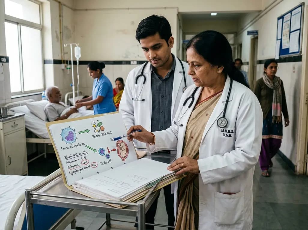
Explore the crucial link between dietary protein and immunocompetence. This review examines how low-protein nutrition shifts metabolic resources away from pathogen defense toward early reproduction, compromising host survival and immunity against severe bacterial infections.
Today

Differential oxygenation during peripheral TA-NRP can cause undetected coronary hypoxemia due to arch vessel clamping. A novel circuit with continuous aortic root monitoring detects desaturation early, facilitating left ventricular unloading and preventing allograft dysfunction and unnecessary heart discard.
Today

A longitudinal study reveals that patients undergoing staged bilateral hip arthroscopy experience progressive contralateral labral deterioration during surgical intervals. However, structural degradation does not compromise 2-year postoperative clinical outcomes or patient-reported satisfaction scores.
Today

A post hoc analysis of the PARM-T2D study shows pemafibrate exerts baseline-dependent effects on LDL-C and particle size in type 2 diabetes and hypertriglyceridemia. It lowers triglycerides and remnants while favorably remodeling atherogenic LDL particles based on initial lipid control status.
Today
