.webp&w=384&q=75)

Loading, please wait...
Loading, please wait...
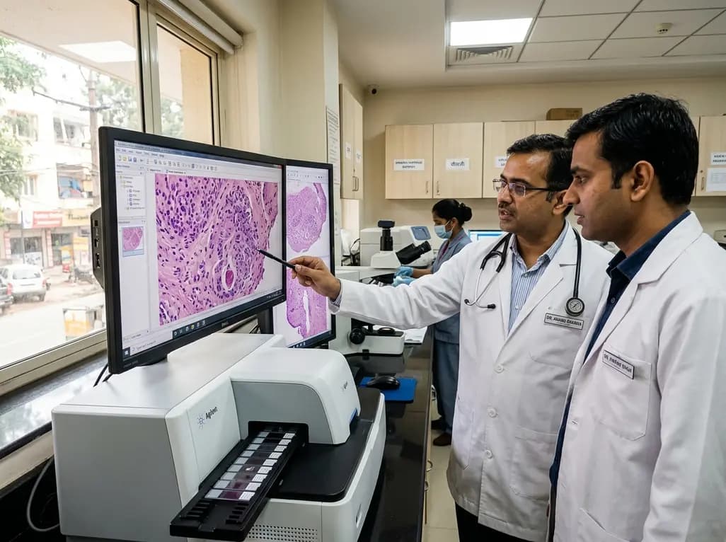
In an era of rapid diagnostic transformation, laboratory medicine is shifting toward automated, computational ecosystems. Agilent Technologies has entered into a definitive agreement to acquire digital pathology software pioneer Proscia for 240 million dollars in cash, alongside up to 30 million dollars in performance milestones. Consequently, this transaction positions Agilent at the forefront of AI-enabled histopathology and tissue-based diagnostics. Modern laboratories face severe staffing constraints and rising specimen volumes. Therefore, the integration of enterprise-grade software with automated staining platforms marks a pivotal shift for clinical diagnostic teams worldwide.
The pathology sector experiences relentless pressure to enhance throughput without compromising analytical rigor. By integrating advanced digital pathology software into its established diagnostic portfolio, Agilent directly addresses the technical bottlenecks that impede conventional glass-slide workflows. Proscia developed its flagship Concentriq platform to serve as an intelligent, vendor-agnostic operating system for clinical and life science laboratories. Furthermore, the platform securely links digital slide scanners, laboratory information systems, and computational algorithms within a centralized workspace. Because the software remains vendor-neutral, diagnostic centers can connect disparate hardware seamlessly. As a result, laboratory directors can modernize existing facilities without replacing their capital scanning investments. In addition, the software facilitates instant remote consultation and centralized diagnostic triaging across multisite health networks. Such operational agility ensures that complex biopsy specimens reach the appropriate subspecialist without shipping physical slides. Ultimately, this acquisition provides clinical teams with scalable computational infrastructure designed to accelerate diagnostic decision-making.
Historically, diagnostic pathology relied heavily on manual microscopy and mechanical glass-slide management. However, physical glass slides present inherent risks, including breakage, staining variability, and protracted transit times between facilities. In contrast, comprehensive enterprise platforms transform high-resolution whole slide images into accessible, searchable diagnostic data. Furthermore, the combined ecosystem enables pathologists to view, annotate, and analyze tissue morphology through high-definition digital interfaces. Laboratory information systems synchronize patient demographic information directly with digitized histology sections. Consequently, diagnostic teams eliminate manual specimen entry errors and enhance specimen traceability throughout the diagnostic pathway. Moreover, automated image management supports seamless case archiving and rapid retrieval for multidisciplinary tumor boards. Clinicians no longer waste valuable time searching through physical storage archives to compare serial biopsies. In addition, integrated software environments allow laboratories to balance daily caseloads evenly among available staff. Pathologists can thus collaborate across regional medical centers to deliver rapid, standardized interpretations for urgent clinical presentations.
A worldwide deficit in trained pathologists threatens diagnostic turnaround times and patient outcomes. Meanwhile, cancer incidence rates continue to rise steadily across aging global populations. Pathologists routinely navigate heavy cognitive burdens while reviewing hundreds of tissue slides each working day. Therefore, digital solutions offer vital relief by organizing complex diagnostic queues according to clinical urgency. In addition, automated algorithms can perform initial slide triage by flagging suspicious cellular features and suspicious margins. Pathologists then prioritize suspected malignant cases before addressing routine screening specimens. Similarly, artificial intelligence algorithms eliminate tedious manual quantification tasks, such as counting mitotic figures or estimating necrosis percentages. Consequently, specialists can devote more time and clinical focus to interpreting ambiguous morphological patterns. Studies confirm that digital workflows significantly reduce overall turnaround times for surgical pathology specimens. Thus, laboratories maintain high diagnostic accuracy even when managing elevated specimen volumes under constrained human resource conditions.
Contemporary cancer therapies depend strictly on accurate biomarker quantification and phenotypic subtyping. However, manual estimation of immunohistochemical expression often yields inter-observer variability among assessing clinicians. For example, quantifying markers such as human epidermal growth factor receptor 2, estrogen receptors, and Ki-67 requires rigorous numerical accuracy. In this regard, computational pathology tools provide objective, highly reproducible pixel-level scoring across digitized tumor microenvironments. Pathologists employ validated algorithms to identify tumor boundaries and evaluate biomarker expression patterns with unprecedented precision. Furthermore, computational intelligence helps clinicians evaluate programmed death-ligand 1 scores to guide immunotherapy selection. As targeted oncology regimens proliferate, oncologists require reliable quantitative data to tailor systemic therapeutic decisions. Integrated software platforms effectively bridge the gap between initial tissue preparation, computational biomarker quantification, and actionable therapeutic guidance. Consequently, treating oncologists receive detailed, standardized diagnostic reports that enable timely, individualized cancer therapy interventions.
In developing healthcare ecosystems like India, specialist oncopathologists remain concentrated primarily within Tier-1 metropolitan centers. Conversely, patients across Tier-2 and Tier-3 cities frequently face prolonged delays when seeking definitive histopathological diagnoses. Therefore, adopting interoperable digital pathology frameworks offers tremendous clinical utility across Indian diagnostic networks. Regional laboratories can scan biopsy specimens locally and transfer digital whole slide images to central academic institutions instantly. Moreover, this telepathology infrastructure eliminates the delays and logistics costs associated with shipping physical tissue blocks across distant states. Telepathology networks also facilitate rapid intraoperative frozen-section consultations during oncological surgeries. As a result, operating surgical teams can determine clean surgical margins in real time, preventing repeat surgical resections. In addition, Indian diagnostic chains can leverage centralized artificial intelligence tools to screen high-volume cervical and oral biopsies efficiently. Through open software platforms, Indian healthcare providers can substantially democratize access to world-class pathology expertise.
Q1: What are the main diagnostic benefits of digital pathology systems?
Digital pathology platforms replace traditional manual glass-slide handling with secure whole slide imaging workflows. Consequently, these systems streamline case distribution, accelerate diagnostic turnaround times, and eliminate the risk of slide breakage. Furthermore, they enable real-time collaboration among distant subspecialists, support integrated artificial intelligence algorithms, and improve measurement accuracy for critical prognostic biomarkers.
Q2: How do artificial intelligence algorithms support practicing pathologists during routine review?
Artificial intelligence algorithms assist pathologists by automating repetitive quantitative tasks, such as assessing cellular proliferation indices and biomarker expression. Additionally, these computational tools can prescreen slides to triage suspicious malignant lesions for urgent evaluation. Pathologists retain full diagnostic oversight while leveraging computational tools to improve diagnostic precision and reduce cognitive fatigue.
Q3: How does enterprise digital software improve cancer diagnosis in underserved regions?
Enterprise software connects peripheral diagnostic laboratories directly with central tertiary cancer centers through secure digital telepathology networks. Consequently, patients in remote or rural communities receive subspecialty expert second opinions without traveling long distances. This approach expedites critical treatment initiation and promotes equal access to advanced cancer diagnostics across diverse geographic populations.
Disclaimer: This content is for informational and educational purposes only. It does not constitute medical advice or replace professional judgment. Refer to the latest local and national guidelines for clinical practice.
References

Read summarized clinical updates, watch expert medical content, and earn CME certifications right from your smartphone.
Open your smartphone camera to scan and install.





Agilent Technologies announced an agreement to acquire digital pathology enterprise leader Proscia for up to $270 million. The strategic move unites laboratory hardware, reagents, and AI algorithms to streamline clinical workflows, reduce diagnostic turnaround times, and expand precision oncology globally.
Today

A comprehensive critical appraisal of the science-to-practice gap in allergology, detailing systemic guideline adherence barriers across ARIA and GINA, health economic disparities, and proven knowledge-translation strategies to optimize real-world patient outcomes.
Today
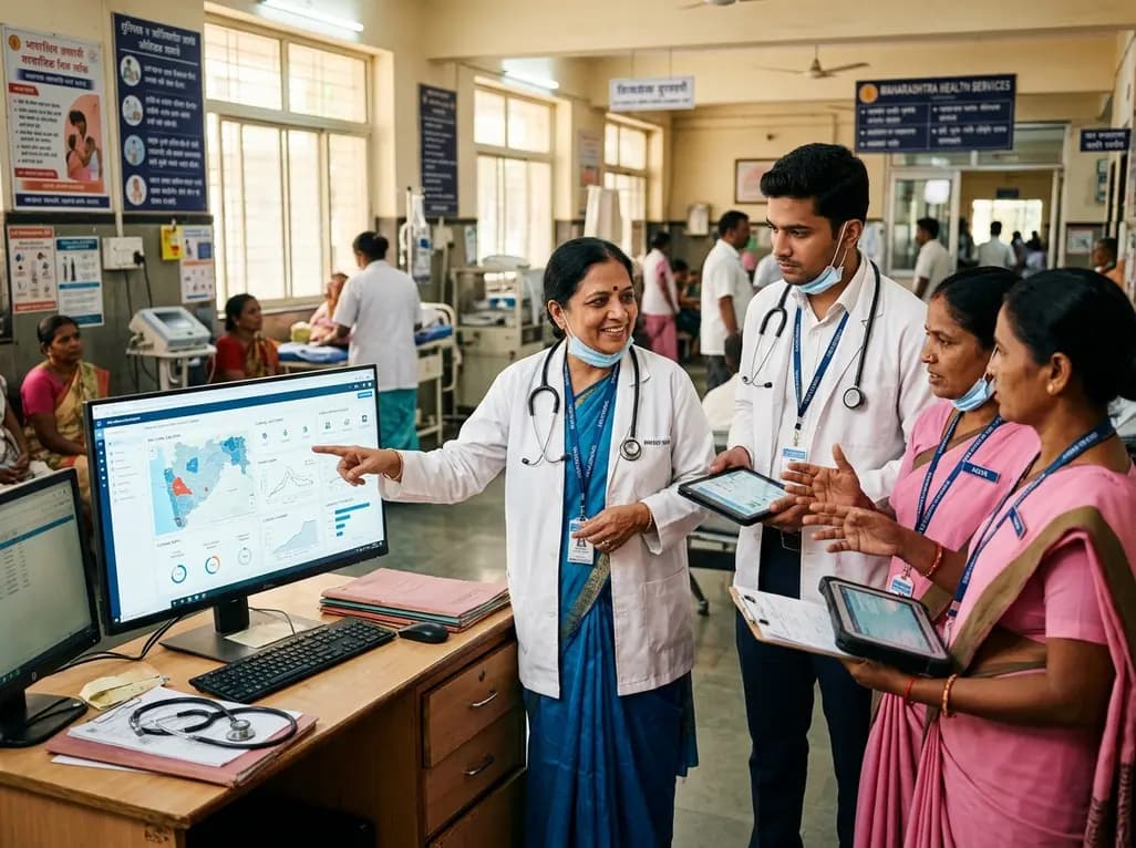
The government of Maharashtra is establishing a novel state-level Living Lab to elevate clinical outcomes, bridge public-private service gaps, and test real-world health interventions. By integrating digital innovations and frontline feedback, the platform fosters sustainable, equitable care models across communities.
Today
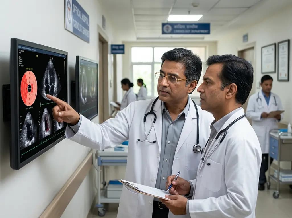
Cardiac involvement is the leading cause of mortality in EGPA. This report highlights an overlap phenotype of lymphocyte-predominant myocardial injury, Takotsubo-like cardiomyopathy, and left ventricular thrombus, emphasizing early multimodality diagnostics and aggressive immunosuppressive biologic therapy.
Today

A landmark biomechanical study demonstrates that sports bra support and running shoe cushioning interact significantly to influence tibial acceleration, breast motion, and comfort in female runners, providing crucial insights for clinical counseling in sports medicine and women's health.
Yesterday
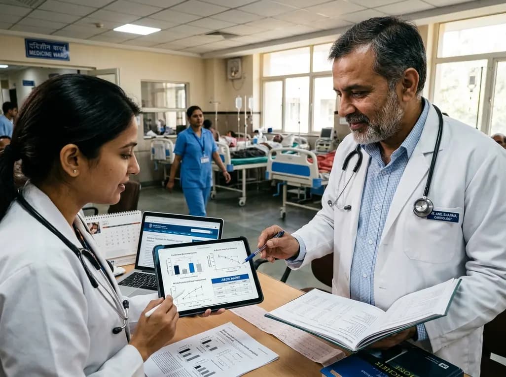
A systematic review and meta-analysis of 5,406 participants reveals that tirzepatide significantly reduces hsCRP by 34.3%, improves cardiorenal biomarkers, and enhances functional capacity and exercise endurance in patients with heart failure.
Today
