.webp&w=384&q=75)

Loading, please wait...
Loading, please wait...
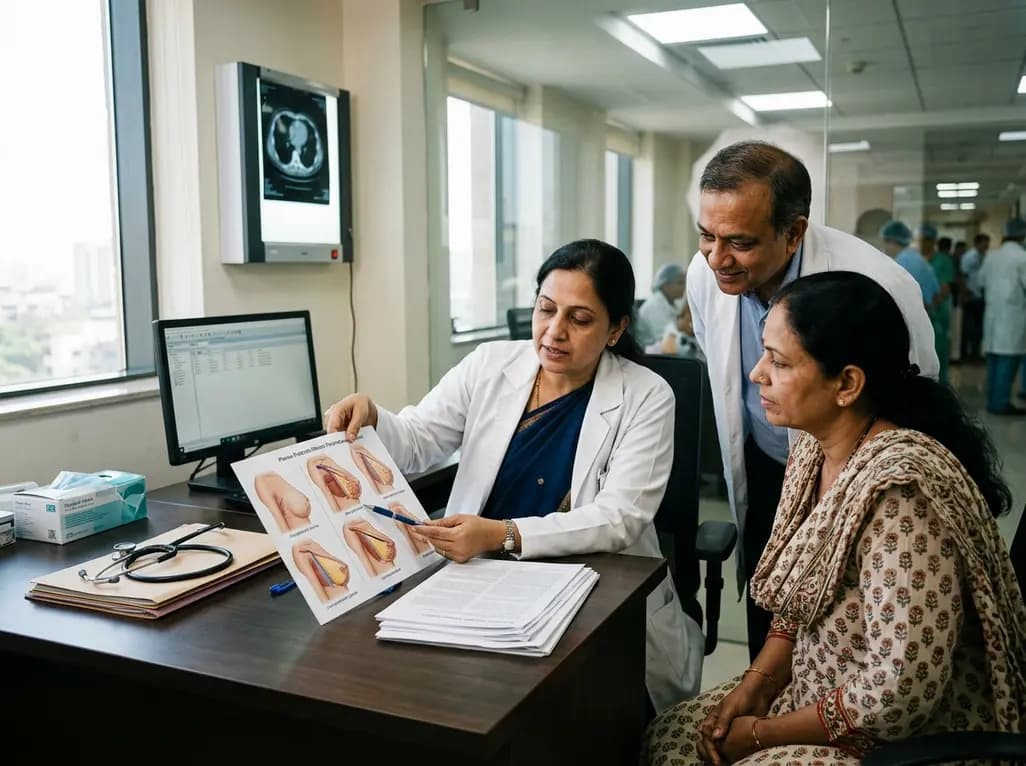
Modern oncologic surgery increasingly balances curative resection with long-term quality of life. Consequently, implant-based breast reconstruction has become the dominant restorative approach following mastectomy worldwide. Concurrently, post-mastectomy radiation therapy significantly lowers locoregional recurrence and prolongs overall survival among high-risk patients. However, ionizing radiation delivers significant physical insults to vulnerable cutaneous and subcutaneous tissues. Radiation induces microvascular thrombosis, chronic inflammatory cascades, and progressive dermal fibrosis. As a result, combining radiotherapy with silicone or saline prostheses creates complex clinical trade-offs. Surgeons and radiation oncologists must weigh oncologic safety against aesthetic failure, chronic pain, and capsular contracture. Historically, many clinical centers viewed radiation as a relative contraindication to immediate prosthetic reconstruction. Nevertheless, modern refinements in surgical technique and radiation physics have fundamentally reshaped this clinical landscape. Clinicians now deploy sophisticated algorithms to preserve anatomical symmetry and maintain reconstructive integrity. Furthermore, multidisciplinary collaboration enables personalized surgical pathways tailored to patient risk profiles. Understanding tissue biology under radiation stress remains vital for every surgical team. Therefore, optimizing procedural timing, biomaterial choice, and anatomical planes represents a critical frontier in modern breast cancer care.
The timing of prosthetic placement represents one of the most vigorously debated decisions in surgical oncology. Surgeons generally choose between immediate direct-to-implant reconstruction, two-stage expander-implant approaches, and delayed reconstructions. Immediate reconstruction preserves the natural skin envelope and provides immediate psychological benefits for the patient. However, delivering radiotherapy to an immediate permanent implant substantially elevates the risk of severe capsular contracture. Furthermore, irradiated tissues exhibit higher rates of infection, skin necrosis, and premature reconstructive failure. In contrast, two-stage procedures allow surgeons to insert a temporary tissue expander during initial oncologic clearance. Radiation oncologists then deliver post-mastectomy radiation therapy either to the inflated expander or to the definitive implant after exchange. Notably, radiating the tissue expander prior to implant exchange protects the definitive device from direct radiation exposure. Consequently, surgeons can perform capsulectomies and exchange damaged tissues during the second operation. Nevertheless, radiation to the expander increases perioperative infection rates during subsequent expansion cycles. Delayed reconstruction avoids radiating prosthetic components entirely by deferring reconstruction until several months after radiotherapy completion. Therefore, multidisciplinary teams must individualize timing based on pathologic staging, patient anatomy, and adjuvant therapy schedules.
The surgical plane selected for device placement significantly influences both mechanical stability and aesthetic durability under radiation. Historically, subpectoral placement served as the gold standard because the pectoralis major muscle provided robust soft-tissue coverage. This muscular barrier protected the underlying device from superficial wound dehiscence and impending extrusion. However, submuscular dissection often causes substantial postoperative animation deformities, chronic chest tightness, and muscle spasm. In addition, irradiating the mobilized pectoralis muscle frequently induces progressive muscular fibrosis and severe contracture. To overcome these distinct morbidities, surgeons have enthusiastically revived prepectoral reconstruction over the past decade. In this technique, the surgeon positions the prosthesis directly over the pectoralis fascia, completely avoiding muscular disruption. Consequently, patients report less postoperative discomfort and completely avoid debilitating dynamic breast distortion. Recent clinical investigations indicate that prepectoral reconstruction achieves complication rates comparable to subpectoral placement, even with post-mastectomy radiotherapy. Nevertheless, prepectoral reconstruction demands well-vascularized, uniform mastectomy skin flaps to prevent delayed ischemic necrosis. Furthermore, thin cutaneous envelopes without muscular padding show higher susceptibility to visible implant rippling. Thus, reconstructive surgeons must carefully evaluate flap thickness and microvascular perfusion when choosing the anatomical plane.
The introduction of biomaterials has revolutionized prosthetic breast reconstruction, especially within the prepectoral space. Surgeons routinely utilize acellular dermal matrices, synthetic meshes, and autologous fat grafting to reinforce soft-tissue support. Biologic matrices, derived from human, porcine, or bovine dermis, provide structural scaffolding and enhance mechanical projection. Furthermore, these matrices promote rapid neovascularization and host cellular infiltration, mitigating inflammatory capsular thickness. Clinical observations suggest that acellular dermal matrix coverage buffers the implant against radiation-induced shear stress. As a result, matrix reinforcement may reduce the incidence of severe Baker grade capsular contracture. However, incorporating foreign matrices also introduces specific biological risks. In the setting of radiation, seroma formation and delayed integration can predispose patients to periprosthetic infection. Moreover, biomaterial costs remain an important practical consideration in resource-sensitive healthcare environments. Autologous fat grafting provides an invaluable adjunct to biomaterials in irradiated reconstructions. Lipotransfer restores lost subcutaneous volume, improves dermal elasticity, and reverses radiation-damaged soft tissue through adipose-derived stem cells. Therefore, combining judicious matrix coverage with secondary lipofilling represents a highly effective strategy to optimize aesthetic outcomes and tissue health.
Technological innovations in radiation oncology have fundamentally changed how clinicians deliver post-mastectomy radiation therapy. Historically, standard two-dimensional radiotherapy delivered imprecise radiation volumes that caused extensive skin fibrosis and unintended cardiac irradiation. Today, modern centers utilize intensity-modulated radiation therapy, volumetric modulated arc therapy, and deep inspiration breath-hold techniques. Consequently, radiation oncologists can contour target volumes with extraordinary precision while sparing adjacent critical organs. Deep inspiration breath-hold significantly displaces coronary vessels away from the irradiated chest wall. Furthermore, advanced inverse treatment planning ensures uniform dose distribution across complex, curved reconstructed breast contours. This dosimetric homogeneity minimizes hot spots, directly decreasing cutaneous toxicity and lowering reconstructive failure rates. In addition, surface-guided radiation therapy allows real-time tracking of patient positioning, preventing inadvertent geographic misses. Surgical oncologists, plastic surgeons, and radiation oncologists must align their treatment strategies through routine tumor board discussions. Preoperative multidisciplinary evaluation ensures that all specialists coordinate adjuvant therapy sequences, expander volumes, and surgical timelines effectively. Ultimately, close interdisciplinary collaboration ensures robust oncologic control while preserving reconstructive integrity and patient satisfaction.
Ionizing radiation induces microvascular damage, progressive tissue ischemia, and severe cutaneous fibrosis around alloplastic implants. Consequently, irradiated patients experience two- to three-fold higher rates of reconstructive failure compared to non-irradiated individuals. Radiation therapy also significantly increases the incidence of severe capsular contracture, delayed wound breakdown, periprosthetic infection, and unplanned reoperations. Multidisciplinary teams must actively counsel patients regarding these heightened surgical risks prior to determining their final reconstructive pathway.
Current clinical literature demonstrates that prepectoral implant reconstruction achieves acceptable safety profiles comparable to traditional subpectoral techniques under radiation. Positioning the implant prepectorally eliminates dynamic animation deformity, reduces postoperative muscle pain, and preserves chest wall mechanics. However, successful outcomes strictly depend on maintaining robust, well-vascularized mastectomy skin flaps and utilizing complete acellular dermal matrix coverage. Surgeons must carefully assess dermal flap perfusion intraoperatively to prevent progressive flap necrosis and device exposure.
Most reconstructive surgeons recommend waiting at least six to twelve months following completion of radiation therapy before performing permanent implant exchange. This critical waiting interval allows acute radiation dermatitis and microvascular inflammation to subside while capillary perfusion gradually stabilizes. Operating too early in irradiated tissue increases the risk of impaired wound healing, severe infection, and surgical breakdown. Delaying surgery allows the surgeon to perform safer capsulectomies and adjunct autologous fat grafting.
Disclaimer: This content is for informational and educational purposes only. It does not constitute medical advice, diagnosis, or treatment recommendations. Clinical decisions should be guided by individualized patient assessments, institutional protocols, and multidisciplinary tumor board consensus. Refer to the latest local and national guidelines for clinical practice.
References

Read summarized clinical updates, watch expert medical content, and earn CME certifications right from your smartphone.
Open your smartphone camera to scan and install.





A comprehensive clinical review evaluating advances in implant-based breast reconstruction during post-mastectomy radiotherapy, focusing on surgical timing, anatomical planes, biomaterials, and complication mitigation.
Today
A large German population-based cohort study reveals that 43% of post-acute infection syndrome cases are recurrences. Patients who experience new respiratory infections without fully recovering from an earlier episode suffer more protracted symptoms and greater functional impairment.
Today
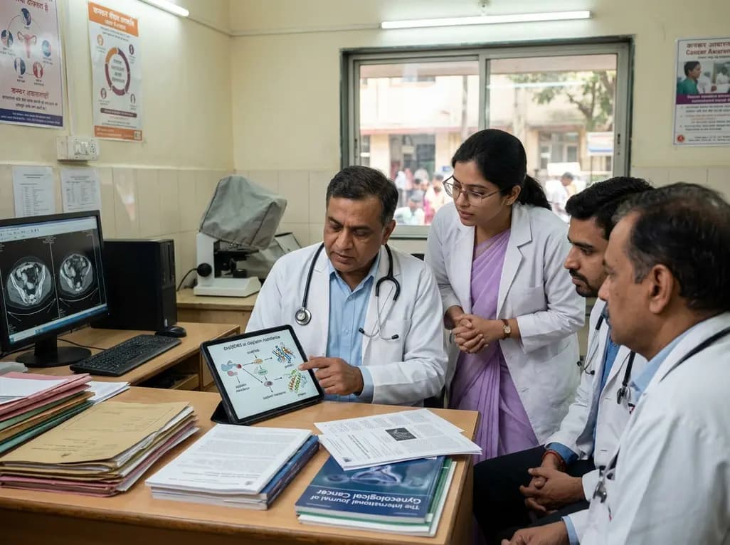
A landmark study reveals that circMCM3 promotes cisplatin resistance in cervical squamous cell carcinoma by recruiting USP49 to stabilize PTBP1 and CCND1. Targeting this newly identified molecular axis presents a promising therapeutic avenue to restore chemotherapy sensitivity and improve gynaecological outcomes.
Today
A 24-week trial shows that patients starting amlodipine/benazepril maintain stable blood pressure across all seasons, whereas those on benazepril/hydrochlorothiazide require significantly more up-titration when initiated in winter (18.9% vs. 5.0%), highlighting amlodipine's superior seasonal hemodynamic stability.
Today

A rare case of a pediatric pathological femoral fracture caused by occult telangiectatic osteosarcoma demonstrates the risks of misdiagnosis after trauma, where intramedullary nailing prompted iatrogenic tumor dissemination and required modular total endoprosthetic reconstruction.
Today
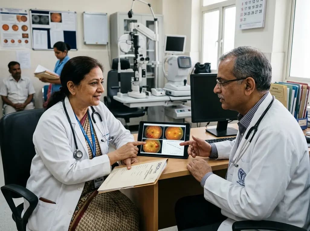
A comprehensive analysis of DRCRnet studies reveals that 15.1% of participants discontinue diabetic retinopathy clinical trials. Identifying baseline demographic and clinical predictors helps ophthalmologists and endocrinologists mitigate real-world treatment dropouts and protect long-term visual outcomes.
Today
