.webp&w=384&q=75)

Loading, please wait...
Loading, please wait...
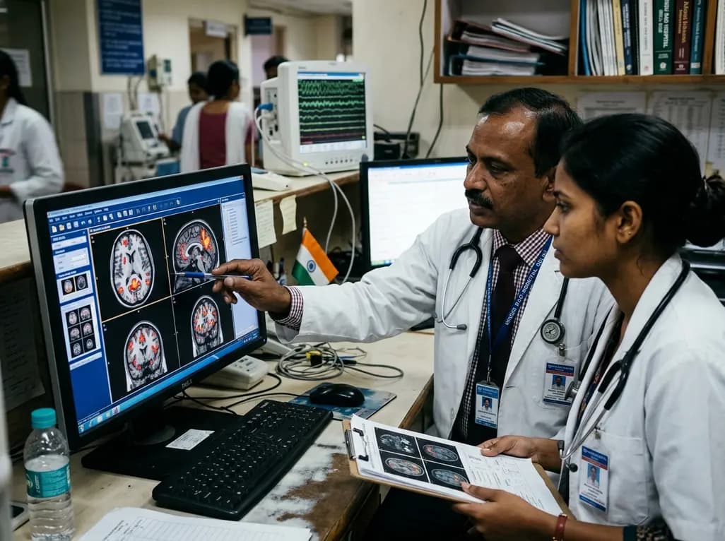
Major depressive disorder in youth frequently presents with severe behavioral complications, among which adolescent non-suicidal self-injury represents an urgent and distressing clinical challenge. Understanding the neurobiological underpinnings of this behavior is essential for improving psychiatric risk stratification and clinical management. While clinicians recognize self-injury as a maladaptive strategy to regulate overwhelming emotional distress, objective neural markers have historically remained difficult to characterize. A recent neuroimaging investigation published in PLoS One offers vital objective insights. By evaluating resting-state functional magnetic resonance imaging, researchers identified distinct functional disruptions that differentiate depressed adolescents engaging in self-injury from those who do not. Consequently, these findings illuminate new avenues for early detection, objective psychiatric assessment, and targeted neuromodulatory interventions.
Non-suicidal self-injury involves deliberate, direct destruction of body tissue without suicidal intent. Common manifestations include intentional cutting, burning, hitting, and skin carving. Epidemiological surveys indicate that nearly 18% of adolescents in community samples report engaging in self-harm. However, within clinical cohorts diagnosed with major depressive disorder, prevalence rates escalate dramatically. Clinicians frequently encounter self-harm as an urgent crisis in pediatric and adolescent psychiatric clinics. Furthermore, the behavior carries substantial psychological morbidity, disrupts educational development, and significantly increases future suicide risk. Despite this immense burden, clinicians often struggle to identify which depressed youth will transition from emotional distress to physical self-injury. Therefore, understanding specific neurobiological mechanisms provides essential guidance for clinical assessment, fostering earlier diagnostic clarity and more targeted therapeutic interventions.
To elucidate the underlying neural architecture, investigators recruited 133 adolescent participants. The cohort comprised 45 adolescents with major depressive disorder who engaged in self-injury, 46 depressed adolescents without self-injury, and 42 age- and sex-matched healthy controls. All participants underwent rigorous resting-state functional magnetic resonance imaging. Researchers initially measured the amplitude of low-frequency fluctuations, which captures regional spontaneous brain activity during rest. Subsequently, regions exhibiting statistically significant group differences served as seed points for functional connectivity analyses across the brain. To ensure clinical validity, researchers deployed a cross-validation prediction framework to link functional brain metrics directly to clinical behavior severity and psychological motivations. Consequently, this analytical strategy ensured that the identified neuroimaging correlates demonstrated strong generalizability.
The functional neuroimaging analysis revealed prominent neurofunctional aberrations localized to key default mode and visual processing regions. Specifically, adolescents diagnosed with depression who engaged in self-injury exhibited significantly increased spontaneous activity in the left precuneus compared to both healthy controls and depressed peers without self-injury. In addition, functional connectivity between the left precuneus and the left cuneus was markedly enhanced. Machine-learning prediction models verified the robustness of these findings, demonstrating highly significant predictive correlations between left precuneus amplitude fluctuations and actual self-injurious behavior. Similarly, functional connectivity between the precuneus and cuneus robustly predicted behavioral severity. Therefore, hyperactive default mode functioning and altered parieto-occipital communication represent critical pathophysiological signatures distinguishing depressed youth engaging in tissue-destructive coping mechanisms.
Beyond identifying structural hubs, the study explored the psychological motivations driving self-injurious actions. In clinical psychiatry, patients frequently report engaging in self-injury to alleviate unbearable emotional turmoil, a psychological mechanism termed intrapersonal negative reinforcement. Notably, elevated functional connectivity between the left precuneus and the left cuneus correlated specifically with negative reinforcement scores. The precuneus plays a foundational role in self-referential processing, autobiographical memory retrieval, and emotional evaluation. Meanwhile, the cuneus participates in visual imagery and integration of external somatosensory inputs. Consequently, heightened connectivity between these regions suggests that intrusive, self-focused negative rumination may trigger intense psychological pain. Physical injury may act as a potent somatosensory disruption that momentarily terminates painful self-reflection, reinforcing repeated maladaptive behavior.
These neuroimaging insights offer profound implications for contemporary adolescent psychiatric care. First, establishing objective brain alterations reduces the social stigma often surrounding self-harm behaviors, framing them as neurobiologically driven symptoms rather than behavioral defiance. Second, resting-state fMRI metrics could eventually serve as objective prognostic biomarkers, helping clinicians identify depressed adolescents at highest risk for self-injury before physical damage occurs. Furthermore, these regional findings delineate clear anatomical targets for novel precision interventions. For instance, non-invasive brain stimulation protocols, such as repetitive transcranial magnetic stimulation targeting parietal default mode nodes, could potentially normalize hyperactive precuneus circuits. Additionally, targeted psychotherapies like dialectical behavior therapy can directly address the identified negative reinforcement pathways.
Managing adolescent self-injury requires a coordinated approach across pediatricians, psychiatrists, clinical psychologists, and primary care physicians. When clinicians understand that aberrant precuneus connectivity drives intense negative rumination, clinical communication can become substantially more empathetic and effective. Healthcare providers should routinely screen depressed adolescents for subtle signs of self-harm, including unexplained scars and somatic complaints. Furthermore, multidisciplinary care teams must collaborate closely with families to establish supportive home environments. Psychoeducation regarding the neurobiological basis of emotional regulation helps caregivers respond with compassionate structure rather than punitive measures. By integrating neurobiological insights with comprehensive clinical support networks, healthcare systems can deliver holistic care that fosters resilience and long-term recovery.
Resting-state functional MRI measures spontaneous low-frequency blood oxygen fluctuations during wakeful rest. By assessing regional activity and functional connectivity across distinct brain networks, neuroimaging identifies heightened precuneus activity and altered parieto-occipital communication, providing objective neurobiological indicators that correlate with self-injurious behavior frequency and underlying psychological distress.
The precuneus represents a central hub of the default mode network responsible for self-referential cognition and autobiographical memory. Hyperactivity within the precuneus fosters excessive negative rumination and emotional pain, which may drive adolescents to inflict physical injury as an immediate somatic mechanism to disrupt distressing internal thoughts.
Adolescents primarily engage in self-injury for intrapersonal negative reinforcement, meaning they use physical pain to escape unbearable negative emotional states or internal numbness. Less frequently, individuals may seek interpersonal positive or negative reinforcement, using self-injury to communicate severe emotional distress or solicit interpersonal support from caregivers.
Disclaimer: This content is for informational and educational purposes only. It is not intended to be a substitute for professional medical advice, diagnosis, or treatment. Always seek the advice of your physician or other qualified health provider with any questions you may have regarding a medical condition. Refer to the latest local and national guidelines for clinical practice.
References
Bai H et al. Altered brain function associated with non-suicidal self-injury in adolescents with major depressive disorder: An rs-fMRI study. PLoS One. 2026. doi: 10.1371/journal.pone.0356601. PMID: 42623393.
Nock MK. Why do people hurt themselves? New insights into the nature and functions of self-injury. Curr Dir Psychol Sci. 2010;19(5):339-344. doi: 10.1177/0963721410383308.
Hooley JM, Franklin JC. Why do people hurt themselves? A new conceptual model of nonsuicidal self-injury. Clin Psychol Rev. 2018;63:44-59. doi: 10.1016/j.cpr.2018.06.002.

Read summarized clinical updates, watch expert medical content, and earn CME certifications right from your smartphone.
Open your smartphone camera to scan and install.





A recent rs-fMRI study reveals altered brain function in adolescents with major depressive disorder engaging in non-suicidal self-injury. Increased left precuneus activity and altered connectivity correlate with self-injury behaviors and negative reinforcement motivations, offering potential clinical biomarkers.
last month

With post-monsoon dengue surges threatening South Asian health systems, physicians must refine triage and management. This guide covers vital warning signs, plasma leakage detection, crystalloid titration, and vector containment strategies to prevent mortality during seasonal arboviral epidemics.
Today

Findings from Project Viva indicate that sustained postpartum weight retention, rather than excessive gestational weight gain alone, directly worsens women's mid-life cardiovascular health, degrading blood pressure and glycemic parameters decades after delivery.
Today

A large UK Biobank study reveals that plasma proteomic signatures capture preclinical organ damage and improve multiorgan risk prediction across early CKM syndrome stages 0 to 2 beyond traditional PREVENT clinical models.
Today

A retrospective CBCT study reveals that superior TMJ space asymmetry correlates moderately with occipital condyle-atlas-axis asymmetry in unilateral TMJ osteoarthritis, highlighting an imaging correlate between jaw biomechanics and upper cervical alignment rather than a standalone diagnostic marker.
Today

A Bayesian multilevel meta-analysis reveals that aerobic training combined with moderate carbohydrate restriction modestly lowers HbA1c in type 2 diabetes. However, sparse data and very low certainty leave incremental benefits over exercise or diet alone unproven, highlighting the need for individualized care.
Today
