

Loading, please wait...
Loading, please wait...
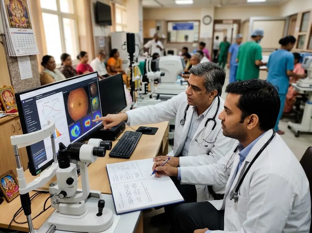
Artificial intelligence is rapidly shifting from narrow, single-task algorithms to versatile generalist architectures across clinical eye care. In particular, ophthalmic foundation models demonstrate remarkable potential because they learn expressive representations from vast archives of unlabelled retinal images. Consequently, these architectures achieve robust diagnostic accuracy with significantly fewer manual annotations than conventional deep learning methods. However, acquiring or building a foundation model represents only the initial technical milestone. The more pressing clinical challenge involves adapting these pre-trained systems to solve specialized diagnostic tasks reliably within local hospital environments.
Pretraining establishes the core visual vocabulary that makes modern architectures so effective. During self-supervised pretraining, the neural network analyzes millions of unlabelled fundus photographs and optical coherence tomography scans. Specifically, algorithms like masked autoencoders learn anatomical landmarks, vascular geometry, and subtle tissue textures without human intervention. Furthermore, this broad feature extraction captures intricate pathophysiological signals that transfer seamlessly across multiple downstream clinical tasks. Clinicians should recognize that the model encodes generalized structural biology rather than isolated disease classifications. Therefore, pretraining establishes a versatile baseline that minimizes the data burden required for novel applications. However, the quality of downstream performance depends heavily on the pretraining domain. If a model encounters a target pathology or imaging modality that diverges sharply from its training cohort, representations may lose fidelity. For instance, models trained predominantly on colour fundus photographs cannot spontaneously interpret complex anterior segment pathology. Hence, researchers must carefully evaluate domain compatibility before initiating any downstream deployment.
Once a base model is selected, researchers must choose an appropriate adaptation method to address their clinical objective. The two most common strategies are linear probing and full fine-tuning. In linear probing, the foundational network remains completely frozen, and developers train only a lightweight classification head on top of extracted embeddings. Consequently, this method requires minimal computing power and prevents catastrophic forgetting. Moreover, linear probing performs exceptionally well when labelled datasets are tiny or when computational resources are severely constrained. In contrast, full fine-tuning updates every parameter across the entire deep neural network. This technique allows the model to alter its internal representations to fit the nuances of a specific disease. Naturally, full fine-tuning often delivers superior diagnostic accuracy on complex tasks such as staging diabetic retinopathy. However, full fine-tuning requires substantial computational hardware and carries a higher risk of overfitting. Additionally, parameter-efficient fine-tuning techniques offer a balanced middle ground, tuning selective adapter layers while maintaining core network stability.
Deciding whether to perform comprehensive fine-tuning or deploy simpler alternatives requires a systematic clinical framework. Clinicians and researchers should never assume that fine-tuning is automatically necessary. Instead, the decision must balance available annotated data, target disease prevalence, and domain alignment. When dealing with rare ocular diseases where labelled cases are scarce, freezing the foundation weights often prevents severe overfitting. Conversely, when large volumes of verified clinical labels exist, full model tuning can optimize diagnostic thresholds. Furthermore, investigators must account for hardware constraints, institutional privacy policies, and software licensing terms. Many proprietary models restrict direct weight access, limiting clinicians to feature extraction through external interfaces. Therefore, teams should always evaluate a supervised baseline alongside any adapted architecture. If a standard convolutional network trained from scratch performs comparably to a fine-tuned foundation model, the additional computational expense may not be clinically justified. Ultimately, teams must match technical complexity to the actual clinical objective.
Rigorous validation methodology determines whether an adapted ophthalmic algorithm can safely assist clinical care. First, researchers must enforce strict patient-level data splitting rather than random image-level partitioning. Splitting images from the same patient across training and testing sets artificially inflates performance metrics and conceals vulnerability. Second, external validation across diverse healthcare facilities remains essential. Algorithms adapted in tertiary academic hospitals frequently underperform when deployed on low-cost fundus cameras in rural screening clinics. Moreover, developers must prioritize probability calibration rather than relying solely on area under the receiver operating characteristic curve. A well-calibrated algorithm provides dependable risk probabilities, enabling eye care teams to establish safe referral cut-offs. Additionally, clinicians require transparent explainability tools, such as visual saliency maps, to confirm that predictions stem from genuine retinal lesions rather than peripheral artefacts. Finally, expert ground truth annotation by multiple independent retinal specialists ensures that the training signal reflects true clinical reality.
The strategic adaptation of foundation models holds profound implications for community ophthalmology in India. Across India, widespread retinal disorders such as diabetic retinopathy and retinopathy of prematurity demand urgent, scalable screening tools. However, community clinics often lack the massive labelled datasets needed to build bespoke models from scratch. In this setting, adapting open-source foundation models through linear probing or efficient fine-tuning provides an invaluable shortcut. Healthcare networks can repurpose robust representations using modest local datasets collected on handheld or non-mydriatic cameras. Furthermore, this approach reduces computational costs, making local on-premise model execution feasible without expensive cloud infrastructure. Nevertheless, researchers must validate these adapted tools on local populations. Rural screening camps frequently encounter significant media haze and mature cataracts that alter image clarity. By following a structured adaptation roadmap, Indian ophthalmologists can deploy trustworthy diagnostic systems that bridge critical rural care gaps.
Linear probing freezes pre-trained foundation weights and trains only a lightweight classification layer using extracted representations. This strategy requires minimal computing power and avoids overfitting on small datasets. Conversely, fine-tuning updates internal model parameters across deep network layers. While fine-tuning provides higher accuracy for complex conditions, it demands substantial computational hardware and careful regularisation to prevent catastrophic forgetting.
Patient-level splitting ensures that all retinal scans from an individual patient remain exclusively within either the training, validation, or testing partition. If images from the same eye appear across partitions, data leakage occurs. This leakage artificially inflates diagnostic performance because the network memorizes unique patient features rather than true pathological signs. Consequently, patient-level partitioning is vital to verify genuine diagnostic reliability across diverse real-world screening cohorts.
Foundation models dramatically reduce the number of expert-annotated images needed to train effective retinal screening tools. In rural Indian outreach programs, clinical resources and specialized ophthalmologists are scarce. By utilizing pre-trained representations, local clinics can rapidly adapt diagnostic algorithms using modest local cohorts captured on portable cameras. This capability lowers technological and economic barriers, enabling accurate, scalable tele-ophthalmology screening directly at primary health centres across underserved districts.
Disclaimer: This content is for informational and educational purposes only and should not be taken as professional medical advice. Always consult a qualified healthcare provider for diagnosis and treatment decisions. Refer to the latest local and national guidelines for clinical practice.
References

Read summarized clinical updates, watch expert medical content, and earn CME certifications right from your smartphone.
Open your smartphone camera to scan and install.





A practical clinical guide to adapting ophthalmic foundation models. Explore the trade-offs between full fine-tuning and linear probing, dataset construction, patient-level splitting, and robust external validation for scalable ophthalmic AI deployment.
Today
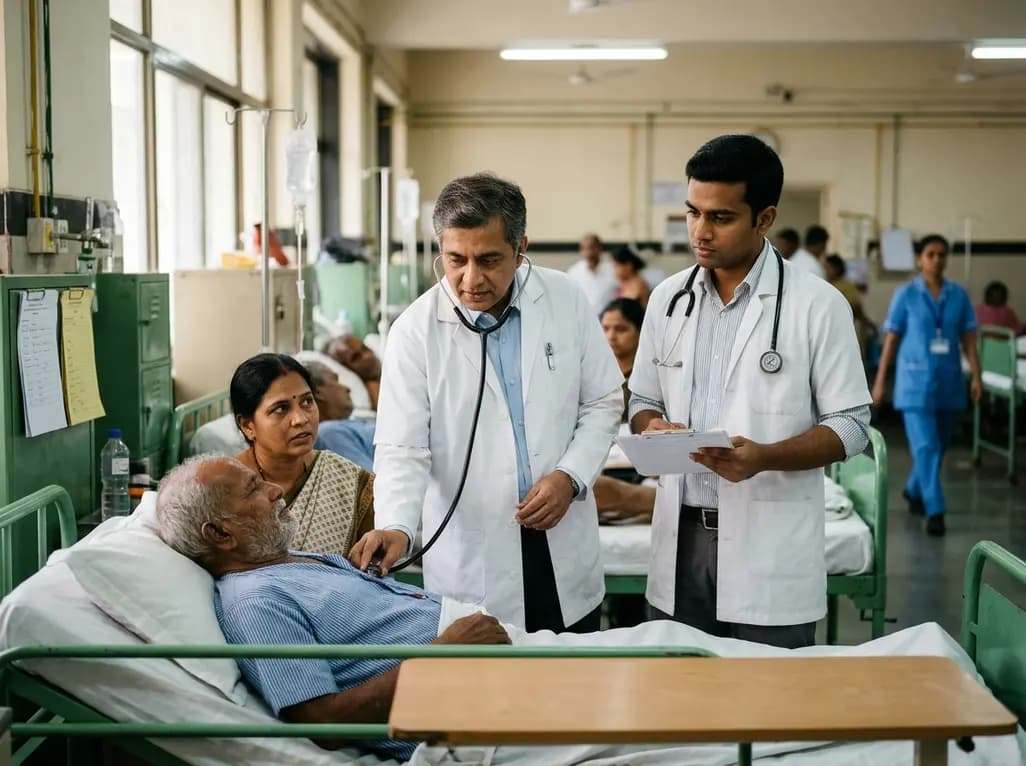
A qualitative study investigates whether traditional bedside clinical examinations still mirror contemporary physician practice. Findings support retaining long cases while reforming short cases toward hypothesis-driven assessments.
Today
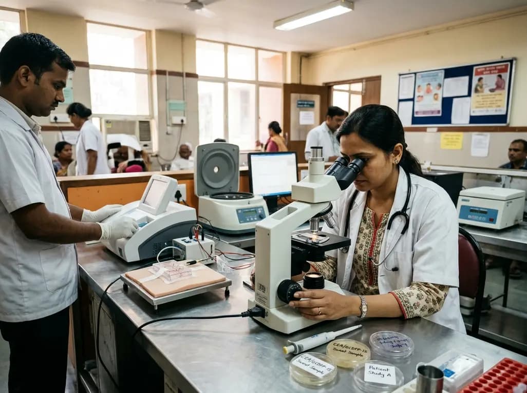
A pilot study evaluated WearDOXXN, a capillary microfluidic sweat patch, for noninvasive breast cancer monitoring by comparing sweat and serum CEA and PIP/GCDFP-15 levels.
Today
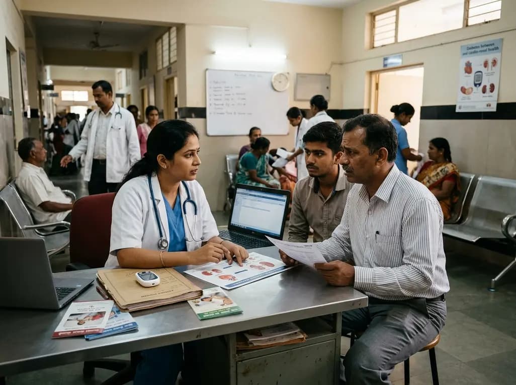
India faces a massive diabetes burden with over 89 million adults affected. Managing blood sugar in isolation fails to prevent severe cardiorenal complications. Clinicians must adopt an integrated metabolic ecosystem that prioritizes early detection, multifactorial risk assessment, and personalized therapies.
Today
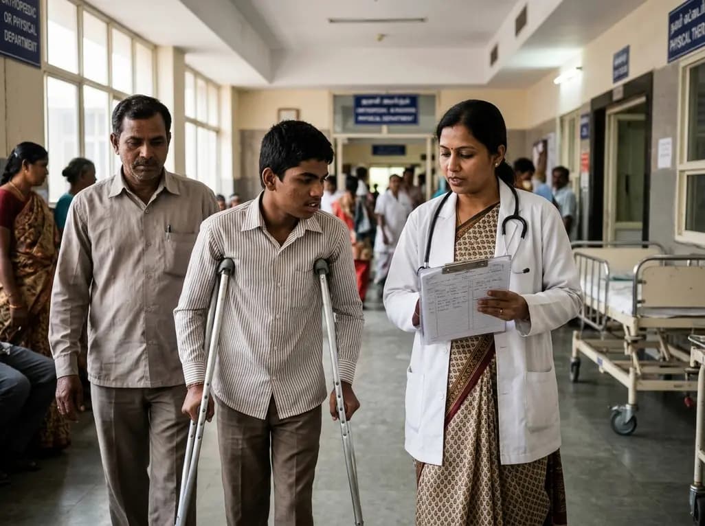
The Gross Motor Function Classification System (GMFCS) stops its validated age bands at 18. Recent evidence emphasizes that evaluating adult cerebral palsy requires purpose-sensitive documentation combining adolescent ratings with dynamic profiles of mobility, fatigue, pain, and community participation.
Today
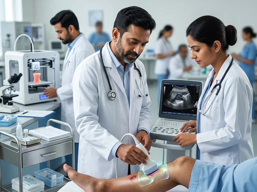
A breakthrough 3D-printed piezoelectric collagen hydrogel activated by ultrasound accelerates diabetic wound repair, demonstrating 98.5% wound closure, robust antibacterial action, and enhanced vascularization without toxic chemical crosslinkers.
Today
