

Loading, please wait...
Loading, please wait...
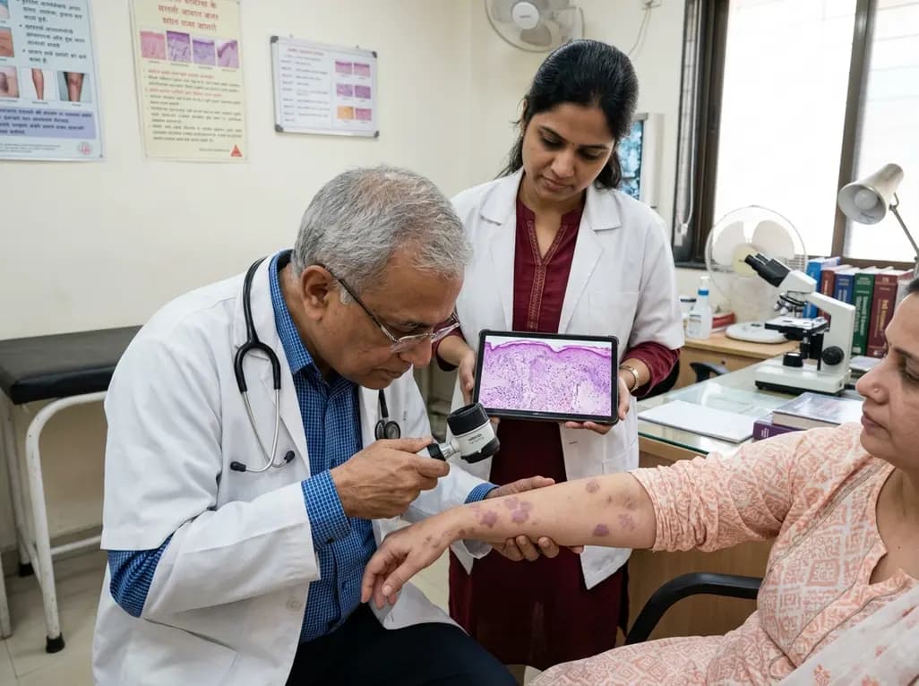
Actinic lichen planus represents a distinctive photosensitive variant of lichen planus that typically develops on sun-exposed anatomical sites. Dermatologists conventionally identify this rare condition in young individuals with darker cutaneous phototypes inhabiting tropical or subtropical environments. However, recent clinical reports show that it can also arise in fair-skinned Caucasian individuals, which frequently creates significant diagnostic dilemmas. Clinicians often misdiagnose these atypical lesions as polymorphic light eruption, chronic photodermatitis, or discoid lupus erythematosus. Therefore, recognizing the broad morphological spectrum of actinic lichen planus remains crucial for prompt intervention. Integrating detailed bedside evaluation with specialized dermatological diagnostic tools ensures accurate disease identification across diverse patient demographics.
Actinic lichen planus historically carries geographic designations such as lichen planus tropicus or lichenoid melanodermatosis. Medical literature predominantly records this entity in children and young adults living across the Middle East, India, and North Africa. Consequently, dermatologists rarely suspect this disorder when examining mature Caucasian patients with Fitzpatrick phototype I or II skin. The exact pathophysiology involves an abnormal cell-mediated cytotoxic response directed against epidermal basal keratinocytes. Specifically, ultraviolet radiation acts as an external trigger that alters epidermal self-antigens in predisposed hosts. Activated T lymphocytes subsequently infiltrate the dermoepidermal junction and release pro-inflammatory cytokines. This inflammatory cascade leads to apoptotic cell death, vacuolar degeneration, and substantial pigmentary incontinence. In addition, warm summer weather and intense ambient sunlight reliably provoke disease flares, establishing a characteristic seasonal pattern. Furthermore, cutaneous lesions often recur annually following recreational or occupational outdoor exposure. Therefore, clinicians must maintain a high index of suspicion whenever patients exhibit recurrent photosensitive eruptions, regardless of geographical origin or baseline ethnic complexion.
Clinicians encounter several discrete morphological variants of actinic lichen planus in routine clinical practice. The classic pigmented subtype usually displays reticulated, macular, or confluent plaques with striking hyperpigmentation. In contrast, annular, plaque-like, and dyschromic variants present distinct clinical appearances that challenge diagnostic acumen. Patients frequently develop these cutaneous lesions on photo-distributed regions, including the forehead, cheeks, lateral neck, and dorsal hands. Moreover, lesions typically exhibit a chronic, relapsing course with pronounced summer exacerbations and spontaneous winter remission. Because actinic lichen planus rarely causes intense pruritus, patients may delay seeking formal dermatological evaluation. Furthermore, systemic involvement, mucosal lesions, and nail changes remain characteristically absent in this condition, unlike classic lichen planus. These atypical features often lead clinicians toward erroneous diagnoses such as chronic actinic dermatitis, melasma, or contact photodermatitis. For instance, middle-aged fair-skinned patients presenting with lateral neck erythema frequently receive treatment for nonspecific photodermatitis. Consequently, unguided topical regimens fail to arrest underlying lichenoid inflammation, permitting progressive dermal melanophage accumulation and stubborn disfigurement.
Dermoscopy serves as an indispensable noninvasive bedside modality for evaluating persistent facial and neck eruptions. When examining pigmented lesions, dermatologists search for specific microstructures that reveal underlying pathology without immediate surgical intervention. In active lesions, dermoscopy distinctly demonstrates Wickham striae, which appear as pearly white reticular, circular, or linear intersecting bands. These striae correspond histopathologically to focal orthokeratotic hypergranulosis and localized epidermal thickening. In addition, clinicians frequently observe diffuse slate-gray or brownish-blue peppering across the lesion background. This characteristic peppering reflects marked pigment incontinence and melanophages nestled within the superficial papillary dermis. Moreover, dermoscopy highlights delicate follicular plugs and surrounding white halos in certain follicular subtypes. In contrast, chronic contact dermatitis and solar lentigines show entirely uniform pigment networks without lichenoid striae. Similarly, discoid lupus erythematosus demonstrates follicular plugging with prominent telangiectasias rather than pearly reticular patterns. Therefore, identifying Wickham striae alongside gray-blue granules strongly supports a lichenoid process and directs targeted skin biopsy.
Histopathologic evaluation of a punch biopsy remains the gold standard for confirming actinic lichen planus. Microscopic sections typically show a well-defined lichenoid interface dermatitis hugging the dermoepidermal interface. Specifically, pathologists identify a dense, band-like infiltrate consisting predominantly of T lymphocytes and histiocytes within the upper dermis. Basal keratinocytes demonstrate extensive vacuolar degeneration and individual apoptotic bodies, commonly termed Civatte or cytoid bodies. Furthermore, the overlying epidermis displays compact hyperorthokeratosis and wedge-shaped hypergranulosis without significant parakeratosis. The papillary dermis consistently harbors abundant melanophages, which accounts for the prominent clinical and dermoscopic pigmentation. In addition, solar elastosis frequently accompanies these lichenoid changes, reflecting substantial cumulative ultraviolet radiation damage. Pathologists must carefully differentiate these features from discoid lupus erythematosus, which shows deeper perivascular infiltrates and basement membrane thickening. Similarly, fixed drug eruptions present with eosinophilic infiltration that remains absent in actinic lichen planus. Thus, histological correlation provides definitive diagnostic clarity when atypical presentations confound bedside assessment.
Managing actinic lichen planus successfully demands a rigorous, multimodal approach focusing on inflammation control and strict photoprotection. Clinicians initiate therapy by prescribing potent or superpotent topical corticosteroids to suppress active dermoepidermal inflammation. However, practitioners must restrict high-potency corticosteroids on the face and neck to avoid epidermal atrophy and telangiectasia. Consequently, topical calcineurin inhibitors, including tacrolimus ointment and pimecrolimus cream, serve as excellent steroid-sparing alternatives for maintenance therapy. In addition, dermatologists frequently incorporate topical retinoids such as adapalene or tretinoin to promote epidermal turnover and disperse post-inflammatory hyperpigmentation. Concurrently, patients must adopt uncompromising sun-avoidance measures, because ultraviolet radiation directly triggers pathogenic cellular signaling cascades. Broad-spectrum sunscreens shielding against both ultraviolet A and B rays represent the cornerstone of long-term prevention. Furthermore, patients should regularly wear wide-brimmed hats, high-collared protective clothing, and sunglasses during peak daylight hours. In severe or treatment-refractory instances, clinicians may carefully consider systemic antimalarials, such as hydroxychloroquine, or short systemic corticosteroid courses to induce rapid remission.
Actinic lichen planus localizes strictly to sun-exposed anatomical areas such as the face, neck, and arms, flaring predominantly during summer months. In contrast, classic lichen planus typically affects flexor surfaces, oral mucosa, and nails with severely pruritic polygonal papules. Furthermore, actinic variants exhibit minimal pruritus, rarely demonstrate the Koebner phenomenon, and leave behind pronounced, persistent post-inflammatory hyperpigmentation.
Dermoscopy provides noninvasive visualization of subsurface cutaneous structures, effectively bridging clinical inspection and histopathology. In actinic lichen planus, dermoscopy reliably reveals pathognomonic Wickham striae alongside slate-gray peppering caused by dermal melanophages. Consequently, these characteristic patterns permit clinicians to distinguish lichenoid eruptions rapidly from chronic photodermatitis, melasma, discoid lupus, or solar lentigines, facilitating timely and targeted punch biopsies without unnecessary delays.
Standard therapy combines anti-inflammatory regimens with rigorous, year-round photoprotection against ultraviolet radiation. Clinicians typically initiate short courses of potent topical corticosteroids, transitioning promptly to topical calcineurin inhibitors like tacrolimus for safe maintenance on delicate facial skin. Additionally, topical retinoids such as adapalene aid pigment clearance. Broad-spectrum daily sunscreen application and protective physical barriers remain mandatory to prevent recurrent seasonal exacerbations.
Disclaimer: This content is for informational and educational purposes only... Refer to the latest local and national guidelines for clinical practice.
References
Ucci L et al. Pigmented Actinic Lichen Planus Mimicking Chronic Photodermatitis in a Fair-Skinned Caucasian Woman: A Case Report. Clin Exp Dermatol. 2026 Oct 22. doi: undefined. PMID: 42908146.
Oba MC, Yuvruk M. Dermoscopy of Actinic Lichen Planus. Dermatol Pract Concept. 2024;14(1):e2024085.
Di Meo N et al. Dermoscopy of Actinic Lichen Planus in Skin of Color. Dermatol Pract Concept. 2024;14(2):e2024101.

Read summarized clinical updates, watch expert medical content, and earn CME certifications right from your smartphone.
Open your smartphone camera to scan and install.





Actinic lichen planus is a rare photosensitive variant of lichen planus. While typical in darker phototypes, it can also affect fair skin, mimicking photodermatitis. Integrating dermoscopy and histology ensures accurate differentiation and prompt management.
Today

Explore vital recurrent stroke predictors in lacunar stroke patients managed with intensive blood pressure lowering. This analysis examines trial findings from SPS3 to uncover residual vascular risks, metabolic factors, and targeted secondary prevention strategies for high-risk neurological patients.
Today

Odisha has established India's first state-level clinical protocol specifically tailored for pregnant women with sickle cell disease. Developed with NHM and UNICEF, the framework provides structured antenatal screening, crisis mitigation, standardized referral pathways, and specialized interdisciplinary care.
Today

India is advancing preventive healthcare by integrating genomics, artificial intelligence, and quantum technologies. With the SANGAM programme sequencing one million Indian genomes, clinicians can unlock personalized therapies, detect cardiometabolic risks early, and curb the burden of diabetes and heart disease.
Today

A randomized controlled trial demonstrates that an eight-week dual-task training intervention significantly improves static and dynamic postural control over standard balance exercises in individuals with chronic ankle instability, addressing critical neurocognitive deficits during athletic and daily activities.
Today

A retrospective cohort study of 7,716 ICU patients from the MIMIC-IV database demonstrates that Gradient Boosting Decision Trees significantly outperform traditional SOFA scores in predicting in-hospital mortality among patients with sepsis and diabetes, with SHAP analysis providing clinical interpretability.
Today
