

Loading, please wait...
Loading, please wait...

Neuro-oncology patients frequently undergo repeated surgical interventions and adjuvant radiation therapy. Consequently, reconstructive surgeons face formidable challenges when managing compromised calvarial soft tissues. Cumulative tissue trauma creates severe dermal thinning and microvascular depletion. Therefore, achieving durable scalp wound reconstruction in this cohort demands reliable techniques that prevent catastrophic complications. When neurosurgeons perform repeat craniotomies, the scarred galea loses compliance. As a result, standard closure generates excessive wound tension, elevating the risk of breakdown, calvarial exposure, and deep intracranial infection. Recent clinical evidence demonstrates that incorporating biological matrices provides effective soft tissue augmentation and tension relief.
Wound healing in the scalp depends on vascular integrity and tissue elasticity. However, prior cranial operations and radiotherapy drastically compromise the surrounding tissue bed. Ionizing radiation causes progressive microvascular endarteritis, diminishing local capillary density and severely reducing cutaneous perfusion. Concurrently, dense fibrosis replaces pliable subcutaneous tissues with rigid cicatrix. When neuro-oncology patients require repeat craniotomies, surgical teams must navigate these poorly vascularized fields. Consequently, closing non-compliant wound edges under high tension triggers focal tissue ischemia. This ischemia frequently precipitates flap dehiscence, necrosis, and subsequent cranial hardware exposure. Moreover, exposed cranioplasty materials or titanium plates invite invasive infections, such as osteomyelitis and meningitis. These life-threatening infections necessitate aggressive debridement, implant removal, and prolonged systemic antibiotic therapy. Such surgical interruptions delay vital antineoplastic therapies, directly compromising oncological survival. Therefore, surgeons must implement reconstructive strategies that reliably relieve tissue tension and reinforce vulnerable cranial soft tissues.
Acellular dermal matrix (ADM) addresses the core mechanical challenges of hostile scalp closures. Derived from decellularized donor tissue, ADM provides an immunologically inert, collagen-rich structural matrix. When implanted beneath the scalp, the material serves two vital biomechanical purposes. First, it facilitates strategic galea tension relief across attenuated fascial margins. Surgeons anchor the matrix directly to native fascial borders, effectively absorbing biomechanical traction and reducing strain on cutaneous suture lines. Second, ADM delivers substantial soft tissue augmentation over compromised calvarial defects. The porous collagen scaffold encourages host fibroblastic infiltration, cellular repopulation, and rapid neovascularization. Over time, recipient blood vessels thoroughly incorporate the matrix without inciting excessive inflammatory responses. Furthermore, this thickened, vascularized biological cushion shields underlying alloplastic hardware, titanium mesh, and bone flaps from progressive erosion. Consequently, the matrix stabilizes high-risk wounds and establishes a dependable foundation for durable scalp healing.
Recent clinical data substantiate the efficacy of ADM in complex neurosurgical cases. A comprehensive retrospective surgical case series evaluated 23 consecutive neuro-oncology patients treated between 2017 and 2022. The study cohort presented with significant clinical vulnerability, displaying a mean age of 57.8 years. Nearly all patients had undergone prior cranial surgery and adjuvant radiotherapy. Furthermore, 34.8% presented with multiple prior craniotomies, and 13.0% had documented histories of surgical site infection. Reconstructive surgeons utilized ADM to accomplish tension-free coverage across extensive defects, with a mean area of 83.0 square centimeters. Over a mean follow-up of 18.2 months, only one patient (4.3%) experienced a complication requiring reoperation for hardware exposure. Every other ADM-assisted closure remained completely healed and intact throughout the study period. Crucially, reconstructed sites maintained structural integrity even during subsequent surgical interventions or additional radiotherapy. These favorable outcomes underscore the reliability of biological matrix reinforcement in heavily pretreated patient cohorts.
Achieving reproducible success with biological matrices requires meticulous surgical technique. Initially, surgical teams elevate compromised scalp flaps within the subgaleal plane, taking extreme care to preserve remaining collateral vessels. Wide undermining mobilizes adjacent tissue flaps, maximizing peripheral compliance around the surgical site. Next, the surgeon evaluates the tissue deficit and quantifies the mechanical tension required for wound closure. When direct approximation generates excessive tension, the surgical team trims an ADM sheet to match the defect. The operator sutures the matrix circumferentially beneath the galea using interrupted absorbable sutures to achieve balanced tension distribution. This biological construct securely bridges the fascial gap, allowing overlying cutaneous margins to close without mechanical strain. Additionally, surgeons ensure close apposition between the matrix and vascularized tissue to promote rapid host angiogenesis. Closed-suction drainage is routinely placed beneath the flap to prevent hematoma or seroma accumulation. Finally, surgeons close the skin with tension-free monofilament sutures.
Microvascular free tissue transfer historically served as the benchmark for large scalp defects in irradiated fields. Free flaps provide reliable vascularized bulk; however, they require prolonged operative durations and sophisticated microsurgical resources. Many neuro-oncology patients with advanced systemic disease cannot comfortably tolerate extended general anesthesia or significant donor-site morbidity. In addition, free flaps create noticeable cosmetic deformities and lack natural hair-bearing potential. In contrast, ADM-assisted closure offers an expeditious, low-morbidity reconstructive solution. The technique completely eliminates donor-site harvesting, thereby shortening operative time and accelerating postoperative mobilization. Furthermore, this approach preserves adjacent hair-bearing scalp, which optimizes both cosmetic appearance and psychological recovery. While extensive calvarial necrosis or massive structural loss still demands free tissue transfer, ADM significantly expands reconstructive options. By reinforcing compromised local flaps, biological matrices allow surgeons to achieve robust primary closure without subjecting vulnerable oncology patients to exhausting microsurgical operations.
Successful integration of biological matrices into neuro-oncological care requires close interdisciplinary coordination. Neurosurgeons, reconstructive surgeons, and radiation oncologists must align treatment schedules to optimize tissue healing before initiating adjuvant therapy. Clinicians must also optimize systemic factors, addressing perioperative glycemic control, nutritional status, and steroid tapering whenever possible. In resource-conscious healthcare environments, the upfront cost of acellular matrices represents a notable consideration for surgical departments. Nevertheless, avoiding severe wound breakdown, chronic hospitalization, and unplanned reoperations yields substantial clinical and economic benefits. Concurrently, ongoing biomedical innovations aim to enhance matrices with antimicrobial properties and angiogenic growth factors to further accelerate vascularization. As clinical adoption grows, prospective trials will help establish definitive guidelines for patient selection and technique standardization. Ultimately, acellular dermal matrices represent a transformative adjunct in modern cranial surgery, protecting vulnerable patients from devastating soft tissue failure.
Prior cranial operations and ionizing radiation severely damage local tissue architecture. Radiation induces microvascular endarteritis, which diminishes blood perfusion and causes dense dermal fibrosis. Consequently, repeat craniotomies produce severe scalp inelasticity. Closing these rigid wounds creates excessive mechanical tension, sharply increasing the likelihood of flap necrosis, dehiscence, and dangerous hardware exposure.
Acellular dermal matrix bridges fascial defects by anchoring directly to native galeal margins. This configuration absorbs mechanical pulling forces, effectively redistributing tension away from superficial cutaneous borders. Furthermore, the porous matrix acts as a biological scaffold that encourages fibroblastic infiltration and host neovascularization, creating durable soft tissue reinforcement over cranial implants.
Yes, clinical evidence indicates that incorporated acellular dermal matrix repairs withstand subsequent radiotherapy and repeat surgeries. Once host cells and capillaries repopulate the collagen scaffold, the matrix integrates permanently into native tissue. Consequently, the augmented soft tissue barrier remains durable, preventing wound breakdown even during subsequent adjuvant oncological treatments.
Disclaimer: This content is for informational and educational purposes only and should not be considered medical advice or substituted for professional clinical judgment. Diagnostic criteria, treatment regimens, and monitoring protocols should always follow established clinical guidelines. Refer to the latest local and national guidelines for clinical practice.
References

Read summarized clinical updates, watch expert medical content, and earn CME certifications right from your smartphone.
Open your smartphone camera to scan and install.





Acellular dermal matrix (ADM) provides vital galea tension relief and soft tissue augmentation in high-risk neuro-oncology patients undergoing repeat craniotomy. This biological adjunct safely mitigates wound breakdown and hardware exposure in previously irradiated, hostile calvarial fields.
Today

A Bayesian component network meta-analysis of 130 RCTs reveals that self-monitoring and gamification provide the clearest positive effects in mHealth physical activity interventions, while social comparison may have adverse psychological trade-offs.
Today

The GESS-AEEM consensus establishes a clear clinical implementation framework for testosterone therapy in postmenopausal women with hypoactive sexual desire disorder, focusing on transdermal dosing, precise laboratory monitoring, and safety communication.
Today
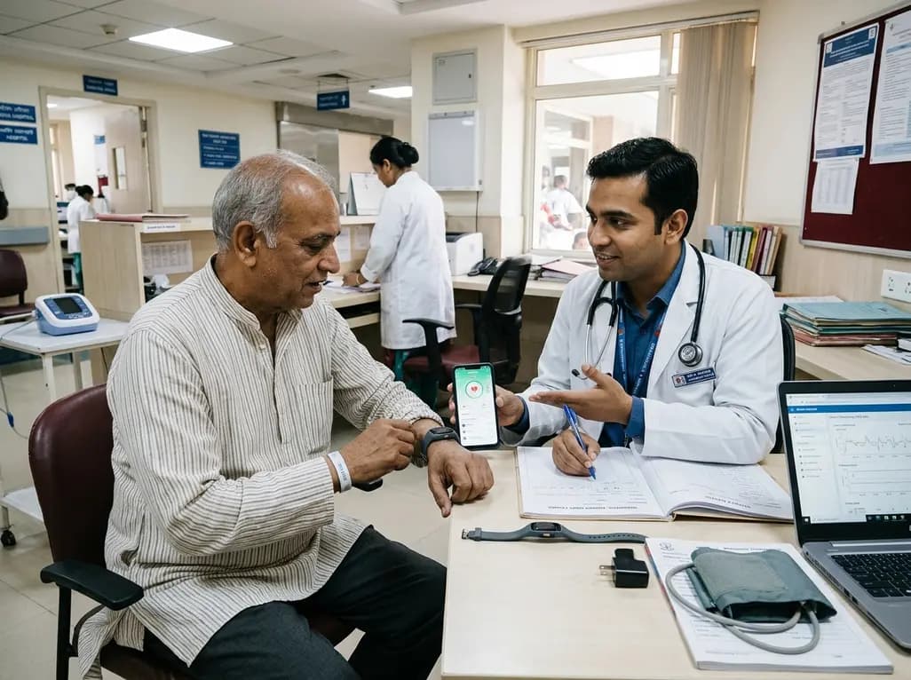
A multicenter observational study evaluated an 8-week digital health monitoring platform using wearable sensors in ambulatory heart failure. While participants achieved high daily wear-time of 11.7 hours and 80% completion, initial recruitment revealed significant technology-related onboarding barriers.
Today
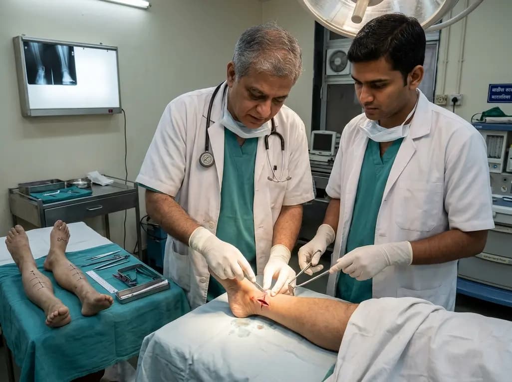
A paired cadaveric study compared triple and quintuple percutaneous Achilles tendon lengthening. While quintuple hemisection showed numerically greater elongation, differences were not statistically significant.
Yesterday
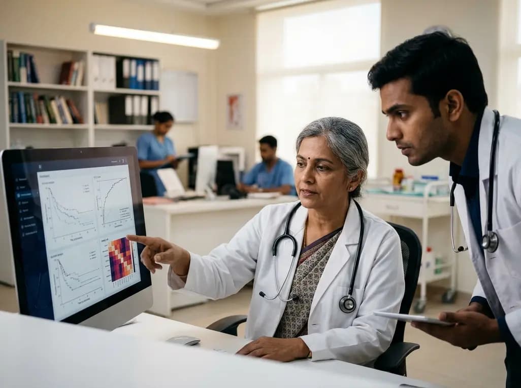
A clinical decision-support framework using health checkup data from 231,683 adults predicts incident obesity with high discrimination (AUROC 0.918). The system integrates temporal survival analysis and risk-factor explanations to prioritize individualized preventive interventions.
Today
