.webp&w=384&q=75)

Loading, please wait...
Loading, please wait...

Modern neuro-oncology emphasizes aggressive cytoreduction while protecting intact neurological pathways. Achieving an optimal high-grade glioma resection remains pivotal because extensive tumor clearance directly prolongs overall survival and delays disease recurrence. In addition, an uneventful postoperative recovery ensures that patients can initiate timely chemoradiotherapy. Consequently, neurosurgeons increasingly leverage intraoperative adjuncts to delineate infiltrative margins and remove residual disease safely.
Malignant gliomas present unique surgical difficulties due to their diffusely infiltrative nature. Therefore, standard white-light illumination rarely differentiates peripheral neoplastic cells from healthy functional brain tissue. When surgeons leave microscopic tumor remnants behind, these residual foci frequently drive early disease progression. Furthermore, aggressive margin clearance near eloquent cortical structures carries a substantial risk of producing permanent neurological deficits.
To overcome these visualization boundaries, neurosurgeons integrate advanced intraoperative guidance tools. For example, oral 5-aminolevulinic acid induces protoporphyrin IX accumulation within malignant cells, creating distinct violet-red fluorescence under blue-violet light. Conversely, intraoperative ultrasound provides real-time acoustic feedback regarding subcortical lesions and tissue depth. Both techniques offer practical, highly accessible alternatives to expensive intraoperative imaging suites.
However, each imaging modality possesses clear diagnostic blind spots when clinicians utilize them in isolation. Fluorescence visualization cannot penetrate beneath opaque cortical surfaces or through pooling blood. Meanwhile, acoustic ultrasound images degrade rapidly during later resection stages due to cavity collapse and saline artifacts. Consequently, relying on a single intraoperative tool often leaves surgical margins uncertain.
A recent surgical investigation evaluated seventy-two consecutive patients who underwent resection for high-grade gliomas between 2019 and 2024. Most cases involved glioblastoma, alongside grade IV astrocytomas, grade III astrocytomas, and grade III oligodendrogliomas. Following primary tumor cytoreduction under standard white light, surgeons applied both optical fluorescence and ultrasound to inspect the residual resection cavity.
Additionally, the operative team collected marginal biopsies to examine histological characteristics and verify neoplastic infiltration. When investigators evaluated 5-aminolevulinic acid independently, it demonstrated a sensitivity of 84.9%. Nevertheless, its specificity reached only 57.8%, reflecting a noticeable rate of false-positive marginal findings. Moreover, peritumoral reactive gliosis and normal vascular structures occasionally emit confounding fluorescent signals that mislead operative decisions.
In contrast, intraoperative ultrasound exhibited an opposing diagnostic pattern across the study cohort. Ultrasound imaging yielded a specificity of 84.5% alongside a sensitivity of 76.0%. Thus, ultrasound effectively confirmed disease-free boundaries but missed subtle infiltrative remnants near cavity walls. These distinct numbers illustrate why isolated imaging methods leave neurosurgeons with diagnostic uncertainty during critical stages of surgery.
Combining optical fluorescence and acoustic imaging dramatically enhanced diagnostic accuracy throughout the surgical cavity. Specifically, when both modalities produced concordant positive results, diagnostic sensitivity rose to 91.0%. Furthermore, this positive-positive combination achieved an impressive specificity of 86.0%. Consequently, concordant findings provide neurosurgeons with reliable confirmation that suspicious marginal tissue contains residual tumor.
Similarly, discordant and doubtful findings yielded valuable intraoperative guidance for surgical decision-making. When tissue exhibited positive 5-ALA fluorescence alongside doubtful ultrasound findings, specificity climbed to 95.2%, while sensitivity was 67.9%. Therefore, even ambiguous ultrasound signals warrant thorough scrutiny when optical fluorescence indicates metabolic activity. Statistical receiver operating characteristic analyses confirmed that dual-modality guidance significantly surpassed both individual techniques.
Additionally, this complementary workflow resolves the physical limitations inherent to each solitary modality. Acoustic waves penetrate deep subcortical structures where optical light cannot reach. Meanwhile, surface fluorescence identifies micro-invasive disease where ultrasound resolution degrades near surgical edges. Thus, combining these technologies provides continuous, comprehensive visualization across superficial and deep tumor interfaces.
Subjective evaluation of ambiguous marginal signals can generate variability between different surgical teams. To eliminate visual bias, investigators developed an automated machine learning tool designated HGGPredictor. This algorithm rapidly synthesizes optical intensity and ultrasound echogenicity scores to evaluate marginal tissue biopsies. Consequently, surgical teams receive quantitative assessments that support swift intraoperative decision-making.
Specifically, the computational tool classifies tissue samples into solid tumor, peripheral infiltration, or normal brain parenchyma. By estimating tumor cell density in real time, the algorithm eliminates diagnostic guesswork at critical anatomical margins. Furthermore, machine learning models refine their predictive accuracy as institutions feed larger clinical datasets into the analytical framework. As a result, digital tools enhance surgical precision without adding unnecessary delays to operative workflows.
Moreover, predictive algorithms provide immense value in high-volume neurosurgical theaters where neuropathology frozen sections require significant processing time. Instead of waiting for preliminary histopathology, neurosurgeons can act upon rapid marginal assessments. Therefore, coupling machine learning with accessible intraoperative modalities transforms conventional surgical resection into an objective, data-driven discipline.
Integrating 5-aminolevulinic acid and intraoperative ultrasound offers immense strategic value for neurosurgical practice across India. High-field intraoperative MRI suites demand massive capital investments, specialized shielded infrastructure, and prolonged operating times. In contrast, portable ultrasound machines and fluorescence-equipped microscopes represent scalable, cost-effective technologies for diverse healthcare centers. Consequently, tertiary hospitals and teaching institutions can readily implement this dual-modality workflow.
Furthermore, surgical teams can integrate this protocol smoothly into standard operative routines without complicating anesthesia protocols. Patients ingest oral 5-ALA approximately three hours before entering the operating room. After debulking the tumor mass under white light, the neurosurgeon places the ultrasound transducer onto the resection bed. Subsequently, switching to blue-light illumination reveals residual superficial fluorescence while ultrasound visualizes deep subcortical extensions.
However, aggressive surgical resection must never compromise eloquent functional pathways. Surgeons must combine multimodal imaging with continuous intraoperative neuromonitoring and subcortical stimulation mapping. Thus, technological guidance protects neurological integrity while achieving maximum cytoreduction. Ultimately, combining optical fluorescence, acoustic imaging, and predictive algorithms establishes a safe, reproducible standard for modern neuro-oncological care.
Combining these modalities substantially increases diagnostic sensitivity to 91% and specificity to 86% when both modalities yield positive findings. Ultrasound detects deep residual tumor beneath the cavity floor, whereas 5-ALA identifies superficial infiltrative margins. Together, they eliminate blind spots and enable safer, more complete high-grade glioma resections.
The HGGPredictor algorithm uses machine learning to classify tissue biopsies taken from the surgical cavity. By analyzing optical fluorescence and acoustic parameters, the tool accurately distinguishes solid tumor, infiltrative margins, and normal brain tissue. This real-time automation guides neurosurgeons objectively during intraoperative decision-making and resection margin assessment.
High-field intraoperative MRI remains expensive and unavailable in many centers across developing healthcare settings. In contrast, intraoperative ultrasound and 5-ALA fluorescence require significantly lower capital investment and integrate easily into existing operating microscopes. Consequently, this combined strategy delivers exceptional diagnostic accuracy and surgical precision without requiring costly specialized infrastructure.
Disclaimer: This content is for informational and educational purposes only... Refer to the latest local and national guidelines for clinical practice.
References

Read summarized clinical updates, watch expert medical content, and earn CME certifications right from your smartphone.
Open your smartphone camera to scan and install.





Combining 5-aminolevulinic acid (5-ALA) fluorescence with intraoperative ultrasound significantly improves diagnostic accuracy during high-grade glioma resection. Furthermore, a novel machine learning algorithm accurately predicts tumor margins, establishing a potent multimodal framework for neuro-oncology.
Today
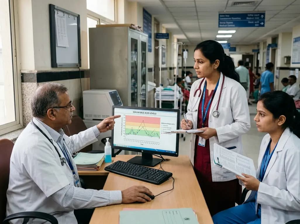
A comprehensive analysis of serum bile acid variability establishes robust reference ranges and reveals how sex, age, BMI, and smoking alter circulating profiles.
Today

A mixed-methods study evaluates a digital perinatal navigator app for high-risk pregnancies, demonstrating high usability, reduced distress, and improved awareness of vital healthcare services.
Today
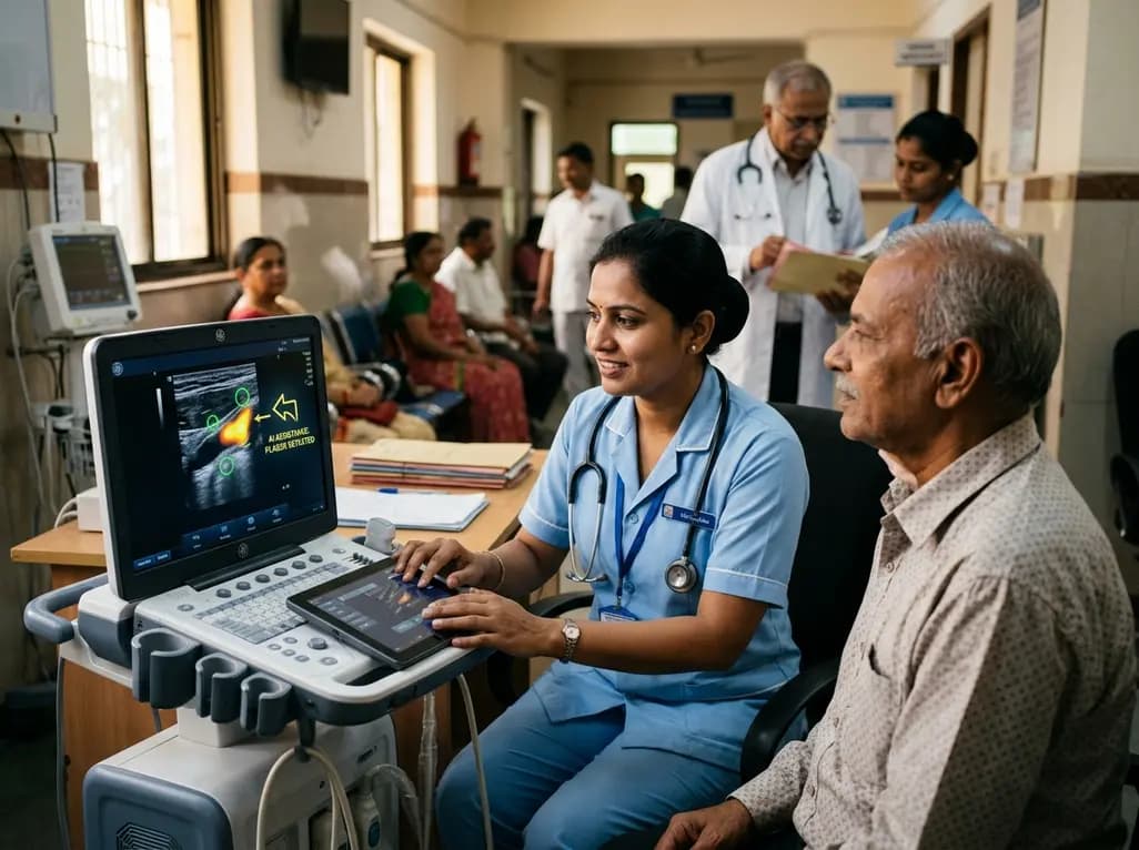
A user-centered study demonstrates that AI-assisted carotid ultrasound enables nonexpert primary care staff to detect subclinical atherosclerosis. With real-time guidance and workflow optimization, this technology supports early cardiovascular risk communication and task-shifting in routine clinical practice.
Today

A hyaluronic acid-modified metal-polyphenol nanocomposite successfully eliminates reactive oxygen species in chondrocytes, halts cartilage breakdown, and promotes tissue anabolism, presenting a novel disease-modifying strategy for early osteoarthritis.
Today

A pilot randomized trial demonstrates that digital wellness applications and medically tailored meals significantly attenuate rapid weight regain following GLP-1 receptor agonist discontinuation, providing valuable transitional support for long-term obesity management.
Today
