

Loading, please wait...
Loading, please wait...

Percutaneous left atrial appendage closure provides vital stroke prophylaxis for patients with non-valvular atrial fibrillation who cannot tolerate chronic anticoagulation. Traditionally, transesophageal echocardiography served as the standard intraprocedural imaging modality for guiding transseptal puncture, appendage sizing, and device deployment. However, interventional teams increasingly adopt 4D intracardiac echocardiography to overcome logistical and clinical limitations associated with transesophageal guidance. Recent real-world evidence indicates that volumetric intracardiac imaging achieves comparable technical success while enhancing catheterization laboratory workflow and avoiding deep general anesthesia. Consequently, structural heart operators can perform complex occlusion procedures with greater autonomy, procedural efficiency, and patient comfort.
Percutaneous occlusion procedures require precise real-time visualization of complex left atrial structures to ensure safe device positioning. For decades, transesophageal echocardiography represented the established modality for guiding transseptal crossing and confirming device stability. Although transesophageal imaging offers excellent resolution, it routinely mandates endotracheal intubation, deep general anesthesia, and continuous airway management. Furthermore, transesophageal probe insertion carries documented risks of esophageal tears, aspiration, and hemodynamic instability, especially in elderly individuals.
Fortunately, recent innovations in volumetric ultrasound catheters have transformed procedural visualization. Interventional cardiologists can now steer four-dimensional intracardiac catheters directly within the cardiac chambers. This technology allows operators to visualize the interatrial septum, left atrial appendage ostium, and adjacent anatomical landmarks with high multiplanar clarity. In addition, real-time volumetric rendering provides dynamic spatial perspective without requiring probe adjustments by a secondary echocardiographer. By navigating the probe from the right atrium or across the fossa ovalis, operators obtain continuous structural feedback. Therefore, catheterization laboratories achieve procedural independence while maintaining meticulous anatomical inspection.
Real-world data demonstrate that volumetric intracardiac ultrasound provides technical efficacy comparable to conventional transesophageal monitoring. In a recent single-operator comparative study evaluating consecutive patients referred for appendage occlusion, device implantation achieved a 94% success rate under intracardiac guidance. In comparison, procedures guided by transesophageal echocardiography achieved a 100% success rate in the historical cohort. Both patient groups exhibited balanced baseline cardiovascular comorbidities and anatomical complexities.
Importantly, interventionalists attribute the minor numerical discrepancy in device deployment to early technical familiarization rather than equipment limitations. As operators progress through their learning curve, procedural confidence and deployment accuracy improve rapidly. Furthermore, volumetric intracardiac echo enables clinicians to evaluate device compression, anchor stability, and residual peri-device leaks in real time. Operators verify standardized release criteria across multi-angle views before releasing the delivery cable. Consequently, switching to intracardiac guidance does not sacrifice procedural rigor or structural closure efficacy. In addition, high-quality volumetric imaging helps clinicians adjust sheath angles quickly to match difficult anatomical orientations.
Patient safety remains a vital priority during structural heart interventions, and intracardiac guidance demonstrates an exemplary clinical safety profile. During the peri-procedural period and initial 30-day post-procedure window, adverse events occurred infrequently across both cohorts. Specifically, the intracardiac guidance group experienced only one non-disabling ischemic stroke and zero procedural deaths. In contrast, investigators observed two patient deaths within 30 days in the transesophageal guidance cohort.
Crucially, neither cohort developed devastating mechanical complications such as acute cardiac perforation, pericardial tamponade, or device embolization requiring surgical rescue. These clinical findings reassure practitioners that intracardiac catheter manipulation does not elevate procedural risks. Moreover, replacing transesophageal imaging completely eliminates the risk of esophageal trauma, oropharyngeal hematoma, and sedation-related hypotension. Because patients avoid systemic paralytics and mechanical ventilation, recovery times decrease significantly. As a result, older patients with extensive comorbidities experience smoother post-intervention monitoring. Thus, real-world registry evidence confirms that volumetric intracardiac ultrasound provides a safe and dependable alternative to invasive transesophageal monitoring.
Transitioning from transesophageal monitoring to intracardiac guidance offers transformative operational advantages for modern cardiac catheterization laboratories. Traditional transesophageal protocols require dedicated cardiac anesthesiologists and expert imaging cardiologists throughout each case. Scheduling conflicts among multiple clinical specialties often create procedural delays and limit daily laboratory capacity. In contrast, intracardiac ultrasound empowers a single operator to perform the procedure and direct intraprocedural imaging simultaneously. Consequently, hospitals experience faster room turnover, streamlined scheduling, and lower anesthesia-related operational expenses.
Equally important, intermediate-term therapeutic efficacy remains robust following intracardiac-guided implantation. At the scheduled 45-day follow-up, 91% of patients in the intracardiac ultrasound group successfully discontinued oral anticoagulation. Similarly, 89% of patients in the transesophageal cohort stopped systemic anticoagulant therapy. These equivalent discontinuation rates verify that intracardiac imaging facilitates secure device sealing and prevents clinically significant residual flow. Therefore, patients attain the definitive stroke prevention benefits of left atrial appendage occlusion while successfully avoiding long-term systemic bleeding risks.
Despite the clear workflow and clinical benefits, implementing volumetric intracardiac guidance requires thoughtful preparation and technical training. Interventionalists must acquire proficiency in steering the ultrasound catheter, recognizing intracardiac views, and maintaining acoustic orientation. In addition, crossing the interatrial septum with an imaging sheath introduces an additional procedural step that demands disciplined catheter hygiene and meticulous anti-coagulation monitoring. Operators typically encounter longer initial fluoroscopy times during their early procedural cases.
However, clinical experience indicates that operators overcome this learning curve quickly after completing a structured series of cases. Preprocedural computed tomography imaging provides valuable anatomical maps that guide intraprocedural transseptal puncture and device sizing. Furthermore, structural heart teams can utilize proctored sessions and simulation modules to accelerate technical mastery. Once the interventional team standardizes sheath setups and console controls, total procedural durations decrease substantially. Ultimately, investing in volumetric intracardiac ultrasound proficiency creates an adaptable, independent procedural environment that improves patient throughput and clinical outcomes across contemporary structural heart programs.
Volumetric intracardiac imaging significantly enhances procedural workflow by eliminating the mandatory requirement for deep general anesthesia and dedicated transesophageal echocardiography personnel. Instead, the primary interventionalist directly controls real-time anatomical imaging from within the catheterization suite. This streamlined approach minimizes room turnover times, reduces scheduling bottlenecks between cardiology and anesthesia departments, and lowers anesthetic complication rates, particularly in frail, elderly patients who face elevated risks from tracheal intubation.
Clinical registries confirm that volumetric intracardiac echocardiography delivers procedural success rates comparable to conventional transesophageal imaging. In real-world comparative evaluations, device deployment success reached 94% with intracardiac guidance versus 100% with transesophageal monitoring. The slight numerical variation primarily reflects the initial operator learning curve rather than device inadequacy. Moreover, intermediate follow-up confirms that over 90% of patients successfully discontinue oral anticoagulation at 45 days in both imaging cohorts.
Intracardiac echocardiography offers notable safety advantages by completely avoiding esophageal instrumentation. Rigid transesophageal probes can cause oropharyngeal trauma, esophageal perforation, and gastrointestinal hemorrhage, especially in anticoagulated patients. Additionally, intracardiac imaging permits procedures under conscious sedation or local anesthesia, reducing hemodynamic instability and vasopressor requirements. Real-world investigations show remarkably low complication rates, with no increase in pericardial effusion, device dislodgement, or major stroke compared to traditional transesophageal-guided left atrial appendage closure.
Disclaimer: This content is for informational and educational purposes only. It is not intended as medical advice, diagnosis, or treatment recommendations. Refer to the latest local and national guidelines for clinical practice.
References

Read summarized clinical updates, watch expert medical content, and earn CME certifications right from your smartphone.
Open your smartphone camera to scan and install.





A real-world evaluation shows 4D intracardiac echocardiography provides 94% success for Watchman FLX left atrial appendage closure with comparable safety and 45-day anticoagulation cessation to TEE while streamlining catheterization laboratory workflows.
Today
Updated national guidelines redefine the management of tuberculosis infection, highlighting targeted screening, IGRA testing, and shorter rifamycin regimens.
Today
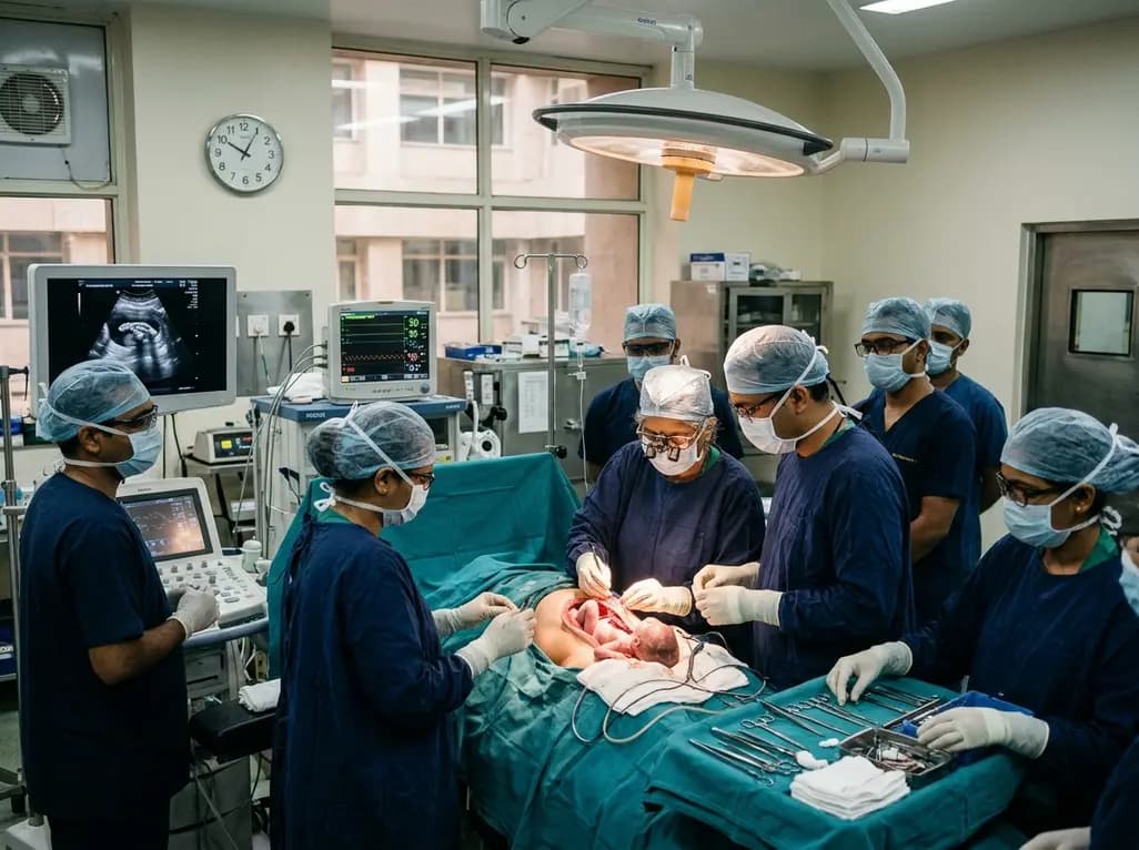
Doctors at AIIMS New Delhi have achieved a historic milestone by performing India's first open prenatal myelomeningocele repair on a 25-week unborn fetus. This landmark multidisciplinary procedure provides vital neuroprotective benefits, lowers hydrocephalus risks, and transforms maternal-fetal medicine nationwide.
Today

A clinical overview of resveratrol supplementation in mitigating exercise-induced muscle damage. This narrative review explores physiological mechanisms, reductions in soreness markers like creatine kinase, dosage strategies, and bioavailability enhancements for sports medicine and nutrition practitioners.
Today

Childhood insulinoma is a rare cause of recurrent hyperinsulinemic hypoglycemia that often eludes early diagnosis. Advanced GLP-1 receptor-targeted PET/CT imaging using 68Ga-exendin-4 complements anatomic MRI to localize occult pancreatic lesions, facilitating focused, curative surgical enucleation.
Today
