The Evolution of Musculoskeletal Assessment
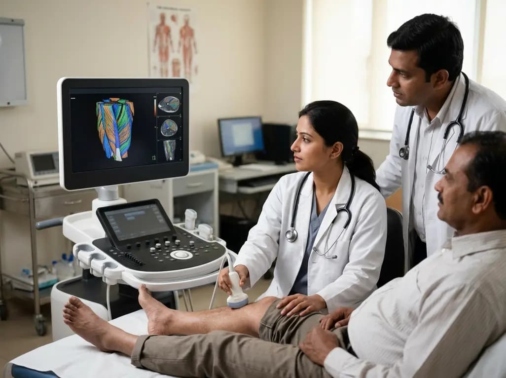
The emergence of 3D ultrasound muscle architecture analysis marks a significant leap in our understanding of human biomechanics and physical therapy. Historically, clinicians relied heavily on two-dimensional imaging to estimate muscle parameters. However, 2D ultrasound often fails to capture the intricate, three-dimensional spatial arrangements of muscle fibers. Consequently, researchers have developed advanced scanning systems to provide a more holistic view of tissue behavior. Understanding how muscle fibers align and change during contraction is vital for diagnosing pathologies and optimizing rehabilitation. Furthermore, this architectural insight helps explain how different muscle compartments contribute to total force production. By utilizing 3D ultrasound muscle architecture, we can now observe the internal dynamics of muscles like the tibialis anterior with unprecedented precision. This capability is particularly relevant in sports medicine, where subtle changes in pennation angles can indicate readiness for return-to-play. Moreover, these imaging advancements allow for more accurate musculoskeletal modeling. Ultimately, these tools empower practitioners to tailor interventions based on the unique physiological responses of each patient. As technology progresses, the transition from 2D to 3D assessments will likely become the standard for comprehensive muscle evaluation.
Limitations of Traditional Imaging Techniques
Traditional 2D ultrasound provides a valuable but limited cross-sectional view of musculoskeletal structures. One major drawback involves the inherent assumption that muscle fibers remain within a single imaging plane during contraction. In reality, fibers often twist or change their orientation in multiple dimensions as they shorten. Additionally, 2D methods frequently struggle to represent the complex geometry of large or deep muscle groups accurately. Therefore, clinicians might overlook critical architectural variations that influence force transmission. To address these issues, the development of automated linear scanning systems has revolutionized the field. These systems move a linear probe systematically over the muscle, capturing a sequence of images that software later reconstructs into a 3D volume. Notably, this approach minimizes operator error and ensures consistent data acquisition across different subjects. Furthermore, 3D imaging allows for the assessment of compartmental differences that 2D scans simply cannot resolve. For example, the tibialis anterior contains superficial and deep regions that behave distinctly. By overcoming the limitations of 2D views, 3D ultrasound muscle architecture offers a more reliable metric for assessing muscle health and function in clinical settings.
Decoding Tibialis Anterior Dynamics
Recent studies focusing on the tibialis anterior have highlighted the profound relationship between activation intensity and structural changes. Twelve participants performed graded isometric dorsiflexion contractions to examine how the 3D ultrasound muscle architecture adapts under load. Specifically, researchers measured pennation angles, fiber lengths, and curvatures at levels ranging from passive rest to 80% of maximum voluntary contraction. The results demonstrated that the pennation angle increases progressively with contraction intensity. For instance, the angle shifted from a passive state of approximately 10.6° to 12.6° during heavy contraction. Simultaneously, fiber lengths decreased significantly as the muscle worked harder to generate force. This physical transformation illustrates the principle of "form follows force," where the muscle's internal arrangement optimizes itself for tension. Interestingly, the study also revealed that fiber curvature increases during contraction, particularly in the deep compartments of the muscle. These findings suggest that the internal pressure and spatial constraints within the muscle belly play a role in fiber shape. Consequently, this data provides a critical baseline for understanding normal muscle behavior during activity, which is essential for identifying abnormal patterns in patients with neuromuscular disorders.
Computational Pipelines in Muscle Analysis
The analysis of 3D ultrasound muscle architecture requires sophisticated computational tools to process vast amounts of imaging data. Researchers in this study utilized two complementary pipelines to ensure a comprehensive assessment. First, a MATLAB-based processing system focused on determining pennation angles and central aponeurosis metrics. This pipeline allowed for a detailed, compartment-specific evaluation of how the muscle's internal connective tissue behaves. Second, a Python-based streamline fiber tracking algorithm calculated fiber lengths and curvatures. By integrating these two distinct approaches, the team achieved a multidimensional view of the tibialis anterior's response to force. Furthermore, the use of automated scanning systems ensures that the data inputs are standardized and high-resolution. This methodological rigor is crucial for producing reproducible results in biomechanical research. Moreover, these computational frameworks enable the creation of digital human models that simulate muscle behavior accurately. As a result, clinicians can use these models to predict the outcomes of surgical interventions or physical therapy programs. The marriage of advanced imaging and automated data processing is paving the way for a new era of evidence-based sports science and orthopedic care.
Clinical Significance of 3D Ultrasound Muscle Architecture
In the context of Indian healthcare, the clinical application of 3D ultrasound muscle architecture holds immense promise for various specialties. Orthopedic surgeons can utilize these insights to evaluate muscle atrophy or recovery following ligament reconstructions. Furthermore, sports medicine specialists can monitor athletes more effectively, using architectural changes as biomarkers for muscle fatigue or injury risk. Specifically, understanding the different behaviors of the superficial and deep compartments of the tibialis anterior can refine gait analysis and orthotics design. For example, patients with foot drop or cerebral palsy may exhibit unique architectural deviations that require targeted rehabilitation. Additionally, the ability to assess muscle function non-invasively and without radiation makes this technology ideal for frequent monitoring. By implementing these 3D protocols, clinicians can provide more personalized care, moving away from a one-size-fits-all approach. Moreover, the integration of these tools into physiotherapy practices in India could enhance the management of chronic musculoskeletal conditions. Therefore, investing in 3D ultrasound training and technology will likely yield significant benefits for patient outcomes across the country. Ultimately, this technology bridges the gap between basic biomechanical research and practical clinical application.
Future Outlook and Biomechanical Modeling
The future of musculoskeletal imaging lies in the continued refinement of 3D ultrasound muscle architecture and its integration with real-time feedback systems. Future research will likely explore dynamic contractions, such as walking or running, to see how architecture changes during rapid movement. Additionally, the development of more portable and cost-effective 3D scanning systems will facilitate wider adoption in smaller clinics. This democratization of technology will allow more practitioners to benefit from high-level biomechanical data. Furthermore, combining 3D ultrasound with electromyography (EMG) could provide a comprehensive picture of both the electrical and mechanical aspects of muscle contraction. Notably, such data is invaluable for the development of advanced prosthetics and bionic limbs that mimic natural muscle behavior. Consequently, the field of rehabilitation engineering will rely heavily on these detailed architectural models. As we refine our understanding of how form follows force, we will become better equipped to restore function and improve the quality of life for individuals with physical impairments. Therefore, the ongoing study of muscle architecture remains a cornerstone of modern medical innovation, driving progress in both diagnostics and treatment.
How does 3DUS improve upon 2D ultrasound for muscle analysis?
3D ultrasound significantly improves upon 2D imaging by providing a volumetric perspective that accounts for the complex spatial arrangement of muscle fibers. While 2D ultrasound is restricted to a single plane, 3DUS captures how fibers twist and orient themselves across multiple dimensions during contraction. This spatial accuracy allows clinicians to measure pennation angles and fiber lengths more reliably, especially when fibers do not lie perfectly within the 2D imaging beam. Consequently, 3DUS offers a much more precise assessment of muscle mechanics and force production capacity.
What is the clinical significance of the pennation angle in sports medicine?
The pennation angle is a critical architectural parameter because it directly influences a muscle\'s force-generating capacity. A larger pennation angle allows for more muscle fibers to be packed into a given volume, which typically increases the maximum isometric force the muscle can produce. In sports medicine, monitoring changes in this angle helps clinicians assess muscle adaptation to training or recovery from injury. Understanding these structural shifts is essential for optimizing performance and designing targeted rehabilitation programs for athletes returning to high-intensity activities.
Why is the tibialis anterior frequently studied using 3D ultrasound?
The tibialis anterior is a primary focus for researchers because it is the main muscle responsible for ankle dorsiflexion and plays a vital role in human gait. Its relatively superficial location makes it accessible for high-resolution 3D ultrasound scanning. Furthermore, the muscle possesses a unique bipennate structure with distinct superficial and deep compartments. Studying the tibialis anterior allows researchers to observe how these different regions contribute to force production, providing valuable data for treating conditions like foot drop and improving gait stability in various patient populations.
Disclaimer: This content is for informational and educational purposes only... Refer to the latest local and national guidelines for clinical practice.
References
Vosse L et al. Form follows force: Activation-dependent, 3D ultrasound-imaging-based analysis of human tibialis anterior's muscle architecture. J Mech Behav Biomed Mater. 2026 Jul 06. doi: undefined. PMID: 42407171.
Sahrmann AS et al. A System for Reproducible 3D Ultrasound Measurements of Skeletal Muscles. IEEE Trans Biomed Eng. 2024 Jul;71(7):2022-2032. doi: 10.1109/TBME.2024.3359854.
Van Hooren B et al. The role of pennation angle and architectural gearing to rate of force development in dynamic and isometric muscle contractions. Scand J Med Sci Sports. 2024;34:e14639. doi:10.1111/sms.14639.
"
}
.webp&w=384&q=75)