.webp&w=384&q=75)

Loading, please wait...
Loading, please wait...
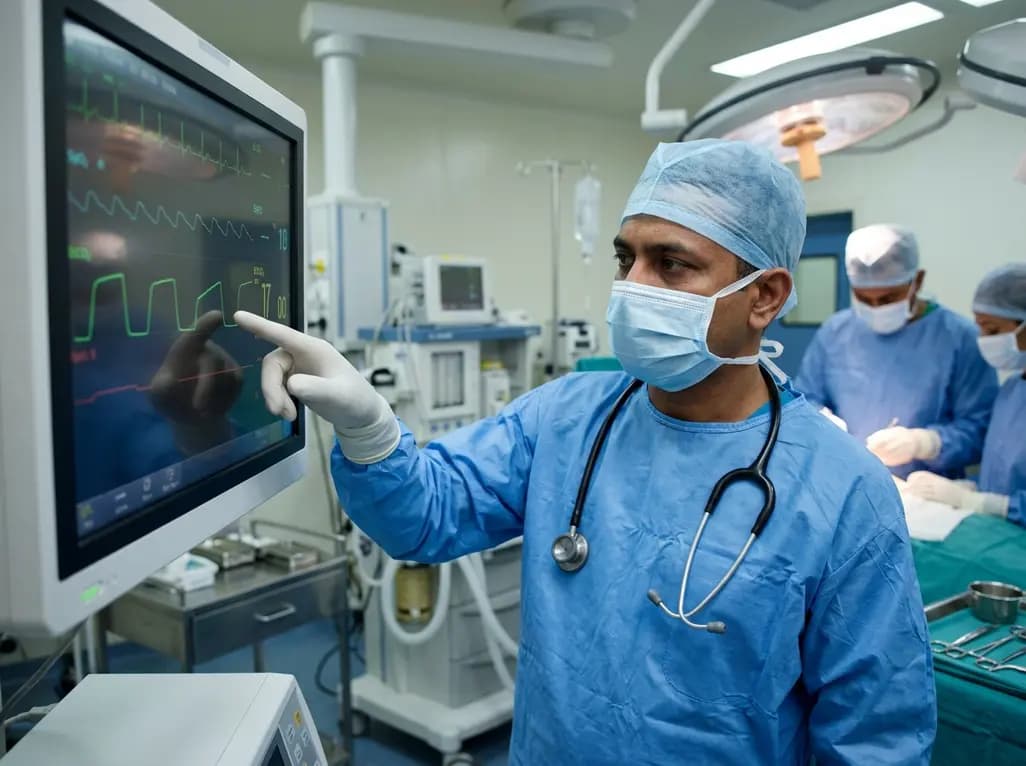
Thoracic surgery represents one of the most challenging environments for anesthesia providers, requiring meticulous control over ventilation and hemodynamics. During procedures like lobectomies or pulmonary artery reconstructions, the risk of impaired blood flow to lung segments is a constant concern. Traditionally, clinicians have relied on invasive measures or intermittent blood gas analysis to monitor these changes. However, emerging research into capnographic pulmonary perfusion assessment is providing a non-invasive alternative that offers continuous, real-time feedback. By analyzing specific waveform morphologies, such as the chair-back capnogram and shifts in the alpha angle, clinicians can now detect perfusion deficits almost instantaneously. This shift toward advanced waveform interpretation marks a significant leap in perioperative safety. Specifically, the study by Shu J et al. provides a robust framework for using these changes to protect patients during complex pulmonary interventions. Consequently, understanding these physiological signals is becoming a standard requirement for modern anesthesiology and surgical practice.
The capnogram is a graphical representation of carbon dioxide concentration over time, and its segments correlate directly with specific physiological phases. The alpha angle, located at the junction of phase II (the expiratory upstroke) and phase III (the alveolar plateau), is particularly sensitive to changes in lung mechanics and blood flow. Under normal conditions, this angle remains relatively stable, reflecting a smooth transition from conducting airway gas to alveolar gas. However, when pulmonary perfusion is compromised, the distribution of ventilation and perfusion becomes significantly heterogeneous. This mismatch alters the rate at which different alveolar compartments empty their carbon dioxide. Furthermore, impaired perfusion often leads to an increase in alveolar dead space, which directly impacts the slope of the expiratory plateau. As a result, the alpha angle tends to increase as the transition becomes less efficient. Monitoring this angle provides a sensitive, albeit often overlooked, window into the hemodynamic status of the lung. By observing these subtle shifts, anesthesiologists can identify early-stage perfusion issues before they manifest as systemic oxygenation failures. Therefore, the alpha angle serves as a primary metric in capnographic pulmonary perfusion assessment, bridging the gap between basic monitoring and advanced physiological diagnosis.
One of the most distinct visual cues identified in recent clinical studies is the emergence of a \"chair-back\" capnogram. This specific morphology occurs when there is a significant impairment in pulmonary artery flow, particularly during surgical manipulation or accidental vessel occlusion. The waveform resembles the silhouette of a high-backed chair, characterized by a sharp initial rise followed by a flattened or slightly dipped plateau. This happens because the lung segments with obstructed blood flow contribute very little carbon dioxide to the exhaled breath. Meanwhile, the well-perfused segments must compensate, but the overall mixture results in a distorted expiratory phase. Additionally, the presence of this pattern usually signals that the pulmonary circulation is no longer unobstructed. Clinicians should view the appearance of a chair-back pattern as an immediate red flag. It often precedes more obvious signs of distress, such as a drop in total end-tidal CO2 or a rise in airway pressures. Moreover, the study by Shu J et al. demonstrates that this pattern is highly reproducible across both animal models and human subjects. Recognizing this visual signature allows the surgical team to halt maneuvers and inspect the vascular integrity of the lung. Consequently, this morphological marker has become a vital tool for preventing long-term ischemic damage during thoracic procedures.
The clinical utility of capnographic pulmonary perfusion assessment extends far beyond simple detection; it fundamentally changes how surgeons and anesthesiologists interact in the operating room. When a surgeon applies traction to the lung or clamps a vessel, the impact on global and regional perfusion is immediately reflected in the capnograph. This allows for a dynamic feedback loop where the surgical approach can be modified in real-time. For instance, if a specific retractor placement causes a chair-back waveform, the surgeon can adjust the device to restore flow. Furthermore, this method is entirely non-invasive, reducing the need for repeated arterial blood sampling and the associated risks of infection or vascular injury. In the context of the Indian healthcare system, where resource optimization is critical, using existing capnography equipment for advanced diagnosis is highly cost-effective. Additionally, the ability to monitor perfusion during one-lung ventilation is particularly valuable. One-lung ventilation inherently creates a shunt, and any further impairment to the dependent lung's perfusion can be catastrophic. By prioritizing capnographic indices, clinicians can ensure that the ventilating lung is optimally perfused. Ultimately, this approach leads to fewer postoperative complications, shorter ICU stays, and improved patient recovery trajectories.
The validation of capnography as a perfusion monitor stems from rigorous experimental phases that compared waveform changes against gold-standard measurements like transit-time flow probes. In animal models, researchers induced varying degrees of pulmonary artery stenosis and measured the corresponding shifts in the alpha angle and ETCO2 levels. These experiments revealed a direct, linear correlation between the severity of perfusion impairment and the degree of waveform distortion. Specifically, as blood flow decreased, the alpha angle increased predictably, and the phase III slope became steeper or more irregular. These animal studies were crucial because they allowed for the isolation of perfusion variables without the confounding factors often present in human surgery, such as underlying chronic obstructive pulmonary disease. Moreover, the transition from animal models to clinical studies confirmed that these findings are highly applicable to human physiology. Clinical trials involving patients undergoing lobectomies showed that the chair-back pattern accurately predicted vascular kinking or thrombosis. This evidence base provides clinicians with the confidence to act on capnographic data. Therefore, the integration of animal-derived benchmarks into clinical practice ensures that the interpretation of these waveforms is grounded in solid physiological evidence rather than anecdotal observation.
Implementing capnographic pulmonary perfusion assessment into routine practice requires a combination of vigilance and education. Anesthesia providers must go beyond simply recording the numerical end-tidal CO2 value and begin actively analyzing the shape of the waveform throughout the procedure. Modern monitors often provide high-resolution displays that make it easier to see subtle changes in the alpha angle or the development of a chair-back morphology. Additionally, setting custom alarms for waveform changes, rather than just numerical thresholds, can enhance safety. Training programs for junior anesthesiologists and surgical residents should emphasize the diagnostic power of the capnogram. For example, case-based learning that features the chair-back pattern can help trainees recognize these emergencies faster. Furthermore, collaboration between the anesthesiologist and the thoracic surgeon is essential. When the anesthesia provider reports a suspicious waveform change, the surgeon should be prepared to verify the position of vascular clamps or the orientation of the lung lobes. This interdisciplinary approach ensures that the information provided by the capnograph is used to its full potential. By adopting these practices, surgical teams can significantly mitigate the risks associated with pulmonary vascular compromise. In conclusion, the capnogram is a sophisticated diagnostic tool that, when used correctly, provides a continuous stream of life-saving data.
A chair-back capnogram is a specific waveform morphology where the expiratory plateau appears distorted, resembling the shape of a chair. This pattern typically emerges when there is a significant obstruction in pulmonary blood flow, such as a clamped or kinked pulmonary artery. It indicates that certain lung segments are not participating in gas exchange, leading to a unique mixture of gases that alters the traditional rectangular shape of the capnograph trace.
The alpha angle, which is the angle between the expiratory upstroke and the alveolar plateau, generally increases during perfusion impairment. As blood flow to the alveoli decreases, the emptying of carbon dioxide becomes less uniform, causing the transition from phase II to phase III to become less sharp. This widening of the angle serves as an early indicator of ventilation-perfusion mismatch and increased alveolar dead space, often appearing before systemic oxygen saturation drops.
While capnographic pulmonary perfusion assessment provides excellent real-time, non-invasive feedback, it does not entirely replace arterial blood gas (ABG) analysis. ABG remains the gold standard for measuring precise partial pressures of oxygen and carbon dioxide, as well as pH and electrolytes. However, capnography is superior for immediate detection of sudden changes, allowing clinicians to intervene quickly. Ideally, both tools should be used complementarily to ensure the highest level of patient safety and diagnostic accuracy.
Disclaimer: This content is for informational and educational purposes only. It does not constitute medical advice or establish a doctor-patient relationship. Clinicians should rely on their professional judgment and the specific clinical context of each patient. Refer to the latest local and national guidelines for clinical practice.
References
Shu J et al. Using capnographic changes to assess pulmonary perfusion impairment during pulmonary surgery: animal experiments and clinical studies. Sci Rep. 2026 Jul 16. doi: 10.1038/s41598-026-62273-0. PMID: 42463824.
Kodali BS. Capnography. 2nd ed. Cambridge University Press; 2023.
Bhavani-Shankar K, Moseley H, Kumar AY, Delph Y. Capnometry and anaesthesia. Can J Anaesth. 1992;39(6):617-632.
"
Read summarized clinical updates, watch expert medical content, and earn CME certifications right from your smartphone.
Open your smartphone camera to scan and install.





New research highlights how specific capnographic changes, such as the chair-back morphology and alpha angle shifts, provide real-time insights into pulmonary perfusion impairment during thoracic surgery. This non-invasive method offers a vital monitoring tool for anesthesiologists and surgeons.
2 months ago
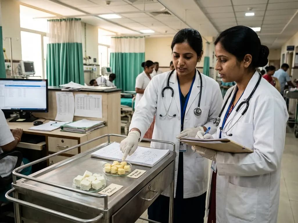
The Food Safety and Standards Authority of India has issued draft regulations prohibiting the sale and manufacture of non-dairy imitation substitutes as paneer. This nationwide regulatory action aims to eliminate consumer deception, protect dietary protein quality, and curtail lipid-related metabolic risks.
Today

A landmark Lancet Oncology Commission reveals profound global disparities in cancer genetic testing and precision medicine. While molecular therapies expand rapidly, access remains heavily concentrated in high-income nations, leaving millions of eligible cancer patients in developing regions without care.
Today
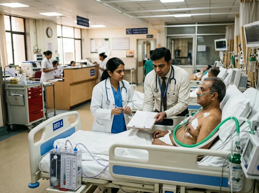
Managing refractory hypoxemia after cardiothoracic surgery becomes perilous when prolonged air leak limits positive-pressure ventilation. This case-based review details how high-flow nasal cannula, paired with awake rehabilitation, enabled successful extubation and avoided invasive re-intubation.
Today
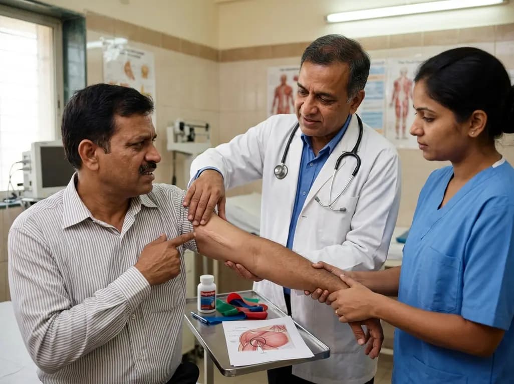
Idiopathic axillary web syndrome (IAWS) is a rare cause of shoulder pain and mobility restriction characterized by palpable axillary cording without prior surgery or trauma. Early diagnosis, nonsteroidal anti-inflammatory therapy, and physical rehabilitation lead to complete resolution of symptoms.
Today

SWEDEPAD-1 trial insights show paclitaxel-coated devices do not improve long-term limb salvage in patients with chronic limb-threatening ischemia and tissue loss. While one-year reinterventions decreased, the devices were linked to a higher risk of major amputation at three months.
Today
