.webp&w=384&q=75)

Loading, please wait...
Loading, please wait...
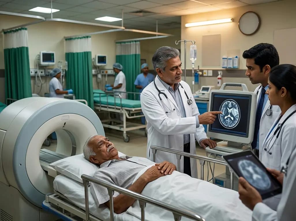
In hyperacute stroke management, rapid differentiation between acute ischaemic stroke and intracerebral haemorrhage remains paramount for initiating time-sensitive revascularization therapies. Traditional high-field magnetic resonance imaging provides exceptional diagnostic accuracy and soft-tissue contrast. However, high operational costs, extensive infrastructure requirements, and fixed physical locations frequently restrict its availability in emergency departments. Consequently, emergency clinicians urgently need accessible, point-of-care neuroimaging alternatives that deliver rapid results at the bedside. Recent technological advancements in low-field MRI stroke diagnosis offer a promising strategy to overcome these long-standing operational barriers. Operating at a field strength of 0.23 Tesla, low-field magnetic resonance systems provide rapid brain imaging directly within emergency triage areas. By eliminating complex helium cooling systems and heavy room shielding, these compact scanners streamline clinical workflows substantially. Furthermore, point-of-care imaging allows clinicians to evaluate acute patients immediately upon hospital presentation without transport delays. As a result, low-field magnetic resonance scanners promise to revolutionize emergency neurovascular triage and expand advanced diagnostic access across diverse healthcare settings.
To enhance tissue contrast on low-field scanners, investigators developed a specialized pulse sequence known as haematoma enhanced inversion recovery. This innovative sequence builds upon fluid-attenuated inversion recovery T1-weighted principles to selectively highlight acute intraparenchymal blood products. Operating with a brief acquisition time of just one minute and seventeen seconds, the sequence minimizes motion artifacts in distressed or uncooperative patients. Initial experimental parameter optimization in preclinical models established an optimal inversion time of 800 milliseconds. This precise parameter setting allows clear visual differentiation between acute intracerebral haemorrhage and healthy brain parenchyma. Moreover, pairing this inversion recovery sequence with diffusion-weighted imaging creates a comprehensive diagnostic protocol. On diffusion-weighted images, both acute ischaemia and acute intraparenchymal blood display high signal intensity due to restricted diffusion. However, on the haematoma-enhanced sequence, intracerebral haemorrhage exhibits marked hyperintensity, whereas ischaemic tissue demonstrates equal signal intensity relative to normal brain tissue. Consequently, this clear contrast provides emergency clinicians with a simple binary visual key to differentiate stroke subtypes quickly.
Before clinical translation, researchers systematically evaluated the novel imaging protocol using established swine models of acute cerebral infarction and acute intracerebral hemorrhage. Swine models were selected due to their anatomical similarity to human cerebral vasculature and brain parenchyma. Scientists acquired hourly serial magnetic resonance images over a continuous 24-hour monitoring period to analyze signal intensity changes over time. These longitudinal evaluations demonstrated consistent signal contrast trajectories throughout the early hyperacute phase. Specifically, signal intensity ratios within acute haematomas remained distinctly elevated compared to surrounding normal brain tissue across all observation timepoints. Consequently, preclinical observations confirmed that imaging contrast characteristics remain stable throughout the critical therapeutic window. Additionally, investigators fine-tuned scanning parameters to maximize signal-to-noise ratios without compromising rapid acquisition speed. The experimental trials proved that 0.23-Tesla scanners reliably produce diagnostic quality images without requiring prolonged scanning times. Furthermore, preclinical testing confirmed robust hardware performance during continuous operation. These experimental findings provided a solid scientific foundation for initiating human clinical trials in acute stroke patients.
Following preclinical success, investigators executed a prospective clinical study involving 60 human patients presenting within 24 hours of symptom onset. The clinical cohort comprised 30 individuals with acute ischaemic stroke and 30 patients with confirmed intracerebral haemorrhage. Standard 3-Tesla magnetic resonance imaging or non-contrast computed tomography served as the reference diagnostic modalities. Two blinded expert radiologists independently reviewed the 0.23-Tesla scans using pre-specified signal criteria to establish diagnostic agreement. Remarkably, the low-field system achieved 100% diagnostic accuracy across all 60 patients evaluated in the clinical study. Inter-rater reliability reached complete agreement, proving the clarity of signal differentiation between stroke subtypes. Furthermore, no adverse events or procedural complications occurred during scanner operation. Notably, the low-field technique successfully identified small parenchymal haematomas, demonstrating high sensitivity even for minor bleeding events. Consequently, these clinical results demonstrate that low-field scanners deliver diagnostic accuracy comparable to high-field units in emergency environments. This high diagnostic fidelity supports integrating low-field systems into routine emergency stroke workflows.
Conventional high-field magnetic resonance systems require dedicated radiofrequency shielding, reinforced flooring, and specialized helium cryogen management. Consequently, these systems are almost universally located in centralized radiology departments situated far from emergency receiving areas. Transporting acutely ill stroke patients from emergency bays to distant radiology suites creates substantial time delays, consuming precious brain tissue. In contrast, 0.23-Tesla low-field scanners possess a small physical footprint and operate using standard wall electrical power. Therefore, healthcare institutions can easily deploy these compact devices directly inside emergency departments, intensive care units, or mobile stroke vehicles. Furthermore, significantly lower equipment and operational costs make advanced magnetic resonance technology financially accessible to community hospitals and rural clinics. By eliminating patient transport outside the emergency department, point-of-care imaging dramatically shortens door-to-needle times for acute thrombolytic therapy. Additionally, low-field systems generate minimal acoustic noise and eliminate severe fringe magnetic fields, greatly reducing projectile risks. As a result, deploying low-field imaging directly at the bedside enhances patient safety while streamlining emergency care.
The clinical success of 0.23-Tesla magnetic resonance imaging marks a significant leap forward in point-of-care neurovascular diagnostics. However, widespread clinical adoption requires validation through prospective multicentre trials involving larger, heterogeneous patient cohorts. Future research must assess diagnostic accuracy across hyperacute timeframes under three hours and evaluate performance in restless or uncooperative patients. Moreover, integrating artificial intelligence algorithms with low-field platforms promises to further transform point-of-care imaging. Deep learning models can automate image reconstruction, reduce background noise, and assist clinicians with automated lesion identification. Additionally, engineers are developing specialized low-field sequences for arterial spin labeling perfusion and magnetic resonance angiography. Combining structural, contrast, and perfusion imaging into a rapid bedside protocol could provide comprehensive neurovascular evaluation within minutes. Consequently, point-of-care low-field magnetic resonance technology is positioned to democratize acute stroke evaluation worldwide. Ultimately, this innovation ensures that patients everywhere receive rapid, accurate diagnostic assessment, improving functional outcomes and expanding equitable access to life-saving stroke interventions.
The 0.23-Tesla MRI uses a combination of diffusion-weighted imaging and haematoma enhanced inversion recovery sequences. Intracerebral haemorrhage displays hyperintensity on both sequences, whereas acute ischaemic stroke shows hyperintensity on diffusion-weighted imaging but remains isointense on the haematoma-enhanced sequence. This distinct signal difference allows clear visual differentiation within minutes.
Low-field MRI units offer a compact footprint, require standard electrical power, and eliminate complex cryogenic cooling. These features enable direct installation inside emergency departments. Bedside imaging eliminates transport delays, dramatically accelerates triage, reduces healthcare costs, and improves access to rapid advanced neuroimaging in resource-constrained medical settings.
Yes, 0.23-Tesla MRI is exceptionally safe for acute stroke patients. Clinical trials reported zero adverse events or complications during scanning procedures. Lower magnetic field strength significantly decreases acoustic noise levels and minimizes projectile hazard risks, making bedside low-field scanning highly tolerable and safe in acute emergency settings.
Disclaimer: This content is for informational and educational purposes only, and does not substitute for professional medical advice, diagnosis, or treatment. Refer to the latest local and national guidelines for clinical practice.
References

Read summarized clinical updates, watch expert medical content, and earn CME certifications right from your smartphone.
Open your smartphone camera to scan and install.





A clinical study demonstrates that a 0.23-Tesla low-field MRI using a novel HEIR sequence achieved 100% diagnostic accuracy in differentiating acute ischaemic stroke from intracerebral haemorrhage within 24 hours, offering a fast, cost-effective point-of-care solution for emergency triage.
Today
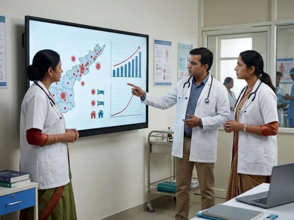
Andhra Pradesh reported 10 new Covid-19 cases, taking the state tally to 49 while deaths remain at four. With 24 patients hospitalized and 16 under home isolation, the Health Department has intensified monitoring. Medical professionals should review regional distribution, diagnostic protocols, and management plans.
Today

An 11-year Swedish registry study of 618 uterine sarcoma patients found that minimally invasive surgery yielded survival comparable to open surgery in early stages. However, adjuvant chemotherapy conferred no survival benefit in localized or advanced disease, highlighting stage and histology as key outcomes.
3 days back

A cross-sectional study evaluates post-intensive care syndrome in cardiac patients 2-4 weeks post-ICU discharge, highlighting cognitive, psychological, and functional impairments and the need for structured multidisciplinary rehabilitation.
3 days back

Anterior cruciate ligament reconstruction failure lacks uniform definition. A narrative review proposes an integrative framework incorporating objective and subjective instability, persistent pain, restricted motion, graft rupture, and secondary meniscal injury to standardize clinical reporting.
3 days back
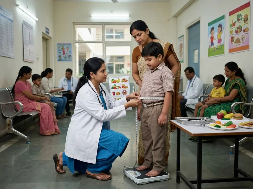
With World Obesity Atlas data warning that over 41 million Indian children are overweight or obese, ICMR and NIN have unveiled a 10-point policy roadmap. The initiative calls for mandatory front-of-pack labeling, HFSS taxes, strict marketing bans, and healthier school environments to curb non-communicable diseases.
Today
