.webp&w=384&q=75)

Loading, please wait...
Loading, please wait...
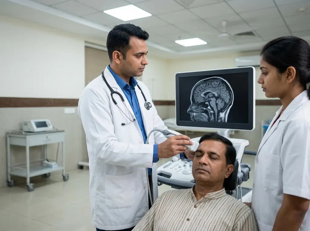
Process engineering and advanced medical diagnostics often require monitoring in environments that are physically or chemically challenging. Conventional ultrasound transducers frequently fail when exposed to extreme temperatures or caustic fluids. This has led to the development of waveguide-based ultrasound inspection, a technique that shields sensitive sensors from harsh conditions while maintaining high-quality imaging. By utilizing waveguides, researchers can transmit acoustic signals from a safe distance, protecting the transducer while capturing critical real-time data. This technology, originally designed for industrial monitoring in solar power plants and battery manufacturing, is now showing remarkable promise for transcranial ultrasound imaging and other complex medical applications where bone or tissue density creates significant barriers.
Historically, ultrasonic testing relied on single-mode waveguides (SMWGs) to provide basic structural health monitoring. While effective at protecting transducers from thermal damage, these early systems were limited by their slow, point-by-point scanning requirements. To overcome these hurdles, engineers have transitioned toward multimode waveguides (MMWGs). These advanced systems exploit multiple wave modes simultaneously, allowing for much faster data acquisition and more detailed imaging. The move toward MMWGs represents a significant leap forward, enabling the visualization of complex internal structures in real time. For medical professionals, this evolution suggests a future where ultrasound can penetrate previously "opaque" anatomical structures with the same efficiency as traditional soft-tissue scans.
One of the primary challenges in both industrial and medical ultrasound is the distortion of sound waves as they pass through different media. In harsh industrial environments, thermal gradients and fluid instabilities can warp wavefronts, leading to blurry images. Similarly, in transcranial ultrasound imaging, the human skull acts as a highly aberrative medium, scattering sound waves and reducing clarity. To address this, researchers are developing sophisticated aberration correction algorithms and advanced modeling approaches. These computational tools analyze the wavefront distortion and mathematically reconstruct the image to restore focus and detail. By refining these algorithms, scientists are making it possible to see through the thickest parts of the skull with unprecedented resolution.
The potential transferability of waveguide technology to the medical field is a significant highlight of recent research. Specifically, the ability to image through the skull without invasive surgery could revolutionize neurology and intensive care. Currently, transcranial ultrasound imaging is often limited to neonates through the fontanel or patients with surgical skull openings. Waveguide-based systems, combined with multimode propagation and aberration correction, offer a non-invasive alternative for monitoring brain health, detecting hemorrhages, or guiding therapeutic interventions. This technology could allow for continuous bedside monitoring in neuro-ICUs, providing real-time data that was previously only accessible through frequent and costly CT or MRI scans.
Optimization is the current frontier for waveguide-based imaging systems. Researchers are focusing on improving wettability and geometry to ensure seamless signal transmission between the waveguide and the target medium. In a medical context, this means refining the interface between the ultrasound probe and the patient’s skin or bone to minimize signal loss. Additionally, the development of computational ultrasound imaging is opening new doors for 3D volumetric scans. These optimizations aim to make the technology more user-friendly and reliable for clinical staff. As these systems become more refined, they will likely integrate into standard diagnostic workflows, offering a safer and more cost-effective way to visualize deep-seated pathologies in high-risk patients.
The journey from industrial structural health monitoring to advanced medical neuroimaging illustrates the versatility of waveguide-based ultrasound. Future research is expected to focus on miniaturizing these systems and further enhancing the speed of aberration correction algorithms. By combining hardware shielding with powerful software processing, the next generation of ultrasound devices will be better equipped to handle the "harsh environments" of the human body, such as dense bone or inflamed tissue. For the medical community in India and beyond, these findings support the continued evolution of point-of-care diagnostics. Ultimately, the integration of waveguide technology into clinical practice could save lives by providing earlier and more accurate detection of neurological conditions.
Waveguide technology acts as a physical buffer between the sensitive transducer and the harsh environment, such as extreme heat or corrosive chemicals. The waveguide transmits the acoustic waves over a distance, allowing the electronics to remain in a controlled, safe temperature zone. This shielding prevents device failure and allows for continuous monitoring in conditions that would otherwise destroy standard medical or industrial ultrasound equipment instantly.
Multimode waveguides (MMWGs) are superior because they utilize multiple wave propagation modes at once, significantly increasing the amount of data captured. While single-mode systems are restricted to slow, point-by-point scanning, MMWGs enable much faster imaging and higher resolution. This speed is critical for real-time monitoring of dynamic processes, such as fluid flow in industrial batteries or blood flow in the human brain during emergency medical procedures.
The human skull is highly irregular in density and thickness, causing sound waves to refract and scatter as they pass through. This distortion, known as aberration, makes it difficult for standard ultrasound to produce a clear image of the underlying brain tissue. Advanced aberration correction algorithms use mathematical models to calculate and reverse this distortion, allowing clinicians to reconstruct sharp, accurate images of the brain despite the presence of bone.
Disclaimer: This content is for informational and educational purposes only. It is not a substitute for professional medical advice, diagnosis, or treatment. Always seek the advice of your physician or other qualified health provider with any questions you may have regarding a medical condition. Refer to the latest local and national guidelines for clinical practice.
References
Othmani C et al. Waveguide-based in-process ultrasound inspection and imaging in harsh environments: advances and challenges. Ultrasonics. 2026 Jul 10. doi: undefined. PMID: 42430861.
Kohtanen E et al. Transcranial Radiation of Guided Waves for Brain Ultrasound. Georgia Institute of Technology. 2021 Jun 08.
Simonetti F. Cryo-ultrasonic imaging of curved components. J. Japanese Soc. NDI, 73, 78-84. 2024.

Read summarized clinical updates, watch expert medical content, and earn CME certifications right from your smartphone.
Open your smartphone camera to scan and install.





Waveguide-based ultrasound imaging is making significant strides in harsh environments and holds immense potential for medical applications, such as imaging through the human skull. This review explores the evolution of multimode waveguides and the development of aberration correction algorithms.
3 months ago

Depemokimab offers twice-yearly dosing for severe eosinophilic asthma and chronic rhinosinusitis with nasal polyps, significantly cutting exacerbations and hospital admissions.
3 days back

A novel copper metal-organic framework hydrogel remodels the uterine microenvironment to stimulate robust endometrial regeneration, reduce intrauterine fibrosis, and restore reproductive fertility, offering a groundbreaking strategy for managing refractory endometrial damage and Asherman syndrome.
3 days back

New research reveals that non-ablative radiotherapy suppresses ventricular tachycardia by increasing conduction velocity via NaV1.5 upregulation and action potential prolongation. However, sustained sodium current elevation and depressed calcium handling highlight the need for careful clinical translation.
5 days back

A single-cell transcriptomic atlas reveals how peripheral nerve injuries cause dorsal root ganglion remodeling. By evaluating proximal nerve crush and resection, researchers identify distinct glial and immune programs that resolve neuropathic pain and promote regeneration.
3 days back

A breakthrough study demonstrates that fullerol-primed extracellular vesicles generated in a 3D SECCS bioreactor attenuate ferroptosis and restore myogenic regeneration in diabetes via the FOXO3 pathway, offering a cell-free therapeutic strategy for diabetic musculoskeletal degeneration.
3 days back
