.webp&w=384&q=75)

Loading, please wait...
Loading, please wait...
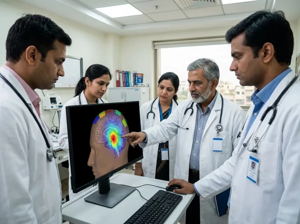
Tumor Treating Fields (TTFields) have emerged as a significant non-invasive therapeutic modality in neuro-oncology. Historically, the FDA has approved this technology primarily for supratentorial glioblastoma. However, the management of infratentorial tumors, specifically brainstem gliomas, remains a profound clinical challenge. These tumors are often surgically inaccessible and resistant to conventional radiotherapy and chemotherapy. Consequently, clinicians are increasingly exploring whether the physical properties of alternating electric fields can be applied to these deep-seated structures. The primary concern has been the feasibility of delivering a therapeutic dose to the brainstem using standard scalp-only transducer arrays. A recent simulation study investigated the potential of TTFields for brainstem gliomas to determine if optimized array layouts could provide sufficient field intensity. This research represents a vital step toward expanding the application of electric field therapy beyond the cerebral hemispheres. By leveraging patient-specific modeling, the study aims to bridge the gap between theoretical dosimetry and clinical application in the posterior fossa.
Understanding how TTFields function is essential for appreciating their potential in the brainstem. These low-intensity, intermediate-frequency alternating electric fields disrupt the highly orchestrated process of mitosis. Specifically, they exert dielectrophoretic forces on polar molecules, such as tubulin and septins, which are critical for spindle formation and cytokinesis. When these processes are interrupted, the cancer cells undergo apoptosis or senescence. In the context of TTFields for brainstem gliomas, the goal is to achieve a local minimum field intensity (LMiFI) of at least 1.0 V/cm, which is widely considered the therapeutic threshold. Achieving this in the infratentorial region requires sophisticated planning. The MAXPOINT platform, developed by Novocure, utilizes finite element calculations to generate detailed maps of electric field distribution. This platform allows for the optimization of scalp-only array layouts by accounting for individual anatomical variations. By simulating how electric currents flow through various tissues like the skull, cerebrospinal fluid, and white matter, researchers can predict the dose delivered to the target tumor volume with high precision.
The research team utilized imaging data from seven patients diagnosed with brainstem gliomas to conduct their feasibility simulation. They integrated MRI and CT scans to create high-resolution patient-specific models. The clinicians defined the Gross Tumor Volume (GTV) based on enhancing lesions on T1 post-contrast MRI. Furthermore, they established a Clinical Target Volume (CTV) by adding a 3 mm peritumoral expansion to the GTV. This approach ensures that microscopic extensions of the tumor are included in the therapeutic planning. The researchers then compared two different array configurations: a standard unplanned layout and an optimized layout generated by the MAXPOINT platform. Using finite element calculations, they generated maps for both local minimum field intensity (LMiFI) and local minimum power density (LMiPD). They employed one-sided paired t-tests to evaluate the statistical significance of the differences between the two layouts. This rigorous methodology allowed the team to determine whether personalized optimization significantly enhances the dose delivered to the brainstem and surrounding posterior fossa structures.
The results of the simulation were highly encouraging for the future of TTFields for brainstem gliomas. The MAXPOINT-optimized layouts achieved significantly higher field intensities across all target regions compared to the standard configurations. Specifically, the median LMiFI in the GTV reached 1.1 V/cm with optimized layouts, compared to 1.0 V/cm with standard ones (p = 0.019). Similarly, the CTV showed a median LMiFI of 1.1 V/cm versus 1.0 V/cm (p = 0.002). Notably, the entire brainstem region experienced a significant increase, with the median LMiFI rising from 1.1 V/cm to 1.3 V/cm. The most substantial improvement occurred in the posterior fossa, where optimized layouts delivered a median LMiFI of 1.5 V/cm, compared to 1.2 V/cm in the standard group. These findings suggest that the therapeutic reference of 1.0 V/cm is not only achievable but can be exceeded with precise planning. Furthermore, the power density measurements (LMiPD) followed a similar trend, reinforcing the conclusion that optimized scalp-only arrays can effectively target deep infratentorial tumors.
In India, where the burden of advanced brain tumors is significant, the development of non-invasive therapies like TTFields is of great interest. Brainstem gliomas in both pediatric and adult populations often carry a poor prognosis due to the critical nature of the surrounding neuroanatomy. Therefore, the ability to deliver therapeutic electric fields using scalp-only arrays without invasive procedures is a major advantage. This study proves that optimized layouts are essential for reaching the 1.0 V/cm threshold in the brainstem. For Indian oncologists and radiologists, this data suggests that if TTFields are adopted for infratentorial cases, personalized treatment planning must be prioritized. Moreover, the study demonstrates that the posterior fossa, despite its depth and surrounding bone structures, is a viable target for electric field therapy. As clinical trials progress, this dosimetric evidence provides a safety and efficacy rationale for exploring TTFields as a combination therapy with existing standards of care. Consequently, this technology might offer a localized treatment option that minimizes systemic toxicity while addressing the infiltrative nature of these gliomas.
While this simulation study confirms the feasibility of delivering therapeutic doses, the transition to clinical practice requires further investigation. Future research should focus on correlating these dosimetric simulations with actual clinical outcomes, such as progression-free survival and overall survival. The study highlighted that TTFields for brainstem gliomas can achieve the necessary intensity, but the biological response of infratentorial tumor cells may differ from those in the supratentorial region. Additionally, researchers must evaluate the tolerability of higher field intensities in the brainstem, which houses vital autonomic centers. The success of the MAXPOINT platform in this study underscores the importance of computational modeling in modern radiotherapy and bio-electric medicine. As technology evolves, we may see more integrated platforms that allow real-time adjustments to array layouts based on tumor response. Ultimately, the goal is to refine these tools to provide a personalized, effective, and safe therapeutic option for patients who currently have very limited choices. This study serves as a foundational pillar for future clinical trials targeting the complex landscape of the brainstem.
The therapeutic reference for Tumor Treating Fields is generally considered to be a local minimum field intensity (LMiFI) of 1.0 V/cm or higher. This study demonstrated that optimized layouts can consistently meet or exceed this threshold in the Gross Tumor Volume and the Clinical Target Volume of brainstem gliomas.
The MAXPOINT platform utilizes patient-specific MRI and CT imaging to perform finite element calculations. This allows the system to optimize scalp-only transducer array layouts by predicting how electric fields will distribute through individual anatomy. It significantly increases the field intensity compared to standard, non-optimized array configurations.
Yes, this simulation study confirms that scalp-only arrays are feasible for treating deep-seated infratentorial tumors. By optimizing the placement of the transducers on the scalp, researchers were able to achieve therapeutic field intensities within the brainstem and the posterior fossa without the need for invasive electrodes.
Disclaimer: This content is for informational and educational purposes only and does not constitute medical advice, diagnosis, or treatment. Always seek the advice of your physician or another qualified health provider with any questions you may have regarding a medical condition. Refer to the latest local and national guidelines for clinical practice.
References
Chen AY et al. Feasibility evaluation of tumor treating fields for brainstem gliomas. J Neurooncol. 2026 Jul 06. doi: 10.1007/s11060-026-05697-y. PMID: 42406140.
Stupp R et al. Effect of Tumor-Treating Fields Plus Maintenance Temozolomide vs Maintenance Temozolomide Alone on Survival in Patients With Glioblastoma: A Randomized Clinical Trial. JAMA. 2017;318(23):2306-2316.
Ballo MT et al. Correlation of Tumor Treating Fields Dosage and Survival Outcomes in Patients With Newly Diagnosed Glioblastoma. JCO Prev Oncol. 2023;7:e2200434.
"
Read summarized clinical updates, watch expert medical content, and earn CME certifications right from your smartphone.
Open your smartphone camera to scan and install.





A new simulation study evaluates the feasibility of Tumor Treating Fields (TTFields) for brainstem gliomas using MAXPOINT optimization. The research demonstrates that scalp-only transducer arrays can achieve therapeutic field intensities in the infratentorial region, offering a potential new treatment path.
3 months ago

Depemokimab offers twice-yearly dosing for severe eosinophilic asthma and chronic rhinosinusitis with nasal polyps, significantly cutting exacerbations and hospital admissions.
3 days back

A novel copper metal-organic framework hydrogel remodels the uterine microenvironment to stimulate robust endometrial regeneration, reduce intrauterine fibrosis, and restore reproductive fertility, offering a groundbreaking strategy for managing refractory endometrial damage and Asherman syndrome.
3 days back

New research reveals that non-ablative radiotherapy suppresses ventricular tachycardia by increasing conduction velocity via NaV1.5 upregulation and action potential prolongation. However, sustained sodium current elevation and depressed calcium handling highlight the need for careful clinical translation.
5 days back

A single-cell transcriptomic atlas reveals how peripheral nerve injuries cause dorsal root ganglion remodeling. By evaluating proximal nerve crush and resection, researchers identify distinct glial and immune programs that resolve neuropathic pain and promote regeneration.
3 days back

A breakthrough study demonstrates that fullerol-primed extracellular vesicles generated in a 3D SECCS bioreactor attenuate ferroptosis and restore myogenic regeneration in diabetes via the FOXO3 pathway, offering a cell-free therapeutic strategy for diabetic musculoskeletal degeneration.
3 days back
