.webp&w=384&q=75)

Loading, please wait...
Loading, please wait...
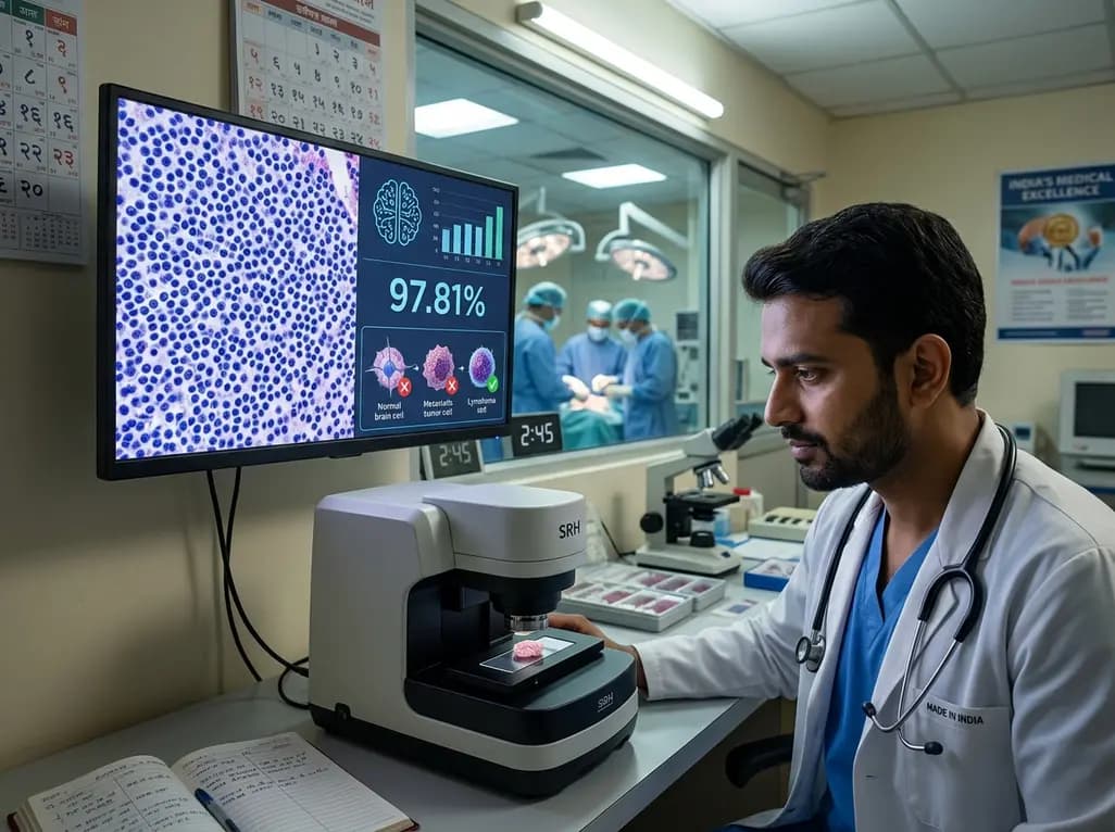
Neurosurgical management of intracranial lesions requires prompt diagnostic guidance during surgery. Distinguishing primary central nervous system lymphoma from other malignant brain lesions represents a critical clinical challenge. Surgeons need accurate real-time tissue assessment because surgical goals differ dramatically between disease entities. Primary CNS lymphoma requires minimal tissue biopsy to avoid unnecessary neurological morbidity, whereas high-grade gliomas necessitate maximal safe cytoreductive resection. Traditional intraoperative diagnosis relies heavily on frozen section analysis, which often suffers from tissue artifacts and prolonged processing times. Recent technological advances in stimulated Raman histology provide a transformative optical imaging alternative for intraoperative neuropathology. Researchers have now integrated this label-free microscopic imaging with deep learning artificial intelligence to enable rapid bedside diagnosis. Consequently, surgical teams can receive diagnostic clarification within minutes inside the operating theatre. This breakthrough technology enhances diagnostic confidence and streamlines decision-making for complex neuro-oncological procedures.
Intraoperative differentiation between primary central nervous system lymphoma and other intracranial neoplasia presents substantial diagnostic hurdles. Specifically, cytological overlap between atypical lymphoid infiltrates, reactive gliosis, and necrotic high-grade gliomas frequently confuses frozen section interpretations. Moreover, frozen section preparation alters fragile cell architecture through freezing artifacts and compression deformities. Pathologists often struggle to establish definitive diagnoses under strict surgical time constraints. Consequently, neurosurgeons face uncertainty when deciding whether to proceed with extensive tumor debulking or terminate the procedure following tissue sampling.
When surgeons mistakenly perform aggressive resection on primary CNS lymphoma, patient morbidity increases without therapeutic benefit. Furthermore, delayed intraoperative diagnosis prolongs anesthesia time, increases surgical risks, and exhausts vital hospital resources. Traditional histopathology requires labor-intensive sectioning and staining, consuming twenty to forty minutes per specimen. Thus, clinical teams urgently require a fast diagnostic modality that preserves tissue integrity while delivering rapid morphological identification. Optical imaging techniques paired with automated computer vision pipelines offer a promising solution to these long-standing intraoperative challenges.
Stimulated Raman histology utilizes non-linear vibrational Raman scattering microscopy to generate high-resolution, label-free tissue images without physical staining. The imaging system captures intrinsic chemical bonds within lipid and protein molecules directly from fresh tissue samples. Subsequently, automated color-mapping algorithms convert optical raw data into virtual digital microscopic slides within three minutes. Because the process requires no frozen sectioning or chemical fixation, clinicians obtain pristine virtual histology effortlessly.
Additionally, this non-destructive technology preserves tissue specimens intact for subsequent downstream molecular testing and next-generation genetic sequencing. Portable bedside Raman microscopes fit seamlessly into operating rooms, allowing surgical personnel to scan biopsy specimens immediately upon collection. Operating teams no longer experience diagnostic delays caused by transporting tissue to central laboratories. Furthermore, digital microscopic images can easily travel via telepathology networks for immediate remote subspecialist consultation. Ultimately, this approach bridges the gap between surgical excision and real-time histopathological analysis, modernizing standard intraoperative workflows.
To optimize diagnostic accuracy, researchers created RapidLymphoma, an advanced deep learning computational pipeline based on self-supervised learning principles. The development team trained this neural network on fifty-four thousand digital microscopic image patches derived from surgical resections and stereotactic biopsies. These diverse training samples represented a broad spectrum of neoplastic central nervous system entities collected across four international medical centers. Final formal histopathological evaluation provided the gold standard diagnostic ground truth for model validation.
Importantly, self-supervised learning allows the network to learn rich structural features directly from unlabeled histology data before fine-tuning for specific diagnostic tasks. The resulting algorithm rapidly evaluates microscopic architecture, cellular density, and nuclear features in under three minutes. In addition, the system generates interpretable visual heatmaps that highlight key diagnostic cytomorphological regions for attending clinicians. Thus, the deep learning model acts as a reliable automated decision-support tool, assisting neurosurgeons and pathologists during high-stakes intraoperative consultations.
In a prospective international multicenter validation trial involving one hundred sixty patients, the automated system achieved outstanding clinical diagnostic accuracy. The deep learning model demonstrated an overall balanced diagnostic accuracy of 97.81 percent when differentiating lymphoma from non-lymphoma entities. Remarkably, the artificial intelligence system achieved 100 percent diagnostic sensitivity for detecting primary CNS lymphoma in this prospective cohort. In comparison, standard frozen section analysis yielded a sensitivity of only 77.77 percent on the same patient sample.
Furthermore, investigators evaluated the platform across two additional independent testing cohorts comprising hundreds of clinical cases. The algorithm achieved balanced accuracy rates exceeding 95 percent when distinguishing lymphoma from IDH-wildtype diffuse gliomas and metastatic brain tumors. Notably, the system maintained consistent diagnostic precision across surgical resection specimens and small needle biopsy cores. Consequently, these robust prospective results confirm that automated microscopic imaging offers superior diagnostic reliability compared to traditional intraoperative frozen section workflows.
Integrating automated optical histology into neurosurgical practice provides profound therapeutic and operational benefits. Real-time differentiation of lymphoma prevents unnecessary radical tumor resections, sparing patients from potential neurological deficits or motor weakness. Once surgical teams confirm a primary CNS lymphoma diagnosis intraoperatively, they can immediately stop surgical debulking and plan prompt systemic chemo-immunotherapy. Conversely, when the system confirms a high-grade glioma or solitary metastasis, surgeons can confidently pursue maximal safe surgical cytoreduction.
Moreover, the platform standardizes diagnostic precision across varied healthcare settings, reducing diagnostic disparities between academic centers and community hospitals. Because visual heatmaps highlight critical histopathological features, neurosurgeons gain immediate actionable feedback even when specialized neuropathologists are unavailable on-site. Subsequently, hospital systems can reduce operating room time, lower overall procedural expenses, and improve surgical safety profiles. Overall, combining advanced optical physics with deep learning represents a transformative paradigm shift in modern neuro-oncological patient management.
Stimulated Raman histology is a label-free optical imaging technology that uses laser-based vibrational scattering to detect lipid and protein chemistry in fresh tissue. Automated software converts raw optical data into virtual digital images mimicking traditional hematoxylin and eosin staining within three minutes, eliminating freezing, sectioning, or chemical staining protocols.
Accurate intraoperative diagnosis directly dictates the surgical strategy for brain tumors. Primary central nervous system lymphoma requires minimal tissue biopsy followed by systemic chemotherapy and immunotherapy. Conversely, gliomas and brain metastases require maximal safe surgical resection. Rapid identification prevents unnecessary brain tissue removal and avoids neurological morbidity.
In a prospective multicenter trial, the RapidLymphoma pipeline achieved a 97.81 percent balanced accuracy and 100 percent sensitivity in identifying primary CNS lymphoma. It outperformed frozen section analysis, which achieved 77.77 percent sensitivity. The system also demonstrated over 95 percent accuracy when differentiating lymphoma from gliomas and metastases.
Disclaimer: This content is for informational and educational purposes only and does not constitute medical advice, diagnosis, or treatment. Always seek the advice of a qualified healthcare provider with any questions regarding a medical condition or clinical management. Refer to the latest local and national guidelines for clinical practice.
References

Read summarized clinical updates, watch expert medical content, and earn CME certifications right from your smartphone.
Open your smartphone camera to scan and install.





Combining stimulated Raman histology with deep learning enables fast, label-free intraoperative detection of primary CNS lymphoma in under three minutes, achieving 97.81% accuracy and outperforming conventional frozen section analysis to guide neurosurgical decision-making.
last month

A breakthrough study using human colonic organoids demonstrates that bacterial serine protease EspP, an EHEC cytotoxin, halts epithelial proliferation and preferentially triggers enteroendocrine cell differentiation, promoting pro-inflammatory chemokine release and immune cell recruitment.
5 days back
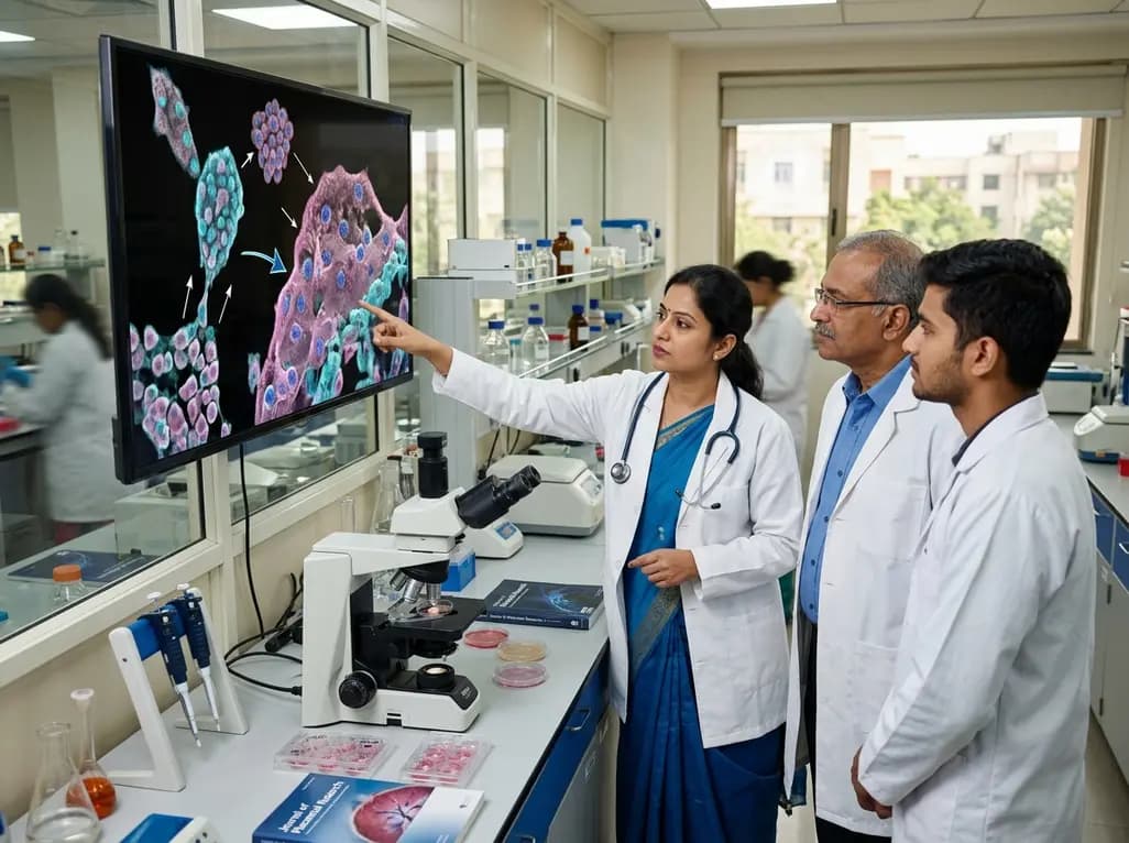
Recent research reveals that downregulating YAP1/TAZ-TEAD via Hippo signaling triggers spontaneous human trophoblast syncytialization in 3D cultures, illuminating placental biology and preeclampsia.
5 days back
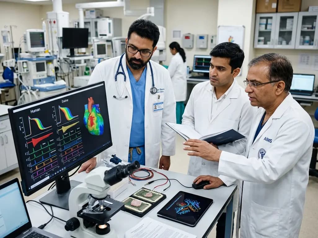
A landmark comparative study evaluates atrial electrophysiology across humans, swine, and rodents. By linking cellular recordings with transcriptomics and in silico modeling, researchers resolve cross-species gaps, explaining translational failures to advance targeted therapies for atrial arrhythmias.
5 days back
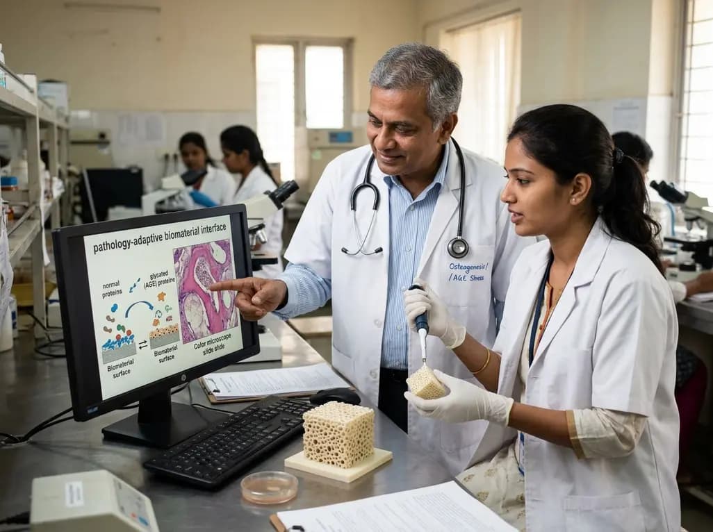
A novel pathology-adaptive surface engineering strategy uses functionalized plasma polymer coatings to selectively modulate AGE adsorption, reducing oxidative stress and restoring bone formation in diabetic and aging microenvironments.
4 weeks back
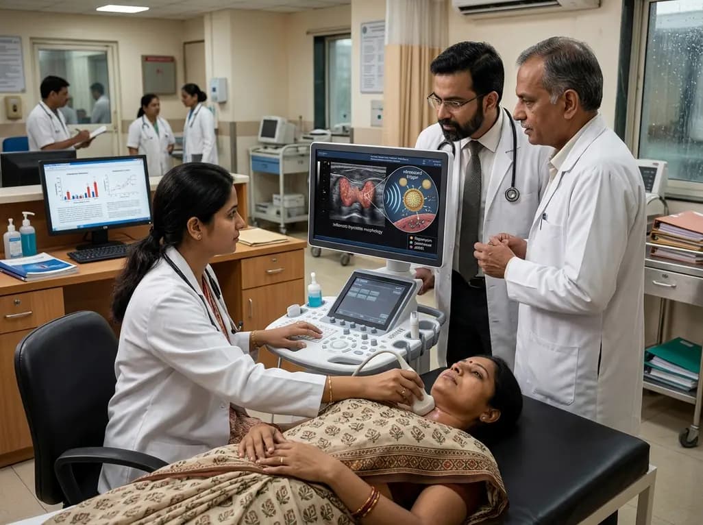
Researchers have developed an innovative ultrasound-responsive nano-contrast agent that co-delivers CIITA-siRNA and rapamycin directly to inflamed thyroid tissue, significantly reducing autoimmune injury and thyroid autoantibodies in preclinical models of Hashimoto thyroiditis.
6 days back
