

Loading, please wait...
Loading, please wait...
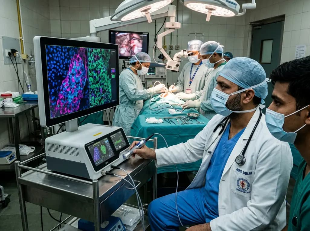
Intraoperative tissue verification is a foundational step during intracranial tumor surgery. Stereotactic neurosurgical procedures carry an inherent risk of sampling error when clinicians target deep lesions. Consequently, operating teams require immediate feedback to ensure that sampled specimens contain diagnostic tissue. Traditional frozen section analysis often takes substantial time, consumes valuable diagnostic material, and causes freezing artifacts. To overcome these procedural hurdles, clinicians increasingly utilize stimulated Raman histology directly within surgical suites. This optical method creates high-resolution digital images from unprocessed, fresh tissue in near real time. When investigators couple this optical technique with artificial intelligence models, surgical teams gain rapid automated diagnostic support. A recent multicenter clinical investigation published in Neurosurgery Focus highlights how optical workflows evaluate routine neurosurgical biopsies while navigating challenges like specimen heterogeneity.
Stereotactic brain biopsies require rigorous precision because neurosurgeons harvest minute cores from deep intracranial locations. Consequently, retrieving non-diagnostic tissue forces repeated passes, which increases the likelihood of intracranial hemorrhage or neurological deficit. Traditional intraoperative neuropathology relies heavily on frozen section consultation or smear preparations. Although conventional frozen sections remain the diagnostic standard, the preparation requires significant labor and introduces mechanical artifacts. Furthermore, freezing often damages delicate tissue architecture, which complicates downstream immunohistochemistry and molecular profiling. Therefore, surgeons need label-free optical evaluation that preserves tissue architecture for genetic sequencing. Advanced optical workflows maintain tissue integrity by avoiding chemical fixation, freezing, and sectioning. As a result, the entire specimen remains intact for subsequent molecular testing. In addition, rapid optical assessment reduces general anesthesia duration, improving overall patient safety.
To understand the clinical utility of stimulated Raman histology, one must examine its optical physics. The system uses two synchronized picosecond lasers that excite intrinsic molecular vibrations within organic compounds. Specifically, the imaging platform detects carbon-hydrogen chemical bonds present within lipids and proteins. By assessing the ratio between lipid-rich and protein-rich structures, the instrument generates high-contrast digital micrographs without exogenous dyes. Furthermore, digital processing assigns pseudo-colors to these signals, producing images that closely mimic standard hematoxylin and eosin staining. Neuropathologists can immediately recognize nuclear atypia, cellular density, microvascular proliferation, and necrotic areas. Consequently, surgical teams assess fresh tissue within minutes inside the operating room. Therefore, the technology removes logistical delays associated with transporting specimens to pathology laboratories. Most importantly, label-free optical imaging leaves the diagnostic sample completely unaltered for post-operative molecular analysis.
Investigators in a clinical trial evaluated 204 optical scans obtained from 89 consecutive neurosurgical patients. The research team processed each scan using Glioma Reveal, a certified deep learning algorithm designed to calculate tumor cell probabilities. Initially, the automated algorithm demonstrated an overall diagnostic accuracy of 80.9% across all analyzed scans. However, diagnostic accuracy rose significantly to 94.1% after secondary neuropathological review resolved initial discordant predictions. For the primary cohort containing gliomas and lymphomas, the algorithm initially demonstrated an 81.5% diagnostic accuracy, 78.8% sensitivity, and 100% specificity. Notably, expert re-evaluation revealed that 80.6% of initial false-negative classifications were morphologically correct within that specific field of view. When researchers accounted for focal geographic variations, the corrected accuracy for gliomas and lymphomas reached an impressive 96.4%.
Spatial sampling discrepancies present substantial hurdles during intraoperative artificial intelligence assessments. Malignant brain tumors exhibit marked internal architectural heterogeneity, containing patches of viable tumor, infiltrative margins, reactive gliosis, and acellular necrosis. In this study, automated models scanned specific regions of interest rather than entire specimen blocks. Consequently, an algorithm might correctly classify an optical scan as non-tumorous if the field of view captures only necrotic debris. However, the broader clinical specimen may still contain diagnostic tumor elsewhere in the tissue block. Thus, scan-level false negatives often reflect true focal absence rather than algorithmic failure. Clinicians must recognize that automated models evaluate only the precise optical focus presented to them. Therefore, surgeons must combine algorithmic readouts with clinical impression and radiographic findings. Relying blindly on automated scores without considering spatial sampling may lead to premature or incorrect surgical decisions.
Although automated diagnostic systems excel at identifying common glial neoplasms, they encounter difficulties when analyzing less common pathologies. In this study, the algorithm demonstrated high diagnostic performance in rare tumors, reaching 100% accuracy following expert pathological review. In contrast, scans from nonneoplastic lesions presented persistent diagnostic obstacles. The automated tool maintained an accuracy of only 62.5% in nonneoplastic cases, even after secondary review by experienced neuropathologists. Inflammatory infiltrates, reactive gliosis, macrophage accumulation, and radiation changes frequently produce cellular patterns that mimic malignant morphology. Consequently, algorithmic models may misinterpret reactive astrocytes as low-grade or high-grade glioma cells. Because automated neural networks train primarily on large datasets of common malignant gliomas, nonneoplastic architectural variations can cause false-positive classifications. Therefore, neuropathologists must continue providing diagnostic oversight whenever biopsy specimens demonstrate ambiguous histological features.
The successful integration of real-time optical histology transforms standard neurosurgical workflows. Future platforms will likely combine label-free imaging with rapid intraoperative molecular testing, such as nanopore sequencing. Furthermore, newer artificial intelligence foundation models are learning to identify distinct methylation signatures directly from optical images. Consequently, neurosurgeons will soon receive both histological confirmation and molecular classifications before patient closure. In addition, these digital imaging platforms facilitate remote telepathology consultations for centers lacking dedicated on-site neuropathology departments. However, hospitals must carefully manage equipment capital expenses and staff training requirements. Surgical teams should view artificial intelligence as an intelligent co-pilot rather than an autonomous diagnostic authority. By combining rapid optical scanning with expert pathological judgment, surgical teams can achieve outstanding diagnostic precision while safeguarding patient safety.
Stimulated Raman histology is a rapid, label-free optical imaging technology that visualizes unprocessed surgical tissue directly in the operating room. By exciting specific chemical bonds in lipids and proteins with synchronized lasers, the system creates high-resolution images. These virtual images closely mimic standard hematoxylin and eosin staining without requiring freezing or chemical dyes.
The Glioma Reveal algorithm utilizes deep convolutional neural networks trained on microscopic images to evaluate tissue samples. It calculates a statistical probability score indicating the presence or absence of neoplastic cells within a defined region of interest. This rapid computational analysis provides neurosurgeons with automated diagnostic guidance within minutes during surgery.
Brain tumors frequently exhibit marked cellular variations, containing necrotic regions, infiltrative margins, and normal brain parenchyma within a single biopsy specimen. If an automated scan captures an acellular or necrotic area, the algorithm correctly identifies no viable tumor cells in that field. However, other portions of the specimen may contain malignant tissue, creating apparent diagnostic discrepancies.
Disclaimer: This content is for informational and educational purposes only and should not be taken as professional medical advice. Always consult a qualified healthcare provider for diagnosis and treatment. Medical knowledge is constantly evolving; therefore, readers should verify information independently. The views expressed do not necessarily reflect the official policy or position of any healthcare institution. Refer to the latest local and national guidelines for clinical practice.
References
Makolli J et al. Artificial intelligence-based tumor detection using stimulated Raman histology in patients undergoing neurosurgical biopsies. Neurosurg Focus. 2026 Nov 01. doi: 10.3171/2026.8.FOCUS26457. PMID: 42967349.
Hollon TC, Pandian B, Adapa AR, et al. Near real-time intraoperative brain tumor diagnosis using stimulated Raman histology and deep neural networks. Nat Med. 2020;26(1):52-58. doi: 10.1038/s41591-019-0715-9.
Reinecke D, Maarouf N, Smith A, et al. Fast intraoperative detection of primary CNS lymphoma and differentiation from common CNS tumors using stimulated Raman histology and deep learning. Neuro Oncol. 2024;26(12):2245-2256. doi: 10.1093/neuonc/noae124.

Read summarized clinical updates, watch expert medical content, and earn CME certifications right from your smartphone.
Open your smartphone camera to scan and install.





Intraoperative stimulated Raman histology paired with deep learning provides rapid, label-free tumor detection during neurosurgical biopsies. A recent clinical study highlights its diagnostic accuracy, the role of scan-level review, and how tissue heterogeneity influences surgical decision-making.
Yesterday

Depemokimab offers twice-yearly dosing for severe eosinophilic asthma and chronic rhinosinusitis with nasal polyps, significantly cutting exacerbations and hospital admissions.
2 weeks back

A novel copper metal-organic framework hydrogel remodels the uterine microenvironment to stimulate robust endometrial regeneration, reduce intrauterine fibrosis, and restore reproductive fertility, offering a groundbreaking strategy for managing refractory endometrial damage and Asherman syndrome.
2 weeks back
A novel magnetoactive hydrogel platform demonstrates that vascular curvature independently guides endothelial mechanosensing. Elevated curvature triggers nuclear YAP translocation, VE-cadherin junction thinning, and eNOS membrane shifts, offering critical insights into arterial tortuosity and aneurysm pathology.
2 days back
A single-cell transcriptomic atlas reveals how peripheral nerve injuries cause dorsal root ganglion remodeling. By evaluating proximal nerve crush and resection, researchers identify distinct glial and immune programs that resolve neuropathic pain and promote regeneration.
2 weeks back
A breakthrough study demonstrates that fullerol-primed extracellular vesicles generated in a 3D SECCS bioreactor attenuate ferroptosis and restore myogenic regeneration in diabetes via the FOXO3 pathway, offering a cell-free therapeutic strategy for diabetic musculoskeletal degeneration.
2 weeks back
