.webp&w=384&q=75)

Loading, please wait...
Loading, please wait...
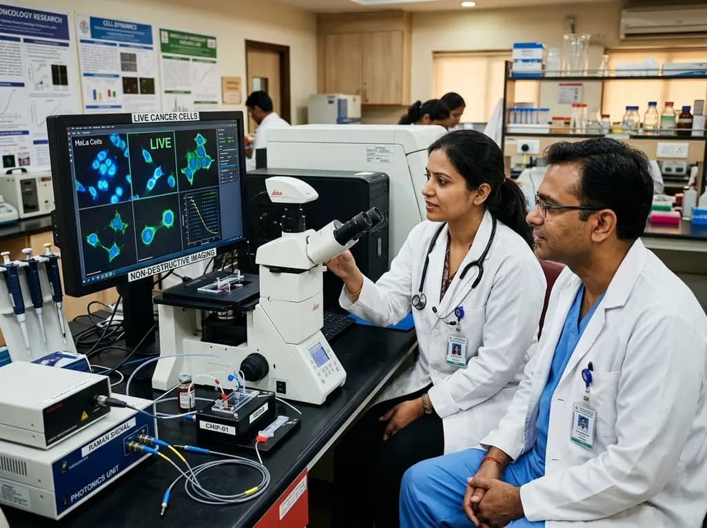
Oncology drug development faces a massive hurdle because traditional laboratory methods rely on fixed, stained, or destroyed samples. Historically, clinical researchers have evaluated cellular behavior through static snapshots taken after biological processes have already concluded. Consequently, these endpoint assays fail to capture how malignant cells continuously evolve, adapt, and develop therapeutic resistance. To address this limitation, advanced real-time cellular imaging technologies are emerging as essential tools for modern biomedical research. By observing living cells continuously without destructive dyes or chemical fixation, scientists gain unprecedented visibility into dynamic biological processes as they unfold in real time.
Modern precision oncology requires deep insights into temporal cellular behavior. Traditional biological autopsies provide valuable genomic and structural data, yet they obscure the transient biochemical changes occurring during drug administration. Furthermore, standard staining methods can disrupt cellular homeostasis or induce artificial stress responses. By eliminating invasive dyes, non-destructive platforms preserve authentic cell signaling pathways. Therefore, monitoring living biology continuously allows medical researchers to track cellular fate trajectories across various therapeutic interventions. Consequently, this innovative approach promises to redefine how drug candidates are evaluated before entering expensive clinical trials.
The core innovation behind label-free observation relies on integrating multiple scientific disciplines into a single analytical platform. Specifically, combining Raman spectroscopy, photonics, microfluidics, and computational biology enables continuous non-invasive measurement of living cells. Raman spectroscopy detects inelastic light scattering, which reflects internal chemical bonds and provides a detailed biochemical fingerprint of the cell. Meanwhile, specialized microfluidic organoid-on-chip systems sustain living tissues in controlled physiological environments for extended durations. As a result, photonics hardware captures high-dimensional chemical data without damaging delicate cellular structures.
Additionally, computational algorithms process these continuous optical streams to generate comprehensive live-cell visual datasets. Researchers often refer to this continuous readout as live-cell cinema because it converts biological motion into readable spectral movies. Unlike traditional assays that measure single time points, label-free photonics platforms track protein, lipid, and nucleic acid shifts second by second. Consequently, scientists can pinpoint exact moments when drug molecules interact with internal target receptors. Moreover, this high-resolution temporal data helps identify early metabolic adjustments before physical structural changes become visible under standard light microscopes.
Accelerating drug discovery requires robust preclinical models that accurately predict clinical outcomes in human patients. Currently, a substantial proportion of oncology drug candidates fail during clinical trials due to unpredicted toxicity or lack of sustained efficacy. However, integrating real-time cellular imaging into preclinical pipelines allows pharmacologists to observe subtle metabolic shifts hours or days earlier than conventional methods allow. Furthermore, researchers can evaluate candidate compounds across diverse patient-derived tumor models to identify ideal therapeutic windows with greater precision.
In addition, continuous monitoring clarifies how cancer populations develop resistance to targeted therapies. When exposed to therapeutic agents, malignant cells frequently activate alternative survival pathways or enter temporary quiescent states. Continuous spectroscopic tracking reveals these adaptive mechanisms in real time, allowing researchers to design rational combination therapies that prevent treatment failure. Furthermore, evaluating drug responses across organoid-on-chip models bridges the gap between two-dimensional cell cultures and complex human physiology. Thus, pharmaceutical developers can prioritize high-potential molecules early, reducing overall development timelines and clinical trial failure rates significantly.
Although cancer research remains the primary driver for live-cell analytical tools, the implications extend far across general medicine. For instance, continuous biochemical tracking offers critical insights into immunology, neurodegenerative disorders, rare metabolic diseases, and regenerative medicine. In neuroscience, observing living neuronal networks helps unravel complex pathological cascades associated with conditions like Alzheimer's and Parkinson's disease. Similarly, immunologists can track real-time interactions between engineered T-cells and target tissue microenvironments during cell therapy development.
Furthermore, non-invasive optical platforms frequently yield valuable diagnostic secondary insights during core biomedical research. For example, highly sensitive photonic sensors developed for cellular monitoring can non-invasively identify hemoglobinopathies, including sickle cell disease and thalassemia. Moreover, tissue engineers utilize continuous monitoring platforms to verify matrix scaffold integration and stem cell differentiation over extended culture periods. Consequently, replacing destructive endpoint testing with live biological observation establishes a versatile foundation for multi-specialty biomedical innovation.
The necessity for advanced drug discovery tools is particularly urgent in nations facing rapidly growing oncological challenges. According to global health estimates from the International Agency for Research on Cancer, India recorded approximately 1.4 million new cancer cases in 2022. Furthermore, projections from national research bodies indicate that India's cancer burden will continue rising over the coming decade. Addressing this public health demand requires accessible precision oncology solutions tailored to complex genomic and clinical profiles across diverse patient populations.
Indian medical oncologists emphasize that integrating real-time biological data with comprehensive genomic profiling and minimal residual disease detection will enhance targeted personalized medicine. Although continuous observation technologies are research tools rather than direct treatments, they significantly strengthen the scientific foundation supporting future therapeutic regimens. By facilitating efficient preclinical drug evaluation, these advanced platforms help streamline global and domestic drug development efforts. Consequently, clinicians remain optimistic that richer biological insights will ultimately translate into improved patient survival and personalized cancer management strategies.
Q1: How does label-free live-cell imaging differ from traditional fluorescent staining?
Traditional fluorescent staining requires adding chemical dyes or biological labels that fix, alter, or ultimately destroy living cells over time. Conversely, label-free live-cell imaging uses inherent optical properties like Raman light scattering to measure cellular biochemistry non-invasively. This allows continuous observation of healthy living biology for extended periods without inducing phototoxicity, cellular stress, or structural destruction.
Q2: Why is real-time observation essential for overcoming cancer drug resistance?
Cancer cells continuously evolve and adapt when exposed to therapeutic agents, often utilizing transient metabolic shifts to survive. Standard static assays only capture cells after treatment has succeeded or failed, missing intermediate adaptive stages. Real-time observation enables scientists to trace subtle biochemical transitions second by second, revealing exact resistance mechanisms and guiding the design of effective combination therapies.
Q3: What role do organoid-on-chip systems play in modern drug discovery pipelines?
Microfluidic organoid-on-chip systems maintain three-dimensional, patient-derived cell clusters in controlled environments that closely mimic human tissue architecture and fluid flow. When combined with real-time optical sensing, these platforms allow continuous monitoring of drug efficacy and toxicity under physiologically accurate conditions. Consequently, they improve preclinical predictions and reduce reliance on animal testing models.
Disclaimer: This content is for informational and educational purposes only. It does not constitute medical advice or replace professional judgment. Refer to the latest local and national guidelines for clinical practice.
References

Read summarized clinical updates, watch expert medical content, and earn CME certifications right from your smartphone.
Open your smartphone camera to scan and install.





Real-time cellular imaging transforms oncology research by allowing scientists to observe living cancer cells continuously without destructive dyes or fixing agents. This novel approach combines microfluidics, photonics, and Raman spectroscopy to accelerate targeted drug discovery and overcome drug resistance.
last month

A randomized trial shows oral DL-3-n-butylphthalide (200 mg thrice daily) significantly slows functional decline in spinocerebellar ataxia type 3 over 12 months, improving SARA and SCAFI scores with favorable safety and zero serious adverse events.
Last week
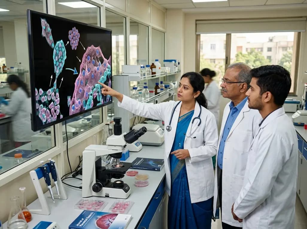
Recent research reveals that downregulating YAP1/TAZ-TEAD via Hippo signaling triggers spontaneous human trophoblast syncytialization in 3D cultures, illuminating placental biology and preeclampsia.
2 weeks back
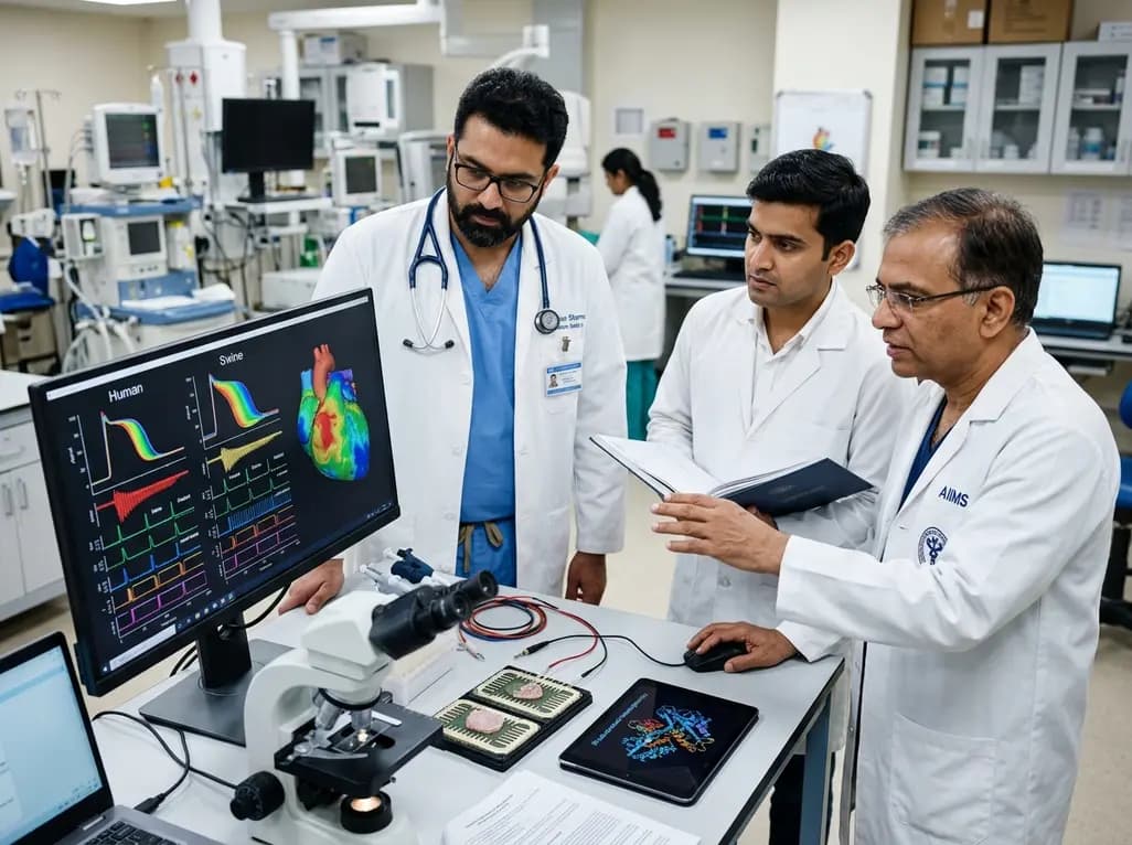
A landmark comparative study evaluates atrial electrophysiology across humans, swine, and rodents. By linking cellular recordings with transcriptomics and in silico modeling, researchers resolve cross-species gaps, explaining translational failures to advance targeted therapies for atrial arrhythmias.
2 weeks back
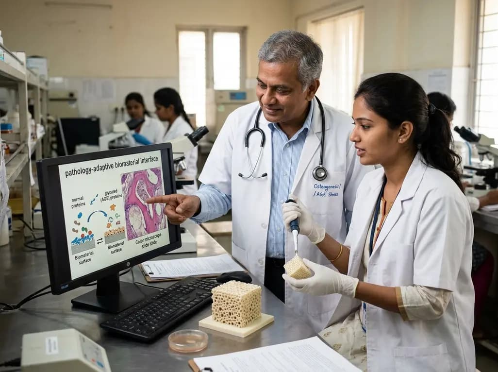
A novel pathology-adaptive surface engineering strategy uses functionalized plasma polymer coatings to selectively modulate AGE adsorption, reducing oxidative stress and restoring bone formation in diabetic and aging microenvironments.
last month
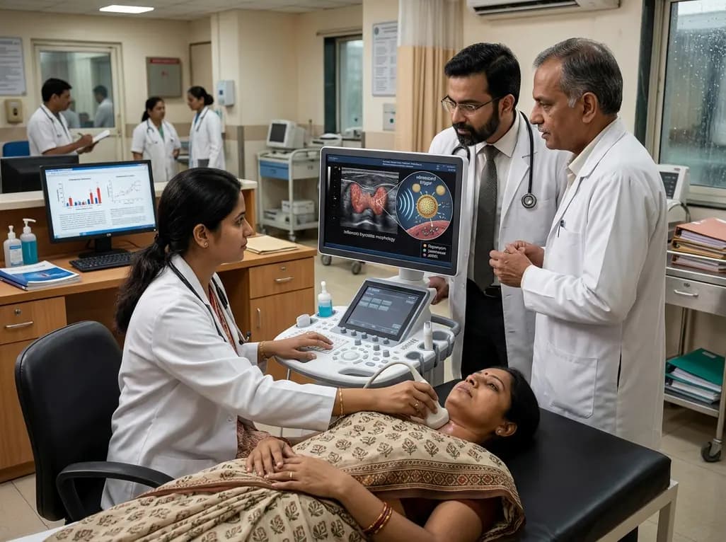
Researchers have developed an innovative ultrasound-responsive nano-contrast agent that co-delivers CIITA-siRNA and rapamycin directly to inflamed thyroid tissue, significantly reducing autoimmune injury and thyroid autoantibodies in preclinical models of Hashimoto thyroiditis.
2 weeks back
