.webp&w=384&q=75)

Loading, please wait...
Loading, please wait...
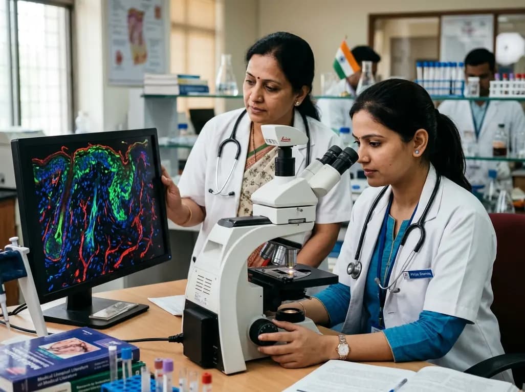
Cutaneous wound healing represents an intricate physiological response that demands tight spatial and temporal orchestration across multiple cell lineages. Historically, clinicians and researchers attributed this coordination primarily to conventional protein-coding genes and noncoding regulatory transcripts. However, high-resolution translatomics has shattered this binary classification by demonstrating that ncRNA-encoded micropeptides actively drive tissue regeneration. These small open reading frame translation products emerge from long noncoding RNAs and circular RNAs, revealing a previously hidden proteome that directly controls cellular repair machinery.
For decades, molecular biologists classified long noncoding RNAs and circular transcripts as purely regulatory molecules. Consequently, scientific investigations focused exclusively on their actions as microRNA sponges, epigenetic modulators, or transcriptional scaffolds. Nevertheless, cutting-edge ribosome profiling and mass spectrometry show that many of these transcripts harbor functional small open reading frames. These hidden sequences actively translate stable peptides typically shorter than one hundred amino acids.
Importantly, these ncRNA-derived micropeptides do not represent nonfunctional translational noise. Instead, they act as potent biological effectors during acute tissue injury. A single genomic locus can therefore yield dual regulatory outputs by generating both a functional RNA transcript and a biologically active micropeptide. Because cutaneous repair requires rapid cellular adaptations, this dual-action mechanism substantially broadens the genomic regulatory bandwidth. Thus, unraveling this hidden proteome provides vital insights into how injured skin coordinates complex survival and regeneration pathways.
Re-epithelialization serves as a fundamental hallmark of successful wound closure. During this dynamic phase, basal keratinocytes at the wound margin must migrate and proliferate across the denuded wound bed. Furthermore, these cells undergo partial epithelial-mesenchymal transition, which temporarily alters their cytoskeletal architecture to enable lateral movement.
Recent investigations show that specific micropeptides directly orchestrate these essential keratinocyte behaviors. Specifically, micropeptides localize to cytosolic protein hubs where they stabilize key transcription factors and cytoskeletal regulators. For instance, peptide effectors can preserve proteins like Twist1 from proteasomal degradation, thereby accelerating epithelial migration and barrier restoration. Additionally, these micropeptides interact directly with junctional protein complexes. Consequently, keratinocytes loosen intercellular adhesions while simultaneously preserving cellular viability. When acute injuries occur, these translational products surge rapidly to promote edge advancement. In contrast, deficient translation or impaired signaling of these micropeptides correlates with delayed re-epithelialization, a defining feature of refractory chronic wounds.
Following initial barrier restoration, fibroblasts remodel the provisional extracellular matrix into a robust structural lattice. During healthy repair, dermal fibroblasts differentiate reversibly into contractile myofibroblasts, synthesize type I and type III collagen, and orchestrate matrix deposition. However, sustained or aberrant myofibroblast activation promotes pathological fibrosis, keloids, and hypertrophic scars.
Within this fibroblastic niche, micropeptides translated from noncoding RNAs serve as critical molecular rheostats. They directly engage transforming growth factor-beta signaling cascades and interact with intracellular chaperone networks. As a result, these micropeptides precisely calibrate the amplitude and duration of myofibroblast activity. Moreover, they regulate the transcription and secretion of matrix metalloproteinases and their endogenous tissue inhibitors. Through this delicate balance, micropeptides ensure adequate mechanical tensile strength while preventing excessive fibrotic contracture. Therefore, targeting micropeptide-driven fibroblast pathways offers a promising strategy to treat both non-healing cutaneous ulcers and disfiguring fibrotic scars.
Timely transition from a destructive inflammatory state to a constructive regenerative environment dictates wound outcomes. Macrophages and neutrophils eliminate pathogens and necrotic debris during early wound stages. Subsequently, resolving macrophages guide endothelial cells to generate new functional capillaries through angiogenesis. In chronic human wounds, persistent inflammation and impaired microvascular perfusion frequently halt this transition.
Emerging research underscores that micropeptides play decisive roles in inflammatory resolution and vascular repair. Specifically, these peptide effectors suppress canonical nuclear factor kappa B activation within wound macrophages, thereby facilitating phenotypic switching from pro-inflammatory M1 states to reparative M2 profiles. Simultaneously, endothelial cells utilize specialized micropeptides to facilitate vascular endothelial growth factor receptor signaling. Consequently, micropeptides promote endothelial sprouting, tube formation, and stable pericyte coverage. By coordinating immune silencing with rapid microvascular revascularization, these endogenous molecules establish a supportive microenvironment essential for clinical tissue survival.
The discovery of micropeptides derived from noncoding RNAs presents transformative therapeutic opportunities for clinical wound management. Globally, chronic diabetic foot ulcers, venous stasis ulcers, and pressure injuries present immense clinical and economic challenges. Conventional topicals and growth factor therapies frequently yield unsatisfactory outcomes due to rapid enzymatic degradation in hostile proteolytic wound beds.
Because micropeptides possess compact secondary structures, synthetic versions demonstrate favorable stability profiles and superior tissue penetration compared to bulky recombinant proteins. Furthermore, clinicians could deliver these molecules through functionalized hydrogels, lipid nanoparticles, or stem cell-derived exosomes. Additionally, tracking endogenous micropeptide expression levels in wound exudates may furnish useful prognostic biomarkers to identify healing trajectories early. However, realizing this translational vision requires substantial further research. Investigators must systematically delineate the precise cellular receptors, half-lives, and off-target profiles of these emerging therapeutic candidates before introducing them into human clinical trials.
Classical growth factors are large, complex proteins encoded by conventional messenger RNAs that bind external cell-surface receptors. In contrast, micropeptides are short sequences translated from noncoding RNAs that primarily function through intracellular protein-protein interactions. Their compact physical structure often grants them superior tissue penetration, lower manufacturing costs, and unique intracellular regulatory capabilities compared to traditional therapeutic proteins.
In diabetic foot ulcers, chronic hyperglycemia, severe oxidative stress, and persistent inflammation disrupt standard RNA translation patterns. Consequently, deficient micropeptide synthesis impairs keratinocyte migration, suppresses angiogenesis, and stalls macrophage switching toward a resolving phenotype. Restoring these depleted micropeptides through targeted delivery platforms can reactivate dormant cellular repair pathways and accelerate closure in recalcitrant diabetic wounds.
Yes, advanced mass spectrometry platforms and specialized targeted immunoassays can detect endogenous micropeptides within wound exudates and tissue biopsies. Because micropeptide levels shift rapidly between the inflammatory, proliferative, and remodeling phases, quantifying them offers substantial diagnostic potential. Clinicians may eventually use these molecular signatures as real-time prognostic biomarkers to identify stalled wounds and tailor therapeutic interventions.
Disclaimer: This content is for informational and educational purposes only... Refer to the latest local and national guidelines for clinical practice.
References
Tang S et al. The Hidden Proteome of Cutaneous Repair: Emerging Roles of ncRNA-Encoded Micropeptides. Am J Physiol Cell Physiol. 2026 Sep 24. doi: 10.1152/ajpcell.90039.2026. PMID: 42781502.
Hu J, Wang Z, Zhao W, Shen G. Non-Coding RNAs: New Players in Skin Wound Healing. Adv Wound Care (New Rochelle). 2020;9(8):467-484.
Yang H, Zhang Y, Du Z, et al. Noncoding RNAs in chronic wound healing: Mechanisms, exosome therapeutics, and translational frontiers. MedComm. 2026;7(3):e70114.
Bhatta A, Atianand M. Functional Micropeptides Encoded by Long Non-Coding RNAs: A Comprehensive Review. Noncoding RNA. 2022;8(3):41.

Read summarized clinical updates, watch expert medical content, and earn CME certifications right from your smartphone.
Open your smartphone camera to scan and install.





Recent discoveries reveal that ncRNA-encoded micropeptides play vital biological roles in cutaneous repair. These tiny peptides modulate keratinocyte migration, fibroblast plasticity, inflammation, and angiogenesis, unlocking novel therapeutic pathways for chronic and non-healing wounds.
Yesterday

Discover how catalytic nanozymes redefine autoimmune disease therapy by establishing a physiological redox set-point to restore immune tolerance without broad immunosuppression.
Yesterday
Recent research reveals that downregulating YAP1/TAZ-TEAD via Hippo signaling triggers spontaneous human trophoblast syncytialization in 3D cultures, illuminating placental biology and preeclampsia.
2 weeks back

A landmark comparative study evaluates atrial electrophysiology across humans, swine, and rodents. By linking cellular recordings with transcriptomics and in silico modeling, researchers resolve cross-species gaps, explaining translational failures to advance targeted therapies for atrial arrhythmias.
2 weeks back

A novel pathology-adaptive surface engineering strategy uses functionalized plasma polymer coatings to selectively modulate AGE adsorption, reducing oxidative stress and restoring bone formation in diabetic and aging microenvironments.
last month

Researchers have developed an innovative ultrasound-responsive nano-contrast agent that co-delivers CIITA-siRNA and rapamycin directly to inflamed thyroid tissue, significantly reducing autoimmune injury and thyroid autoantibodies in preclinical models of Hashimoto thyroiditis.
2 weeks back
