.webp&w=384&q=75)

Loading, please wait...
Loading, please wait...

Moyamoya disease is a progressive cerebrovascular disorder characterized by stenosis or occlusion of the terminal internal carotid arteries. Surgical bypass remains the primary therapeutic intervention to prevent recurrent ischemic or hemorrhagic events. Quantitative evaluation of surgical success historically relied on qualitative angiographic scoring systems. However, recent advances in digital subtraction angiography allow precise objective measurements of cerebral perfusion and vessel remodeling. Understanding external carotid artery collateralisation and arterial residual volume after moyamoya disease revascularisation provides invaluable clinical insights. Vascular surgeons and neurologists can better predict long-term clinical outcomes by analyzing these hemodynamic metrics.
Cerebral revascularisation establishes new blood flow channels from the external carotid artery system to ischemic cortical tissue. Both direct bypasses and indirect synangiosis procedures rely on postoperative angiogenesis to restore cerebral perfusion. Researchers recently evaluated the collateralisation angiogenesis ratio using advanced quantitative digital subtraction angiography. This objective metric measures how effectively the external carotid artery branches sprout new capillaries into the brain parenchyma. In addition, quantitative assessment helps clinicians track longitudinal structural remodeling in surgical candidates.
Standard qualitative evaluations often produce significant inter-observer variability among neurointerventional specialists. Conversely, quantitative angiography offers reproducible numerical parameters to evaluate neoangiogenesis postoperatively. Consequently, clinicians can track subtle improvements in regional cerebral microcirculation across long follow-up intervals. Higher collateralisation angiogenesis ratios correlate directly with robust clinical recovery and reduced risk of secondary stroke. Furthermore, early identification of poor angiogenesis enables proactive clinical management. Neurologists can optimize antiplatelet therapy or consider supplemental indirect surgical procedures when collateral development remains inadequate.
Intracranial arterial residual volume represents the residual functional lumen within severely stenotic intracranial vessels. Digital subtraction angiography quantifies this volume across various stages of disease progression. Notably, clinical investigations confirm a strong negative correlation between intracranial arterial residual volume and Suzuki stage. Patients with advanced ischemic or hemorrhagic Moyamoya disease exhibit significantly lower arterial residual volumes. Suzuki staging reflects progressive occlusion of the internal carotid artery terminus and middle cerebral artery. Therefore, diminishing arterial residual volume directly mirrors advancing parenchymal ischemia and vascular compromise.
Interestingly, clinical studies demonstrate that intracranial arterial residual volume does not correlate directly with baseline modified Rankin Scale scores. Patients with low residual vascular volumes may maintain stable functional scores temporarily due to transient compensatory mechanisms. However, structural vascular reserve remains severely depleted, leaving these patients vulnerable to catastrophic ischemic events. In addition, the presence of fetal-type posterior cerebral arteries significantly preserves intracranial arterial residual volume. Fetal-type variants provide robust collateral flow from the posterior circulation, mitigating anterior ischemic burden. Recognizing these anatomical variations allows clinicians to stratify perioperative stroke risk more accurately.
Achieving robust external carotid artery collateralisation depends on several distinct preoperative anatomical and clinical parameters. Multivariate regression analyses highlight intracranial arterial residual volume and Suzuki staging as strong independent predictors of postoperative collateral development. Specifically, higher intracranial arterial residual volume correlates positively with greater external carotid artery collateralisation following surgical intervention. Furthermore, patients presenting with advanced Suzuki stages demonstrate higher rates of compensatory angiogenic sprouting. Severe ischemia acts as a powerful biological stimulus for local growth factors like vascular endothelial growth factor.
Consequently, ischemic cerebral tissue recruits external carotid arterial branches more aggressively post-bypass. Conversely, mild vascular stenosis generates insufficient hypoxic drive, resulting in sluggish postoperative collateral development. Understanding these independent predictors guides personalized surgical planning for patients with Moyamoya disease. For example, surgeons can select combined direct and indirect bypass techniques for patients with high angiogenic potential. Alternatively, direct bypass alone may be preferred when immediate flow restoration is essential. Furthermore, quantitative angiographic metrics provide clear benchmarks to evaluate procedural efficacy over time.
Comprehensive postoperative care after moyamoya disease revascularisation requires systematic hemodynamic and clinical monitoring. Neurologists must balance blood pressure management to prevent both hyperperfusion syndrome and ischemic stroke. Early post-bypass hyperperfusion can cause cerebral edema or intracranial hemorrhage, particularly in regions with rapid revascularisation. Therefore, tight perioperative blood pressure control remains vital during the initial postoperative recovery phase. In addition, quantitative DSA protocols performed at scheduled follow-up intervals offer clear visualization of collateral development.
Long-term medical management focuses on optimizing microvascular patency and preventing secondary thrombotic complications. Low-dose aspirin is frequently prescribed to preserve bypass graft patency and facilitate collateral vessel maturation. Moreover, controlling systemic cardiovascular risk factors such as hypertension, diabetes, and dyslipidemia optimizes overall endothelial function. Clinicians should educate patients regarding subtle neurological symptoms that signal transient ischemic attacks or hypoperfusion. Regular multidisciplinary follow-up ensures timely detection of graft failure or contralateral disease progression. Consequently, structured surveillance protocols significantly improve functional independence and overall quality of life for Moyamoya patients.
Integrating quantitative angiographic metrics into routine clinical workflows represents a significant advancement in cerebrovascular care. Traditional qualitative grading scales often fail to detect subtle differences in surgical collateral development. By contrast, digital subtraction angiography-based quantitative analysis provides precise, reproducible measurements of collateralisation angiogenesis ratios and arterial residual volumes. These numerical parameters allow clinicians to establish objective baseline measurements and track longitudinal progress reliably. Consequently, clinical decision-making becomes more precise and evidence-based across diverse patient populations.
Furthermore, quantitative metrics assist surgical teams in refining procedural selection and surgical technique. Identifying patients with low angiogenic potential allows neurosurgeons to modify indirect revascularisation procedures or combine them with direct bypass options. In addition, standardized quantitative evaluations facilitate multi-center clinical trials by providing uniform outcome measures. As image processing software becomes more automated, quantitative DSA will likely become standard practice in comprehensive stroke centers. Ultimately, adopting these objective angiographic metrics enhances risk stratification, optimizes surgical strategies, and improves long-term outcomes for patients undergoing moyamoya disease revascularisation.
The collateralisation angiogenesis ratio is a quantitative digital subtraction angiography metric. It measures the extent of new blood vessel growth arising from the external carotid artery following cerebral revascularisation. This ratio provides clinicians with an objective numerical value to evaluate surgical success and postoperative microvascular perfusion improvement.
Suzuki staging demonstrates a significant negative correlation with intracranial arterial residual volume in Moyamoya disease. As Suzuki stage increases, indicating more severe steno-occlusive arterial disease, intracranial arterial residual volume decreases substantially. This reduction reflects progressive loss of native intracranial vessel diameter and overall vascular lumen volume.
Quantitative DSA metrics eliminate observer subjectivity associated with traditional grading scales. They provide precise numerical data on vessel volume and collateral angiogenesis. These objective measurements help clinicians predict patient outcomes, monitor longitudinal vessel remodeling, identify insufficient bypass development, and tailor long-term medical management strategies effectively.
Disclaimer: This content is for informational and educational purposes only and does not constitute medical advice, diagnosis, or treatment. Always seek the advice of a qualified healthcare provider with any questions you may have regarding a medical condition. Refer to the latest local and national guidelines for clinical practice.
References

Read summarized clinical updates, watch expert medical content, and earn CME certifications right from your smartphone.
Open your smartphone camera to scan and install.





A quantitative study evaluates external carotid artery collateralisation and arterial residual volume after revascularisation in Moyamoya disease. Findings reveal strong correlations between Suzuki stage, vascular reserve, and post-bypass angiogenesis, offering vital insights for surgical and clinical monitoring.
Today
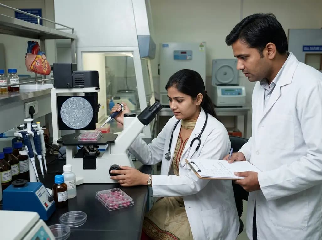
Researchers at SGPGIMS Lucknow have isolated and cultured human heart valve interstitial cells from donor tissues. This milestone advances understanding of rheumatic heart disease pathways like TGF-beta/SMAD3 and ERK1/2, laying the foundation for tissue-engineered heart valves and drug testing.
Today

A study identifies a critical hypospadias gene-environment interaction. Research shows that the risk gene DNAH8 and DEHP exposure combine to disrupt steroidogenesis and mesenchymal progenitor cell differentiation, significantly increasing the risk of severe urethral malformations in male fetuses.
6 days back
A pre-clinical study reveals that elevated serum pro-N-cadherin levels correlate strongly with severe cardiac fibrosis and diastolic dysfunction following radiation exposure, promising a potential early biomarker for radiation-related heart disease.
4 days back
Researchers at Kyushu University have uncovered a novel compound, lipoic acid trisulfide (LASSS), that enhances hepatocyte growth factor (HGF) signaling and protects against nitration-induced protein dysfunction, presenting a potential breakthrough for age-related muscle atrophy and sarcopenia.
3 days back

Deepinder Goyal's healthtech startup Temple is raising capital at a $500 million valuation and pricing its temporal wearable at Rs 80,000. Worn on the side of the forehead, the device tracks microvascular blood flow and real-time Entropy to monitor physiological stress, metabolic expenditure, and recovery.
Today
