.webp&w=384&q=75)

Loading, please wait...
Loading, please wait...
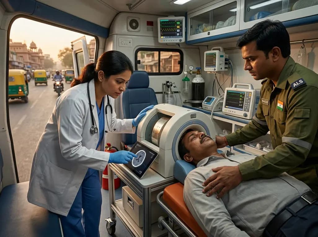
Stroke remains a dominant global health challenge, representing a leading cause of both long-term disability and mortality. In the clinical management of acute stroke, the concept of the "golden hour" is paramount, as every minute of delayed treatment results in the loss of millions of neurons. Traditionally, definitive diagnosis and treatment initiation have been confined to the hospital setting. However, recent advancements are shifting this paradigm toward the prehospital environment. Lightweight MRI stroke care has emerged as a potentially transformative solution for hyperacute management. By bringing advanced neuroimaging directly to the patient in an ambulance, clinicians can significantly reduce the time to reperfusion therapies. The landmark study by Barker JL et al. highlights how these portable devices are bridging the gap in emergency medical services.
Historically, prehospital stroke care relied almost exclusively on clinical assessment scales, such as the Cincinnati Prehospital Stroke Scale. While these tools are useful for screening, they cannot differentiate between ischemic and hemorrhagic strokes, which require diametrically opposed treatments. To address this, many healthcare systems introduced Mobile Stroke Units (MSUs) equipped with computed tomography (Scan) scanners. These units have proven highly effective in accelerating the administration of intravenous thrombolysis. Nevertheless, traditional CT-based MSUs face significant logistical challenges. They are exceptionally heavy, expensive to maintain, and require specialized personnel to operate and interpret images. Furthermore, the radiation exposure associated with CT, though small, remains a consideration in repeated imaging. Consequently, researchers have sought lighter and more versatile alternatives that can offer high-quality soft-tissue visualization. This search led to the development of ultra-low-field magnetic resonance imaging. Unlike their high-field counterparts, these portable systems do not require heavy shielding or specialized cooling infrastructure. As a result, they offer a more flexible platform for emergency medical services. By integrating these systems into standard-sized ambulances, healthcare providers can now envision a future where high-fidelity diagnostic data is available within minutes of patient contact.
The primary innovation driving lightweight MRI stroke care is the use of ultra-low-field (ULF) magnets, typically operating at 0.064 Tesla. Traditional clinical MRI scanners utilize 1.5T or 3.0T magnets, which are massive and necessitate dedicated copper-shielded rooms to prevent electromagnetic interference. In contrast, ULF-MRI systems utilize permanent magnets that are significantly lighter and consume very little power. Specifically, these devices can operate using a standard electrical outlet or even high-capacity batteries within a vehicle. Moreover, the lack of a high-intensity fringe field eliminates the risk of metallic projectiles, making the environment much safer for both paramedics and patients in a confined space. This portability allows the scanner to be mounted on a specialized docking system within an ambulance. Furthermore, sophisticated software algorithms and artificial intelligence are now used to reconstruct high-quality images from the lower signal-to-noise ratio inherent in ULF systems. These advancements ensure that even with a weaker magnet, the diagnostic quality remains sufficient to identify major pathologies like intracranial hemorrhage or large vessel occlusions. Therefore, the technological barrier that once made MRI in a moving vehicle impossible has been largely overcome. This breakthrough paves the way for a more decentralized model of stroke care that can be deployed across diverse geographical regions.
Clinical feasibility is the cornerstone of any new medical technology, and the Barker JL et al. study provides compelling evidence for the efficacy of portable MRI. Specifically, the research demonstrates that ultra-low-field MRI can accurately detect acute ischemic lesions as small as several millimeters in diameter. In addition to identifying infarcts, these portable scanners excel at detecting intracranial hemorrhage, which is a critical contraindication for thrombolytic therapy. During the study, the diagnostic decisions made using portable MRI data closely matched those derived from standard high-field hospital scanners. Furthermore, the integration of diffusion-weighted imaging (DWI) sequences into these portable units provides a level of physiological detail that CT scanners cannot match. Notably, DWI is highly sensitive to early cytotoxic edema, allowing for the detection of ischemia within minutes of symptom onset. This capability is particularly beneficial for "wake-up strokes" where the exact time of onset is unknown. By utilizing the mismatch between different MRI sequences, clinicians can better estimate the age of the infarct and determine treatment eligibility. Consequently, the use of MRI in the prehospital setting does not just speed up the existing workflow; it enhances the precision of the diagnosis itself. This high-definition approach to prehospital care ensures that patients receive the most appropriate intervention as early as possible.
While the technical results are promising, several barriers must be addressed to ensure the widespread adoption of lightweight MRI. One primary concern involves the impact of vehicle vibration and external electromagnetic noise on image quality. Since an ambulance is a dynamic environment, specialized stabilizing hardware and active noise-cancellation techniques are required to maintain image integrity. Additionally, the transmission of large imaging files to hospital-based neurologists requires robust telemedicine infrastructure. In many regions, including parts of India, the availability of high-speed 5G or satellite connectivity is crucial for real-time consultation. Furthermore, training paramedics and emergency medical technicians to operate the device and ensure patient safety is a significant undertaking. However, the cost-benefit analysis often favors these portable systems over traditional MSUs. The initial capital investment for a lightweight MRI unit is significantly lower than that of a mobile CT scanner. Moreover, the lower operational costs and lack of specialized radiation shielding make it a more sustainable option for low- and middle-income countries. Therefore, despite the initial hurdles, the potential for these devices to democratize access to advanced neuroimaging is profound. As the technology matures, we can expect to see more modular and user-friendly iterations that are tailored for rapid deployment in urban and rural settings alike.
Looking ahead, the integration of lightweight MRI into the prehospital ecosystem will likely redefine the standard of care for stroke patients. Future systems may combine portable neuroimaging with automated AI-driven analysis, providing immediate triage results even before the ambulance reaches the hospital. Furthermore, the ability to monitor patients during long-distance transfers between community clinics and comprehensive stroke centers will enhance safety and outcomes. Ultimately, this technology represents a shift from the "drip and ship" model to a more comprehensive "diagnose and treat" approach in the field. By maximizing the efficiency of the prehospital phase, we can significantly reduce the long-term burden of stroke on families and healthcare systems. The continued refinement of lightweight MRI stroke care will be essential in achieving this goal and ensuring that life-saving interventions are available to all, regardless of their proximity to a major medical center.
Standard hospital MRI scanners use powerful superconducting magnets, typically 1.5 or 3.0 Tesla, which require liquid helium cooling and specialized shielding. In contrast, lightweight MRI systems utilize ultra-low-field magnets, often around 0.064 Tesla. These systems are significantly lighter, portable, and do not require cooling or a shielded room. While they have a lower signal-to-noise ratio, advanced software and AI algorithms are used to generate diagnostic-quality images suitable for acute triage.
Yes, lightweight MRI is highly effective at detecting intracranial hemorrhage, which is essential for determining if a patient can safely receive clot-busting medications. Studies have shown that ultra-low-field MRI has high sensitivity and specificity for identifying blood within the brain. This capability allows paramedics and physicians to differentiate between an ischemic stroke and a hemorrhagic stroke in the prehospital setting, ensuring that patients receive the correct life-saving treatment immediately.
While CT is excellent for detecting blood, MRI provides superior visualization of soft tissue and can detect ischemia much earlier through diffusion-weighted imaging. Additionally, lightweight MRI systems are more cost-effective, lighter, and safer because they do not use ionizing radiation or high-intensity magnetic fields that attract metal objects. This makes them easier to install in standard ambulances and allows for more frequent monitoring of the patient's condition during transport without cumulative radiation risks.
Disclaimer: This content is for informational and educational purposes only and does not constitute medical advice, diagnosis, or treatment. Always seek the advice of a physician or other qualified health provider with any questions you may have regarding a medical condition. Refer to the latest local and national guidelines for clinical practice.
References
Barker JL et al. Lightweight Magnetic Resonance Imaging for Hyperacute Stroke Care in the Prehospital Setting. Stroke. 2026 Jul 21. doi: 10.1161/STROKEAHA.125.052982. PMID: 42478381.
Sheth KN, et al. Assessment of Brain Injury Using Portable, Low-Field Magnetic Resonance Imaging at the Bedside of Critically Ill Patients. JAMA Neurology. 2020;77(11):1391-1397.
Deoni SC, et al. The Moveable MRI: A New Paradigm for Pediatric and Prehospital Neuroimaging. Journal of Magnetic Resonance Imaging. 2022;56(4):1120-1132.
"
Read summarized clinical updates, watch expert medical content, and earn CME certifications right from your smartphone.
Open your smartphone camera to scan and install.





A breakthrough study discusses the use of lightweight, low-field MRI for prehospital stroke care, offering a portable alternative to CT-equipped mobile stroke units for faster diagnosis and treatment during the critical golden hour.
3 months ago

Depemokimab offers twice-yearly dosing for severe eosinophilic asthma and chronic rhinosinusitis with nasal polyps, significantly cutting exacerbations and hospital admissions.
4 days back

A novel copper metal-organic framework hydrogel remodels the uterine microenvironment to stimulate robust endometrial regeneration, reduce intrauterine fibrosis, and restore reproductive fertility, offering a groundbreaking strategy for managing refractory endometrial damage and Asherman syndrome.
5 days back

New research reveals that non-ablative radiotherapy suppresses ventricular tachycardia by increasing conduction velocity via NaV1.5 upregulation and action potential prolongation. However, sustained sodium current elevation and depressed calcium handling highlight the need for careful clinical translation.
Last week

A single-cell transcriptomic atlas reveals how peripheral nerve injuries cause dorsal root ganglion remodeling. By evaluating proximal nerve crush and resection, researchers identify distinct glial and immune programs that resolve neuropathic pain and promote regeneration.
5 days back

A breakthrough study demonstrates that fullerol-primed extracellular vesicles generated in a 3D SECCS bioreactor attenuate ferroptosis and restore myogenic regeneration in diabetes via the FOXO3 pathway, offering a cell-free therapeutic strategy for diabetic musculoskeletal degeneration.
5 days back
