.webp&w=384&q=75)

Loading, please wait...
Loading, please wait...
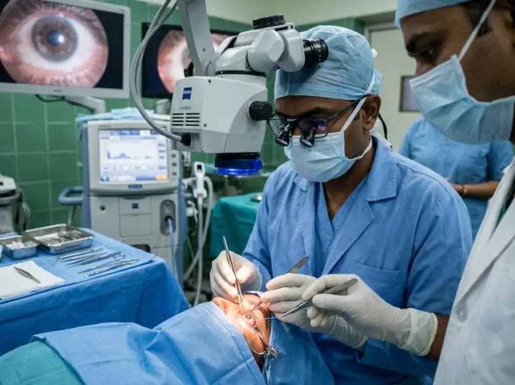
Clinicians often encounter complex cases of lens dislocation or aphakia requiring stable surgical solutions. Recently, the L-suture technique IOL fixation has gained attention for its efficacy and structural integrity. This modified approach provides a reliable method for transscleral fixation, ensuring long-term stability for patients with compromised capsular support.
A recent case series involving 35 eyes demonstrated significant improvements in best-corrected visual acuity. Furthermore, the study highlighted that the L-suture method offers superior maximum tensile strength when compared to the traditional Z-suture technique. This increased strength reduces the risk of suture breakage or lens tilt over time. Consequently, patients experience better visual outcomes with minimal complications.
Moreover, researchers observed that the corneal endothelial cell density remained stable postoperatively. This indicates that the procedure is gentle on intraocular structures. Because the technique utilizes a specific suturing pattern, it minimizes IOL tilt, which is crucial for high-quality vision. Additionally, the follow-up data suggests that the procedure is safe for long-term use in diverse clinical scenarios.
While various techniques exist for scleral fixation, the L-suture stands out due to its mechanical properties. Notably, the in vitro tests confirmed that the L-suture could withstand higher stress levels than its predecessors. This finding is particularly relevant for active patients who require durable lens placement. Therefore, surgeons might consider this technique as a primary option for secondary IOL implantation.
The L-suture technique utilizes a specific geometry that enhances tensile strength. This design provides better fixation and reduces the likelihood of lens displacement compared to the Z-suture method.
Yes, clinical data shows a minimal decrease in corneal endothelial cell count following the procedure. This indicates that the technique is safe and well-tolerated by the eye.
Disclaimer: This content is for informational and educational purposes only. It does not constitute medical advice or a professional relationship. Always seek the advice of a qualified healthcare provider for medical concerns. Refer to the latest local and national guidelines for clinical practice.
References
Alimu S et al. L-Suture Technique for Transscleral Intraocular Lens Fixation. Retina. 2026 Jun 22. doi: 10.1097/IAE.0000000000004911. PMID: 42320017.
American Academy of Ophthalmology. Management of Malpositioned Intraocular Lenses. Preferred Practice Patterns. 2024.

Read summarized clinical updates, watch expert medical content, and earn CME certifications right from your smartphone.
Open your smartphone camera to scan and install.





A study on the L-suture technique for transscleral IOL fixation shows significant improvements in visual acuity and superior tensile strength compared to the Z-suture method. This case series involving 35 eyes highlights the technique's safety and efficacy in managing lens dislocation and aphakia.
4 months ago

Depemokimab offers twice-yearly dosing for severe eosinophilic asthma and chronic rhinosinusitis with nasal polyps, significantly cutting exacerbations and hospital admissions.
3 days back

A novel copper metal-organic framework hydrogel remodels the uterine microenvironment to stimulate robust endometrial regeneration, reduce intrauterine fibrosis, and restore reproductive fertility, offering a groundbreaking strategy for managing refractory endometrial damage and Asherman syndrome.
3 days back

New research reveals that non-ablative radiotherapy suppresses ventricular tachycardia by increasing conduction velocity via NaV1.5 upregulation and action potential prolongation. However, sustained sodium current elevation and depressed calcium handling highlight the need for careful clinical translation.
5 days back

A single-cell transcriptomic atlas reveals how peripheral nerve injuries cause dorsal root ganglion remodeling. By evaluating proximal nerve crush and resection, researchers identify distinct glial and immune programs that resolve neuropathic pain and promote regeneration.
3 days back

A breakthrough study demonstrates that fullerol-primed extracellular vesicles generated in a 3D SECCS bioreactor attenuate ferroptosis and restore myogenic regeneration in diabetes via the FOXO3 pathway, offering a cell-free therapeutic strategy for diabetic musculoskeletal degeneration.
3 days back
