

Loading, please wait...
Loading, please wait...
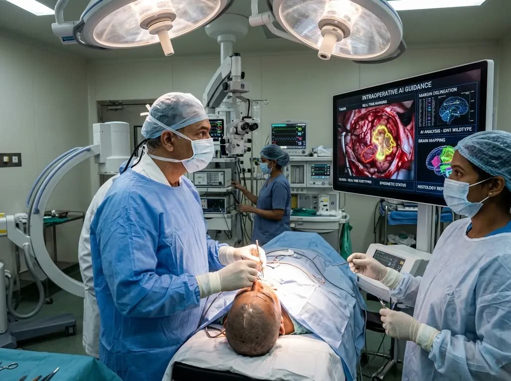
The 2021 World Health Organization classification of central nervous system tumors established an essential shift toward integrated molecular neuro-oncology. Consequently, molecular alterations such as IDH mutations, 1p/19q codeletions, and DNA methylation profiles now direct tumor classification and therapy. However, routine molecular assays still take several days or weeks to return actionable findings to clinicians. Intraoperative artificial intelligence has emerged as an innovative clinical solution to overcome this temporal mismatch. Modern computational platforms extract critical diagnostic data directly from tissue samples inside the surgical suite. By interpreting optical scans, digital pathology, and genomic sequencing instantaneously, algorithms assist neurosurgeons at the definitive point of surgical decision-making. Therefore, surgical teams can achieve safer cytoreduction and protect vital functional brain tissue.
Historically, surgical teams relied on intraoperative frozen section analysis to differentiate neoplastic tissue from normal brain parenchyma. Although frozen sections provide rapid morphological clues, the technique frequently produces freezing artifacts, cellular distortion, and sampling ambiguity. Furthermore, standard optical microscopy cannot identify key genomic alterations, creating diagnostic uncertainty during critical resections. As a result, neurosurgeons often balance the risk of leaving residual invasive tumor against the catastrophic danger of damaging eloquent neurological pathways. Modern intraoperative artificial intelligence bridges this historical gap by analyzing digital tissue representations in real time. Advanced machine learning algorithms recognize subtle cellular textures and nuclear architectures that escape human visual assessment. Consequently, these computer vision tools infer underlying genetic markers and predict tumor biological behavior before the patient leaves the operating table. In addition, machine learning models integrate seamlessly into modern surgical microscopes, clinical monitors, and digital pathology scanners. This rapid feedback loop empowers operating teams to adjust surgical boundaries dynamically during maximal safe resections. Ultimately, bridging molecular diagnostic knowledge and real-time intraoperative execution revolutionizes surgical neuro-oncology. Surgeons now obtain definitive biological answers in minutes rather than waiting weeks for final molecular reports.
Standard cryosection histopathology remains an indispensable intraoperative tool, yet technical artifacts constantly complicate accurate tissue evaluation. To address this persistent limitation, biomedical engineers trained convolutional neural networks to examine whole slide cryosection images automatically. For instance, the Cryosection Histopathology Assessment and Review Machine, known as CHARM, systematically evaluates frozen sections from diverse glioma populations. This sophisticated model achieved outstanding receiver operating characteristic areas under the curve ranging between 0.88 and 0.93 across glioma subtypes. Furthermore, CHARM reliably classifies IDH mutation status and distinguishes high-grade astrocytomas from lower-grade counterparts within minutes. In parallel, specialized architectures like LGNet demonstrate remarkable precision in differentiating primary central nervous system lymphoma from high-grade gliomas. This distinction carries paramount surgical importance, because confirmed lymphoma requires medical therapy rather than aggressive surgical debulking. Additionally, these artificial intelligence models capture microenvironmental nuances, including localized necrosis and heightened cellularity adjacent to invasive boundaries. Pathologists and surgeons thus receive objective diagnostic verification without delaying surgical flow. Consequently, deep learning elevates traditional frozen sections into rich genomic prediction tools.
Fluorescence-guided surgery has transformed high-grade glioma resection by highlighting malignant boundaries under specialized illumination. Specifically, 5-aminolevulinic acid induces selective intratumoral accumulation of fluorescent protoporphyrin IX. Nevertheless, conventional visual inspection of fluorescence remains subjective and frequently fails to reveal low-grade infiltration or heterogeneous margins. To enhance this intraoperative capability, researchers paired hyperspectral optical imaging with supervised machine learning classifiers. Hyperspectral sensors capture hundreds of distinct spectral bands beyond human vision, recording subtle variations in tissue autofluorescence and absorption profiles. Furthermore, advanced algorithms analyze these dense spectral signatures to predict tumor type, histological grade, and IDH mutation status with accuracies approaching 93%. As a result, neurosurgeons obtain objective tissue classification without obtaining destructive physical biopsies. In addition, this combined optical modality detects marginal residual tumor cells that standard white-light illumination consistently misses. Consequently, the surgical team can differentiate hypercellular tumor cores from edematous infiltrating zones in real time. Machine learning algorithms continuously refine spectral interpretation across different illumination environments, providing consistent diagnostic guidance throughout long surgical procedures.
DNA methylation profiling represents the gold standard for central nervous system tumor classification, but traditional arrays require weeks of processing. Recently, portable long-read nanopore sequencing platforms introduced rapid epigenetic profiling directly to laboratory teams. For example, researchers developed the Rapid Nanopore Brain Intraoperative Classification pipeline, known as ROBIN, to process sequencing data in real time. This sophisticated workflow implements adaptive sampling and integrates three independent methylation classification algorithms simultaneously. Consequently, the ensemble architecture delivers accurate epigenetic diagnoses in under two hours while the patient remains in surgery. Prospective multi-institutional trials confirmed that ROBIN achieves 90% diagnostic concordance with final comprehensive neuropathological assessments. Moreover, the platform detects somatic copy number aberrations and structural genetic variants without necessitating separate bioinformatics pipelines. Therefore, neurosurgeons obtain verified methylation subclass information mid-operation, enabling decisive alterations in surgical strategy. If sequencing identifies a benign or radio-sensitive entity, the team can immediately avoid high-risk functional territory resections. Furthermore, continuous algorithm optimization reduces hands-on library preparation steps, making rapid sequencing increasingly practical for hospital laboratories worldwide.
Stimulated Raman histology provides label-free, sub-micron optical imaging of fresh, unprocessed surgical tissue specimens. By interrogating the intrinsic chemical vibrations of lipids, proteins, and nucleic acids, stimulated Raman histology generates virtual hematoxylin and eosin equivalents within two minutes. Recently, artificial intelligence foundation models trained on massive multi-center Raman datasets revolutionized intraoperative margin evaluation. In rigorous comparative studies, Raman foundation models outperformed traditional magnetic resonance imaging sequences, including FLAIR and contrast enhancement, in identifying infiltrative tumor margins. Furthermore, these artificial intelligence models demonstrated superior sensitivity compared to 5-aminolevulinic acid fluorescence during microscopic border inspection. At the molecular level, deep learning classifiers achieve greater than 90% diagnostic accuracy across primary glioma subtypes. In addition, morphometric profiling algorithms analyze single-cell nuclear architecture across optical virtual slides. These quantitative morphometric features reliably distinguish IDH-mutant glioma cells from aggressive IDH-wildtype tumors. Consequently, the technology provides interpretable cellular phenotyping alongside rapid automated diagnostic predictions. Neurosurgeons can thus interrogate resected cavity walls repeatedly to confirm true negative margins.
Despite remarkable algorithmic milestones, intraoperative artificial intelligence faces notable hurdles before achieving universal operating room deployment. Currently, neurosurgery lacks extensive multi-center prospective randomized clinical trials proving that real-time algorithmic classification directly improves patient overall survival or neurological preservation. Furthermore, high capital equipment costs for stimulated Raman histology scanners and rapid sequencing devices limit accessibility across community hospitals. Regulatory approval frameworks also demand rigorous validation across diverse patient demographics, hardware platforms, and slide preparation artifacts. Additionally, surgical departments must establish standardized computational workflows, secure server connections, and multidisciplinary training protocols for operative teams. Looking toward the future, investigators are developing unified multimodal artificial intelligence models. These expansive architectures combine intraoperative optical imaging, digital frozen sections, molecular sequencing data, and preoperative neuroimaging into a singular diagnostic framework. Consequently, multimodal algorithms will deliver unprecedented intraoperative clarity, guiding bespoke surgical resections tailored to individual tumor biology. In conclusion, continued interdisciplinary collaboration among neurosurgeons, pathologists, and computer scientists will translate these artificial intelligence innovations into standard surgical oncological care.
Intraoperative artificial intelligence models analyze diagnostic tissue representations directly within the operating room, bypassing standard multi-week laboratory workflows. By processing frozen section whole-slide images, stimulated Raman histology scans, or rapid nanopore sequencing reads, algorithms infer critical genetic alterations like IDH mutations and DNA methylation classes. Consequently, surgical teams receive definitive molecular classifications within minutes, allowing neurosurgeons to tailor resection aggression dynamically while preserving vital functional brain tissue during the operation.
Stimulated Raman histology generates virtual digital pathology images from fresh, unfixed tissue without requiring freezing, sectioning, or chemical staining. It utilizes intrinsic chemical vibrational signatures of cellular lipids and proteins to render diagnostic virtual hematoxylin and eosin slides within minutes. Furthermore, this method eliminates classic freezing artifacts and tissue destruction, allowing artificial intelligence foundation models to evaluate intact cytoarchitectural patterns and detect residual microscopic infiltration directly at surgical cavity margins.
Universal clinical adoption remains constrained by the absence of multi-center randomized controlled trials demonstrating improved progression-free survival or functional patient outcomes. Furthermore, the substantial financial cost of proprietary optical scanners and sequencing devices poses severe adoption barriers for many healthcare centers. Additionally, hospitals must navigate complex regulatory clearances, implement robust computational infrastructure, and establish rigorous training programs to ensure seamless collaboration among operating neurosurgeons, pathologists, and bioinformaticians during live surgeries.
Disclaimer: This content is for informational and educational purposes only and should not be construed as medical advice or used for treatment or diagnosis without consulting a healthcare professional. All healthcare providers must make decisions in accordance with their professional judgment, institutional protocols, and specific clinical circumstances. The views expressed are based on research published to date and may change with new evidence. Mention of specific tests, technologies, or products does not imply endorsement. Refer to the latest local and national guidelines for clinical practice.
References
Goff NK et al. Intraoperative artificial intelligence for brain tumor surgery: from tissue diagnosis to real-time surgical guidance. Neurosurg Focus. 2026 Nov 01. doi: 10.3171/2026.8.FOCUS26439. PMID: 42967344.
Nasrallah MP, Zhao J, Tsai CC, et al. Machine learning for cryosection pathology predicts the 2021 WHO classification of glioma. Med. 2023;4(8):526-540. doi: 10.1016/j.medj.2023.06.002.
Deacon S, Cahyani I, Holmes N, et al. ROBIN: A unified nanopore-based assay integrating intraoperative methylome classification and next-day comprehensive profiling for ultra-rapid tumor diagnosis. Neuro Oncol. 2025;27(9):noaf103. doi: 10.1093/neuonc/noaf103.

Read summarized clinical updates, watch expert medical content, and earn CME certifications right from your smartphone.
Open your smartphone camera to scan and install.





Intraoperative artificial intelligence bridges the gap between delayed molecular pathology and brain tumor surgery, delivering real-time histological subtyping, margin delineation, and epigenetic classification directly in the operating theatre to optimize neurosurgical outcomes.
Yesterday
Depemokimab offers twice-yearly dosing for severe eosinophilic asthma and chronic rhinosinusitis with nasal polyps, significantly cutting exacerbations and hospital admissions.
2 weeks back
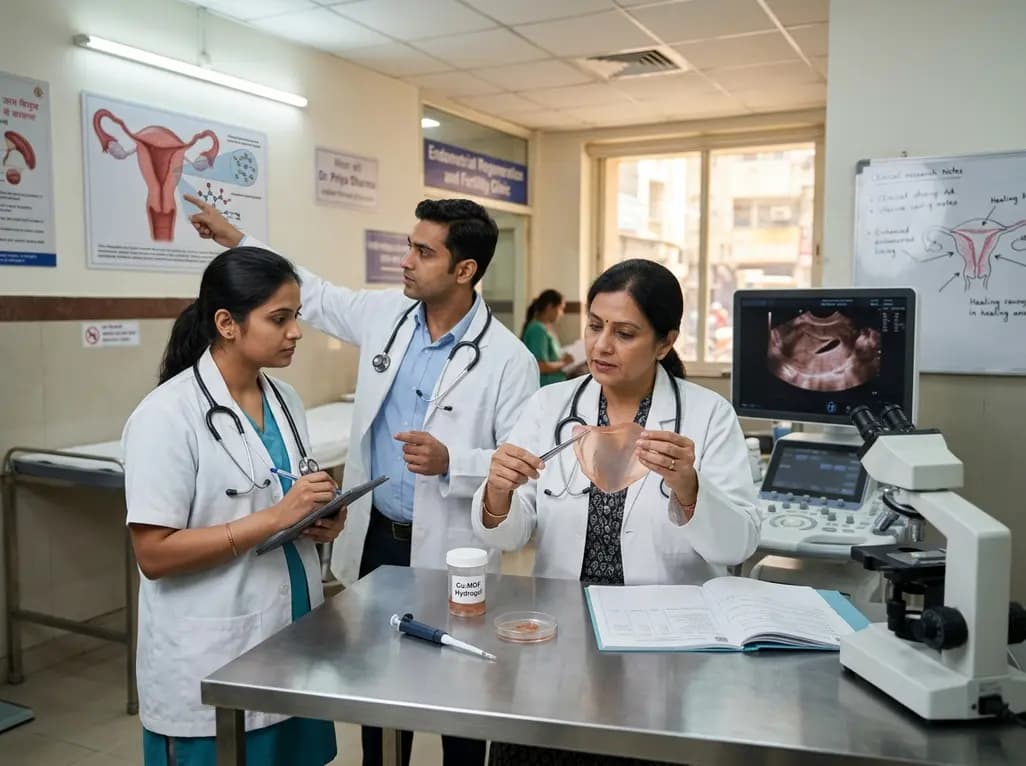
A novel copper metal-organic framework hydrogel remodels the uterine microenvironment to stimulate robust endometrial regeneration, reduce intrauterine fibrosis, and restore reproductive fertility, offering a groundbreaking strategy for managing refractory endometrial damage and Asherman syndrome.
2 weeks back
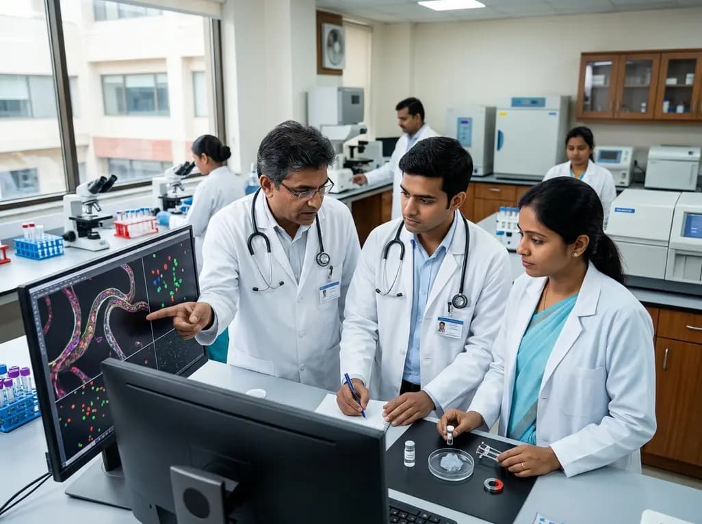
A novel magnetoactive hydrogel platform demonstrates that vascular curvature independently guides endothelial mechanosensing. Elevated curvature triggers nuclear YAP translocation, VE-cadherin junction thinning, and eNOS membrane shifts, offering critical insights into arterial tortuosity and aneurysm pathology.
2 days back
A single-cell transcriptomic atlas reveals how peripheral nerve injuries cause dorsal root ganglion remodeling. By evaluating proximal nerve crush and resection, researchers identify distinct glial and immune programs that resolve neuropathic pain and promote regeneration.
2 weeks back
A breakthrough study demonstrates that fullerol-primed extracellular vesicles generated in a 3D SECCS bioreactor attenuate ferroptosis and restore myogenic regeneration in diabetes via the FOXO3 pathway, offering a cell-free therapeutic strategy for diabetic musculoskeletal degeneration.
2 weeks back
