.webp&w=384&q=75)

Loading, please wait...
Loading, please wait...
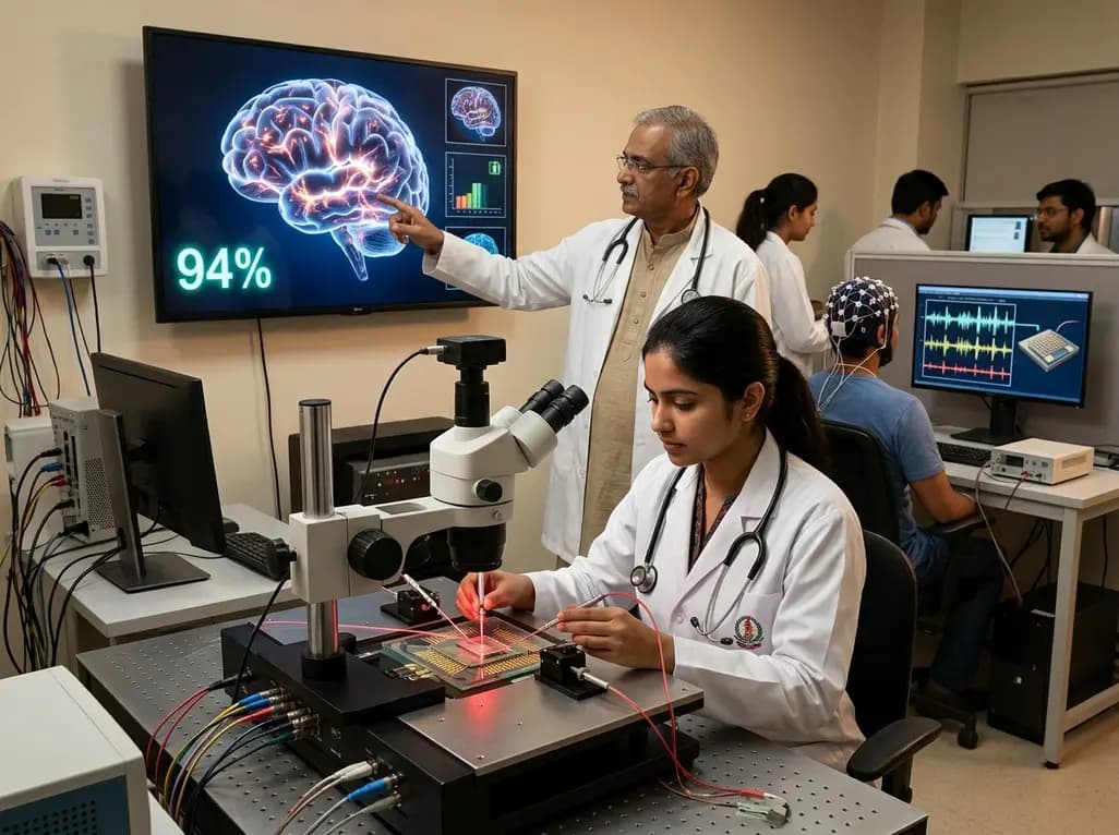
The field of clinical medicine is currently witnessing a transformative shift as the boundaries between bio-engineering and neuro-diagnostics continue to blur. At the heart of this evolution is the development of brain-inspired hardware designed to mimic the unparalleled efficiency of the human central nervous system. Recently, a groundbreaking study by Dhara S et al. has introduced a sophisticated optoelectronic synapse (OES) based on \u03b3-phase Indium (III) Selenide (InSe). This innovation represents a major milestone for neuromorphic computing in neurology, particularly in addressing the chronic challenge of energy consumption in artificial intelligence systems. By leveraging specific electronic defects within the metal lattice, researchers have created a device that not only functions like a biological synapse but does so with a power footprint that is remarkably lower than existing electronic architectures. For clinicians and researchers in India, understanding these hardware advancements is crucial, as they form the backbone of next-generation medical imaging, brain-computer interfaces, and real-time neuro-monitoring tools. As AI becomes more deeply integrated into hospital workflows, the transition from power-hungry software models to energy-efficient neuromorphic hardware will likely define the future of sustainable healthcare technology.
To understand the clinical potential of this technology, one must first appreciate the materials science that enables its synaptic behavior. The researchers utilized Indium Selenide (InSe) thin films, which undergo a critical transition from an amorphous state to a crystalline \u03b3-phase during thermal annealing. This structural evolution is not merely a physical change; it induces specific Indium (In) vacancies within the material. These vacancies act as electronic traps that facilitate the slow release of charges, a phenomenon known as persistent photoconductivity. Specifically, this mechanism allows the device to exhibit a long decay time of approximately 214 seconds, which is essential for emulating the temporal dynamics of biological memory. Furthermore, the device successfully demonstrates core synaptic characteristics, including paired-pulse facilitation, short-term memory, and long-term potentiation. Consequently, these features allow the artificial synapse to learn and forget information in a manner that closely parallels human cognitive processes. Moreover, the crystallization of the InSe film enhances its electrical conductivity by nearly ten times, providing a stable and responsive medium for processing complex neural signals. This high level of control over charge dynamics ensures that the OES can maintain synaptic plasticity, which is the fundamental requirement for any adaptive learning system used in medical diagnostics.
One of the most significant barriers to the widespread adoption of AI in clinical settings is the massive energy requirement of traditional Von Neumann computing architectures. Modern diagnostic algorithms typically run on power-intensive GPUs that require significant cooling and electrical infrastructure. Conversely, the human brain operates on a mere 20 watts of power while performing trillions of operations. The Indium Selenide OES reported by Dhara S et al. bridges this gap by achieving a synaptic energy consumption of approximately 180 attojoules (aJ) per spike. To put this in perspective, this value is well below the energy range typically observed in biological systems, which often operate in the femtojoule range. This ultralow energy consumption is primarily due to the enhanced conductivity and efficient charge-trapping mechanisms inherent in the crystalline \u03b3-phase. Resultantly, this breakthrough makes it possible to design \"always-on\" medical sensors and implantable devices that do not require bulky batteries or frequent recharging. Additionally, by reducing the heat generated during computation, these devices are safer for direct integration with biological tissues. Therefore, the advancement of neuromorphic computing in neurology through such low-power hardware is essential for developing sustainable, long-term neuro-prosthetics and wearable monitoring systems for chronic conditions like epilepsy or Parkinson's disease.
Beyond energy efficiency, the practical utility of any artificial synaptic device is measured by its computational accuracy in real-world tasks. The InSe-based optoelectronic synapse was tested using artificial neural network simulations to perform the MNIST handwritten digit recognition task, achieving an exceptional accuracy of 94%. Furthermore, the researchers applied the device architecture to a U-Net structure-based network for semantic segmentation, reaching an accuracy of 85%. For radiologists and neurologists, U-Net structures are particularly relevant because they are the gold standard for medical image segmentation, such as identifying tumors in MRI scans or delineating vascular structures in CT angiography. Notably, the ability of a neuromorphic chip to handle these complex visual tasks with high precision suggests that we are moving closer to real-time, on-device medical image analysis. Instead of sending raw imaging data to a centralized server, future bedside devices could utilize these low-power synapses to provide instantaneous diagnostic feedback. This localized processing capability would significantly reduce latency, which is critical during emergency surgical procedures or in acute stroke management where every second counts. Consequently, the integration of high-accuracy neuromorphic hardware into clinical vision systems could fundamentally improve the speed and reliability of computer-aided diagnosis.
The successful replication of synaptic functionality using optoelectronic stimuli opens new pathways for developing bidirectional brain-computer interfaces (BCIs). Traditional BCIs often rely on electrical stimulation, which can sometimes lead to tissue damage or signal interference. In contrast, optoelectronic systems use light as a trigger, offering a non-invasive or minimally invasive alternative for modulating neural activity. The persistent photoconductivity and long decay times observed in the InSe device are particularly useful for maintaining neural states without continuous external input. This could lead to more naturalistic neural prosthetics that can store and process information locally, mimicking the feedback loops of the human spinal cord and peripheral nerves. Furthermore, the ability to achieve learning and forgetting behavior means these devices can adapt to the user's changing neural patterns over time, a process known as neuroplasticity. Additionally, the remarkably low power requirements ensure that such implants could operate for years on minimal energy, perhaps even harvesting power from the body's own thermal or mechanical energy. As research continues to refine these materials, we can expect to see a new class of bio-electronic devices that are more compatible with the human body's electrical and optical signaling pathways, ultimately restoring lost sensory or motor functions with unprecedented efficiency.
As India continues to strengthen its position in the global semiconductor and biotechnology sectors, the development of neuromorphic hardware presents a strategic opportunity for local innovation. The implementation of such advanced technology in the Indian healthcare system could help bridge the gap in specialist availability by providing high-level diagnostic tools to rural and underserved areas. However, the transition from laboratory prototypes to clinical-grade medical devices requires rigorous validation and a robust regulatory framework. Specifically, the Central Drugs Standard Control Organisation (CDSCO) will need to establish guidelines for the safety and efficacy of AI-driven hardware that interacts directly with neural tissue. Moreover, ethical considerations regarding data privacy and the autonomy of brain-inspired systems must be addressed as these technologies become more prevalent. Nevertheless, the findings from Dhara S et al. provide a strong scientific foundation for the next decade of neuro-technological growth. By focusing on energy efficiency and high accuracy, this research aligns with the global movement toward sustainable and accessible AI. Looking ahead, the synergy between clinicians and materials scientists will be the key to unlocking the full potential of neuromorphic systems, ensuring that they are not only technically superior but also clinically relevant and safe for patient use.
Indium vacancies in the crystalline lattice of Indium Selenide act as electronic defects that capture and slowly release charge carriers. This process mimics the way biological synapses manage neurotransmitter release and reuptake. By delaying the return of the material to its baseline state, these vacancies enable persistent photoconductivity, which allows the device to \"remember\" a light stimulus long after it has ceased, effectively simulating short-term and long-term memory in artificial systems.
A consumption of 180 attojoules per synaptic event is significant because it is thousands of times lower than the energy used by current digital AI hardware. For medical wearables, this translates to extremely long battery life and reduced heat emission. This efficiency allows for always-on monitoring of vital neurological signals, such as EEG patterns, without the discomfort of device heating or the logistical challenge of frequent battery replacements in implantable neuro-sensors.
U-Net structures are specialized neural networks used for pixel-level image segmentation, which is vital for identifying small lesions or anatomical structures in neuro-imaging. By integrating these structures into neuromorphic chips like those using Indium Selenide, medical devices can perform high-accuracy image analysis locally. This reduces the need for external data processing, enables faster diagnostic results during surgery, and maintains high precision even in low-power, portable imaging equipment used in field medicine.
Disclaimer: This content is for informational and educational purposes only and does not constitute medical advice or professional consultation. The technology described is currently in the research and development phase and is not yet approved for routine clinical use. Doctors and healthcare providers should refer to the latest local and national guidelines for clinical practice and medical device usage.
References
Dhara S et al. Amorphous to Crystalline Transition and Indium-Vacancy Mediated Synaptic Functionality With Ultralow Energy Consumption in \u03b3-Phase Indium (III) Selenide. Small. 2026 Jul 15. doi: 10.1002/smll.74597. PMID: 42454463.
Morris A. AI gets a cerebellum - Northwestern Now. Nature Communications. 2026 Jul 10. https://news.northwestern.edu/stories/2026/07/ai-gets-a-cerebellum/.
Bakhit B et al. Modified Hafnium Oxide Memristors for Low-Power Neuromorphic Computing. Science Advances. 2026 Apr 23. doi: 10.1126/sciadv.adi9876.
"
Read summarized clinical updates, watch expert medical content, and earn CME certifications right from your smartphone.
Open your smartphone camera to scan and install.





Researchers have developed an ultralow-energy optoelectronic synapse using Indium Selenide, mimicking brain function at just 180 aJ per spike. This breakthrough in neuromorphic computing in neurology achieves 94% accuracy in pattern recognition, offering transformative potential for BCI and medical imaging.
3 months ago

Depemokimab offers twice-yearly dosing for severe eosinophilic asthma and chronic rhinosinusitis with nasal polyps, significantly cutting exacerbations and hospital admissions.
4 days back

A novel copper metal-organic framework hydrogel remodels the uterine microenvironment to stimulate robust endometrial regeneration, reduce intrauterine fibrosis, and restore reproductive fertility, offering a groundbreaking strategy for managing refractory endometrial damage and Asherman syndrome.
4 days back

New research reveals that non-ablative radiotherapy suppresses ventricular tachycardia by increasing conduction velocity via NaV1.5 upregulation and action potential prolongation. However, sustained sodium current elevation and depressed calcium handling highlight the need for careful clinical translation.
6 days back

A single-cell transcriptomic atlas reveals how peripheral nerve injuries cause dorsal root ganglion remodeling. By evaluating proximal nerve crush and resection, researchers identify distinct glial and immune programs that resolve neuropathic pain and promote regeneration.
4 days back

A breakthrough study demonstrates that fullerol-primed extracellular vesicles generated in a 3D SECCS bioreactor attenuate ferroptosis and restore myogenic regeneration in diabetes via the FOXO3 pathway, offering a cell-free therapeutic strategy for diabetic musculoskeletal degeneration.
4 days back
