

Loading, please wait...
Loading, please wait...
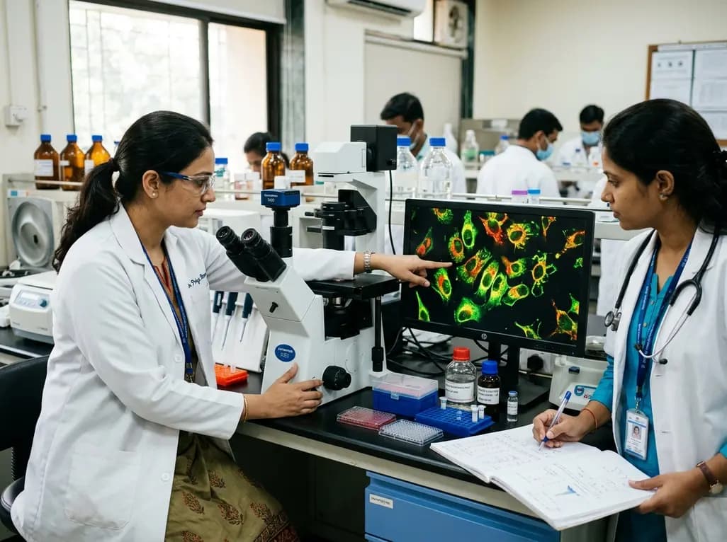
Understanding intracellular microenvironments remains a cornerstone of modern cancer biology. Recently, researchers from the Tata Institute of Fundamental Research in Hyderabad and international partner institutions engineered novel fluorescent sensors to track acidity levels inside malignant cells. Aberrant proton dynamics directly influence malignant proliferation, drug resistance, and metastatic dissemination. Therefore, mapping organelle pH with high precision provides pivotal diagnostic and therapeutic insights. These newly synthesized molecular probes display distinct spectral emissions depending on their sub-cellular localization. Consequently, the technology equips investigators with an unprecedented tool to visualize organelle physiology in real time.
The research team developed two distinct dual-colour molecular systems. One sensor pairs pyrene with a rhodamine dye to produce a red-blue fluorescent signal. Meanwhile, the second sensor couples naphthalimide with rhodamine to yield a red-green emission profile. Both designs incorporate a piperazine unit that directs the molecules toward target organelles. Furthermore, these sensors exploit sophisticated photophysical phenomena. They operate through electron transfer, charge transfer, and resonance energy transfer mechanisms between fluorophores. Under acidic conditions, the rhodamine core undergoes ring opening to produce a strong red signal. In contrast, neutral or basic surroundings preserve the blue or green emission of the partner fluorophore. The red-green sensor demonstrates four distinct pKa values across testing conditions. Consequently, this broad responsive range permits accurate acid-base measurements from neutral states down to highly acidic thresholds. In addition, the probes can detect pH variations well beyond pH 6. Thus, researchers can track fine physiological shifts without losing optical sensitivity.
Malignant cells rely on dysregulated proton gradients to sustain unchecked growth. Specifically, intracellular organelles maintain starkly divergent internal environments to support their enzymatic functions. Mitochondria operate within a relatively alkaline to neutral pH range between 7.5 and 8.0. In contrast, lysosomes maintain an intensely acidic lumen between pH 4.5 and 5.0 to degrade biomolecules. Disruption of these delicate gradients frequently indicates cellular stress, apoptosis, or malignant transformation. The newly reported sensors address this diagnostic challenge by homing to both compartments simultaneously. Inside mitochondria, the sensors emit predominantly blue or green fluorescence due to prevailing alkaline conditions. However, when localized within acidic lysosomes, the probes generate overlapping blue-red or green-red emission signatures. Therefore, microscopists can observe compartmental identity and acidity within a single cultured cell. HeLa cervical cancer cell experiments confirmed that the probes distribute accurately without inducing acute cytotoxicity. As a result, the sensors provide clear optical demarcation between metabolic powerhouse organelles and digestive catabolic centers.
Quantitative fluorescence microscopy often faces significant technical challenges in living specimens. Traditional intensity-based probes frequently suffer from concentration discrepancies, uneven tissue penetration, and rapid photobleaching. Consequently, fluctuations in excitation laser intensity can distort experimental readings. To overcome these limitations, the investigators engineered these sensors to function ratiometrically. Ratiometric analysis evaluates the ratio between two emission wavelengths rather than measuring raw luminescence intensity alone. Because both fluorophores exist on the same molecular scaffold, external variables affect both optical channels equally. Therefore, calculating the emission intensity ratio effectively cancels out errors arising from unequal probe loading. Furthermore, this method eliminates inaccuracies caused by varying cellular thickness and sensor distribution. Researchers tested this calibration rigorously in both controlled laboratory buffers and cultured cancer cell lines. The resulting spectral curves demonstrated consistent and reproducible pH tracking across varied conditions. Thus, ratiometric quantification provides biomedical scientists with dependable measurements that standard single-emission dyes cannot deliver.
Currently, cell biologists rely heavily on commercial markers such as MitoTracker and LysoTracker to stain intracellular structures. Although these commercial reagents highlight organelle positions effectively, they rarely offer quantitative information regarding local proton concentrations. In contrast, the dual-colour probes developed in this study serve two essential diagnostic functions at once. They identify the spatial position of the organelle and simultaneously quantify its surrounding acidity. When researchers compared the new probes against standard commercial markers in HeLa cells, the experimental sensors exhibited comparable localization fidelity. Moreover, the red-green sensor extended the effective imaging window beyond conventional ranges. This expanded dynamic scope allows investigators to record microenvironmental shifts that previous dyes failed to detect. In addition, the chemical synthesis incorporates stable heterocyclic linkers that minimize nonspecific intracellular precipitation. Consequently, these molecular sensors remain soluble and optically stable throughout prolonged imaging protocols. Researchers can therefore monitor real-time acidification dynamics during drug exposure without requiring multiple complementary dyes.
The development of these responsive molecular sensors carries profound implications for preclinical cancer pharmacology. Many antineoplastic therapies, including lysosomotropic agents and mitochondrial inhibitors, directly disturb organelle acidity. For instance, chloroquine and bafilomycin disrupt lysosomal acidification to inhibit protective autophagy in therapy-resistant tumors. By utilizing dual-colour ratiometric probes, oncologists can directly visualize whether prospective drugs hit their target pathways in living cells. Furthermore, altered proton extrusion represents a classic hallmark of aggressive cancer phenotypes under the Warburg effect. Monitoring localized pH shifts enables investigators to study how tumors adapt to hypoxic and acidic conditions. In addition, this platform offers a robust screening assay for assessing targeted drug delivery systems. Many smart nanocarriers exploit acidic lysosomal environments to trigger drug release inside malignant cells. Accordingly, precise pH monitoring helps researchers optimize these nanomedicines before advancing to clinical trials. Ultimately, these innovative chemical tools bridge laboratory spectroscopy with practical translational oncology.
Q1: How do these novel fluorescent sensors determine organelle acidity?
The novel dual-colour sensors integrate specific fluorophores with piperazine moieties. Consequently, they migrate selectively to distinct organelles like mitochondria and lysosomes. When local proton concentrations shift, the rhodamine core undergoes reversible structural alterations. Therefore, the probe emits distinct fluorescent spectra in neutral versus acidic compartments. This mechanism allows investigators to map intracellular pH variations with high spatial accuracy across living cancer cells.
Q2: Why is ratiometric imaging superior to traditional intensity-based fluorescence?
Conventional single-wavelength dyes often succumb to photobleaching and concentration discrepancies. In contrast, ratiometric sensors measure the ratio of emission intensities at two different optical wavelengths. Consequently, variations in laser excitation power, dye uptake, or cellular thickness cancel out mathematically. Thus, researchers obtain highly reproducible quantitative data regarding organelle acidity. This capability substantially enhances the reliability of imaging assays during preclinical oncological evaluations and dynamic pharmacodynamic investigations.
Q3: How does monitoring cancer cell pH assist oncological research?
Malignant cells frequently exhibit profound metabolic reprogramming and altered proton extrusion. Consequently, tumor organelles demonstrate disrupted pH homeostasis that drives invasion, chemoresistance, and immune evasion. By monitoring these microscopic shifts, oncologists and biochemists can evaluate how prospective antineoplastic agents affect cellular physiology. Furthermore, tracking acidity enables earlier detection of therapeutic response and facilitates the rational design of acid-activated nanoparticle delivery systems.
Disclaimer: This content is for informational and educational purposes only. It does not constitute medical advice or replace professional judgment. Refer to the latest local and national guidelines for clinical practice.
References

Read summarized clinical updates, watch expert medical content, and earn CME certifications right from your smartphone.
Open your smartphone camera to scan and install.





Scientists have developed innovative dual-colour fluorescent sensors capable of tracking real-time pH variations within cellular organelles such as mitochondria and lysosomes. Tested in HeLa cells, these ratiometric molecular probes provide clinicians and researchers with enhanced precision for studying cancer biology.
Yesterday

Depemokimab offers twice-yearly dosing for severe eosinophilic asthma and chronic rhinosinusitis with nasal polyps, significantly cutting exacerbations and hospital admissions.
Yesterday

A novel copper metal-organic framework hydrogel remodels the uterine microenvironment to stimulate robust endometrial regeneration, reduce intrauterine fibrosis, and restore reproductive fertility, offering a groundbreaking strategy for managing refractory endometrial damage and Asherman syndrome.
Yesterday

New research reveals that non-ablative radiotherapy suppresses ventricular tachycardia by increasing conduction velocity via NaV1.5 upregulation and action potential prolongation. However, sustained sodium current elevation and depressed calcium handling highlight the need for careful clinical translation.
3 days back

A single-cell transcriptomic atlas reveals how peripheral nerve injuries cause dorsal root ganglion remodeling. By evaluating proximal nerve crush and resection, researchers identify distinct glial and immune programs that resolve neuropathic pain and promote regeneration.
Yesterday
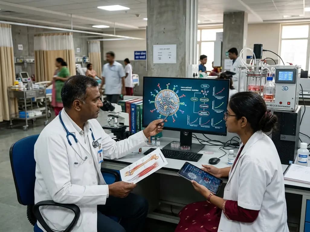
A breakthrough study demonstrates that fullerol-primed extracellular vesicles generated in a 3D SECCS bioreactor attenuate ferroptosis and restore myogenic regeneration in diabetes via the FOXO3 pathway, offering a cell-free therapeutic strategy for diabetic musculoskeletal degeneration.
Yesterday
