.webp&w=384&q=75)

Loading, please wait...
Loading, please wait...
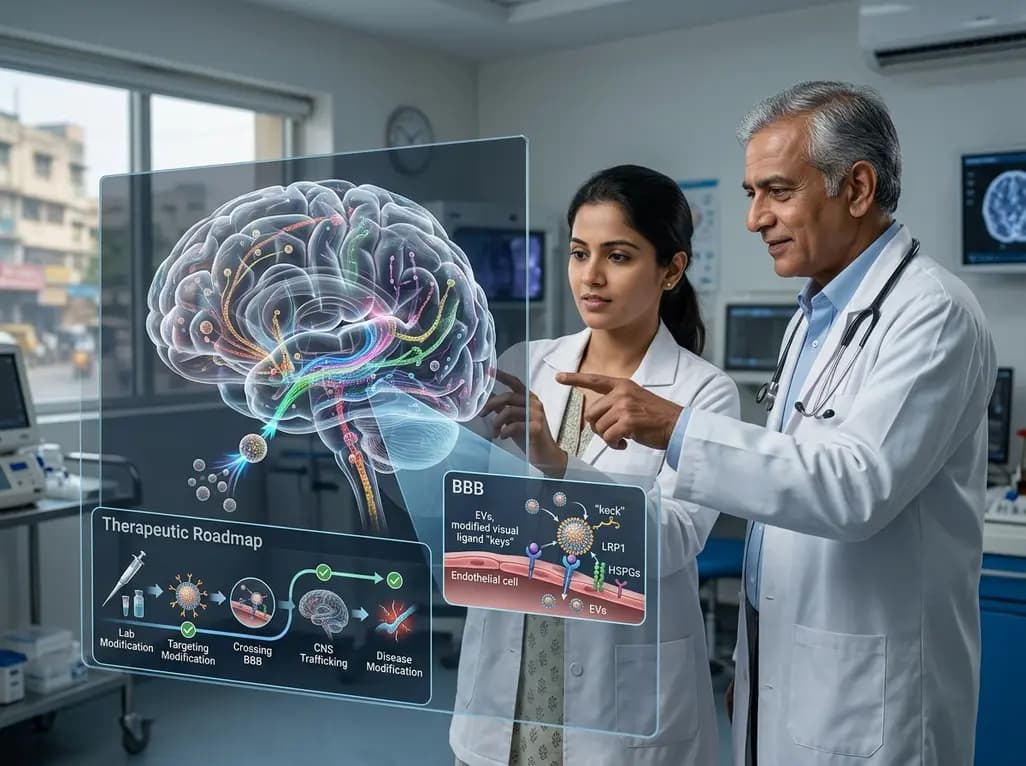
Extracellular vesicles (EVs) represent a sophisticated biological system for intercellular signaling within the central nervous system (CNS). These membrane-bound particles facilitate the exchange of lipids, proteins, and nucleic acids between various brain cells, including neurons, glia, and the vascular endothelium. In the context of extracellular vesicles CNS research, understanding how these particles navigate the complex neural environment is essential for developing next-generation treatments. Current evidence suggests that EVs do not move randomly; instead, they follow specific molecular pathways defined by their surface ligands and the protein corona they adsorb from biological fluids. This inherent targeting capability makes them attractive candidates for therapeutic delivery, provided we can decode the rules governing their transport across the blood-brain barrier (BBB) and their eventual intracellular fate.
Effective brain targeting by extracellular vesicles CNS depends on a highly regulated sequence of molecular interactions. Initially, EV ligands and their associated protein coronas engage specific receptor modules on target cells. This interaction is not merely a docking mechanism; it dictates the selection of endocytic routes and determines whether the cargo will be degraded or delivered to the cytosol. For instance, heparan sulfate proteoglycans (HSPGs) and members of the LDL receptor family, most notably LRP1, play crucial roles in regulating the handling of proteopathic proteins like tau and amyloid-β. By manipulating these pathways, researchers hope to design EVs that can bypass lysosomal degradation and achieve rare but necessary cytosolic delivery. Transitioning from natural biology to engineered precision remains a primary goal in neuro-nanomedicine.
The blood-brain barrier remains the most significant physiological hurdle for any CNS-targeted therapy. Extracellular vesicles CNS studies have highlighted the importance of endothelial HSPGs, LRP1, and the transferrin receptor (TfR) in supporting receptor-mediated uptake at the BBB. To enhance this process, bioengineers are developing engineered ligands, such as rabies virus glycoprotein-derived peptides and Angiopep-2, which can be conjugated to the EV surface. These modifications aim to exploit natural transcytosis pathways to ferry therapeutic cargo from the systemic circulation into the brain parenchyma. While these strategies show promise in preclinical models, the transition to human clinical application requires a deeper understanding of how disease states, such as neuroinflammation, alter these transport mechanisms and potentially lead to the dissemination of toxic seeds.
Once an EV successfully enters a target cell, its intracellular journey determines the ultimate therapeutic readout. Most vesicles are channeled toward lysosomal degradation or recycling back to the extracellular space. For nucleic acid delivery, a major kinetic barrier is endosomal escape, the process by which cargo exits the endosome to reach the cytoplasm. Research into extracellular vesicles CNS delivery confirms that while EVs are naturally better at this than many synthetic nanoparticles, the efficiency remains low. Improving this step is critical for the success of RNA-based therapies in neurodegenerative conditions. Scientists are currently exploring how integrin and tetraspanin nanoclusters influence the avidity of EV-cell interactions, which might be key to unlocking more efficient delivery routes.
Translating the complex biology of extracellular vesicles CNS into safe and manufacturable therapeutics requires rigorous design and assay standards. We must synthesize principles across various conditions, including Alzheimer’s disease, Parkinson’s disease, and glioblastoma, to identify common therapeutic targets. Standardizing the isolation and characterization of EVs is necessary to ensure batch-to-batch consistency and regulatory compliance. Furthermore, we must distinguish between the beneficial signaling of healthy EVs and the potentially harmful role of vesicles in spreading neuroinflammation. As we outline this therapeutic roadmap, the focus remains on creating platforms that are not only effective but also scalable for the global pharmaceutical market, ensuring that breakthrough discoveries in the lab eventually reach the bedside of patients with CNS disorders.
The interaction between extracellular vesicles CNS and the brain's immune system is a double-edged sword. Phosphatidylserine readers and complement proteins often shape the "myeloid sink," where specialized immune cells like microglia capture and neutralize circulating EVs. This capture can either dampen therapeutic efficacy or, if properly harnessed, be used to modulate neuroinflammation. Integrin and ICAM-1 nanoclusters on the EV surface influence organotropism and can promote immune suppression in the context of brain tumors or autoimmune demyelination. Understanding these immune dynamics is vital for ensuring that EV-based drugs are not prematurely cleared from the system and that they do not trigger adverse inflammatory responses, which are already heightened in most chronic central nervous system diseases.
Extracellular vesicles offer several advantages over synthetic carriers, including lower immunogenicity, inherent ability to cross the blood-brain barrier, and the capacity to carry complex biological cargo. Because they are derived from natural cells, they possess surface proteins that facilitate specific cellular targeting and endocytic uptake. This makes them highly efficient at navigating the complex microenvironment of the brain, potentially improving the delivery of proteins and nucleic acids to diseased neurons.
Receptor modules such as LRP1 are critical regulators of protein homeostasis in the brain. They are involved in the clearance of toxic aggregates like amyloid-β and tau. In the context of EV-based therapy, targeting these receptors allows for more precise delivery of therapeutic agents to the specific cells involved in disease pathology. By engineering EVs to bind with high affinity to LRP1, researchers can enhance the clearance of proteopathic seeds and reduce neuroinflammation.
The most significant technical hurdle is achieving efficient endosomal escape. While EVs can enter cells through endocytosis, the majority of their nucleic acid cargo often remains trapped within endosomes and is eventually degraded by lysosomes. To be effective, the RNA must reach the cytosol where it can function. Developing engineering strategies that trigger the release of cargo from the endosomal compartment is the focus of current research to enable successful gene therapies for the CNS.
Disclaimer: This content is for informational and educational purposes only... Refer to the latest local and national guidelines for clinical practice.
References
Baker B et al. Brain targeting and trafficking of extracellular vesicles in central nervous system diseases: a therapeutic roadmap. Nanomedicine (Lond). 2026 Jul 11. doi: 10.1080/17435889.2026.2698782. PMID: 42434808.
Mizrahy S, et al. Targeting Central Nervous System pathologies with nanomedicines. Minded Cofund. 2024.
Solanki H, et al. Advancing Extracellular Vesicle Therapeutics into Clinical Trials for Central Nervous System Disorders. Athenaeum Scientific Publishers. 2024.

Read summarized clinical updates, watch expert medical content, and earn CME certifications right from your smartphone.
Open your smartphone camera to scan and install.





Extracellular vesicles (EVs) are emerging as pivotal players in central nervous system (CNS) signaling and drug delivery. This article outlines the complex mechanisms of brain targeting, the role of receptor modules like LRP1 and HSPGs, and the barriers to translating EV biology into effective CNS therapeutics.
3 months ago

Depemokimab offers twice-yearly dosing for severe eosinophilic asthma and chronic rhinosinusitis with nasal polyps, significantly cutting exacerbations and hospital admissions.
2 days back

A novel copper metal-organic framework hydrogel remodels the uterine microenvironment to stimulate robust endometrial regeneration, reduce intrauterine fibrosis, and restore reproductive fertility, offering a groundbreaking strategy for managing refractory endometrial damage and Asherman syndrome.
3 days back

New research reveals that non-ablative radiotherapy suppresses ventricular tachycardia by increasing conduction velocity via NaV1.5 upregulation and action potential prolongation. However, sustained sodium current elevation and depressed calcium handling highlight the need for careful clinical translation.
5 days back

A single-cell transcriptomic atlas reveals how peripheral nerve injuries cause dorsal root ganglion remodeling. By evaluating proximal nerve crush and resection, researchers identify distinct glial and immune programs that resolve neuropathic pain and promote regeneration.
3 days back

A breakthrough study demonstrates that fullerol-primed extracellular vesicles generated in a 3D SECCS bioreactor attenuate ferroptosis and restore myogenic regeneration in diabetes via the FOXO3 pathway, offering a cell-free therapeutic strategy for diabetic musculoskeletal degeneration.
3 days back
