.webp&w=384&q=75)

Loading, please wait...
Loading, please wait...
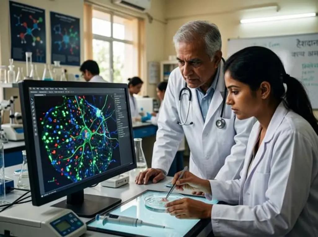
Expansion microscopy mapping is transforming how researchers visualize biological specimens at the molecular level. This technique physically enlarges samples using expandable hydrogels, effectively bypassing the diffraction limit of light. Furthermore, recent innovations integrate this process with label-free modalities like stimulated Raman scattering and mass spectrometry. Consequently, scientists can now map complex chemical signatures within single cells without relying on external dyes.
The primary benefit of this approach lies in its ability to enhance spatial resolution using existing hardware. For instance, researchers can resolve subcellular details that were previously invisible. Additionally, multimodal imaging allows for the simultaneous detection of diverse molecular species. These combined capabilities facilitate a more comprehensive understanding of spatial biology. However, maintaining the structural integrity of specimens during the expansion process remains a technical challenge.
Experts anticipate that this coupling will revolutionize pathology and clinical diagnostics in India. Moreover, the integration of AI-driven analysis will likely accelerate the interpretation of high-resolution data. Therefore, these advancements provide a robust framework for investigating cellular environments in health and disease. Because this method is accessible, it empowers more laboratories to conduct high-end spatial omics studies.
It is a technique that physically enlarges biological specimens to improve the effective resolution of standard microscopes, allowing for detailed molecular mapping.
Label-free imaging enables the detection of inherent chemical signatures in a sample without the need for fluorescent markers or expensive labels.
Disclaimer: This content is for informational and educational purposes only. It does not constitute professional medical advice, diagnosis, or treatment. Always seek the advice of your physician or other qualified healthcare provider with any questions you may have regarding a medical condition. Refer to the latest local and national guidelines for clinical practice.
References

Read summarized clinical updates, watch expert medical content, and earn CME certifications right from your smartphone.
Open your smartphone camera to scan and install.





Expansion microscopy enhances spatial resolution by physically enlarging biological specimens. Recent breakthroughs integrate this with label-free and multimodal imaging for single-cell molecular mapping, enabling researchers to detect complex chemical signatures and advance the field of spatial biology.
4 months ago

Depemokimab offers twice-yearly dosing for severe eosinophilic asthma and chronic rhinosinusitis with nasal polyps, significantly cutting exacerbations and hospital admissions.
3 days back

A novel copper metal-organic framework hydrogel remodels the uterine microenvironment to stimulate robust endometrial regeneration, reduce intrauterine fibrosis, and restore reproductive fertility, offering a groundbreaking strategy for managing refractory endometrial damage and Asherman syndrome.
4 days back

New research reveals that non-ablative radiotherapy suppresses ventricular tachycardia by increasing conduction velocity via NaV1.5 upregulation and action potential prolongation. However, sustained sodium current elevation and depressed calcium handling highlight the need for careful clinical translation.
6 days back

A single-cell transcriptomic atlas reveals how peripheral nerve injuries cause dorsal root ganglion remodeling. By evaluating proximal nerve crush and resection, researchers identify distinct glial and immune programs that resolve neuropathic pain and promote regeneration.
4 days back

A breakthrough study demonstrates that fullerol-primed extracellular vesicles generated in a 3D SECCS bioreactor attenuate ferroptosis and restore myogenic regeneration in diabetes via the FOXO3 pathway, offering a cell-free therapeutic strategy for diabetic musculoskeletal degeneration.
4 days back
