.webp&w=384&q=75)

Loading, please wait...
Loading, please wait...
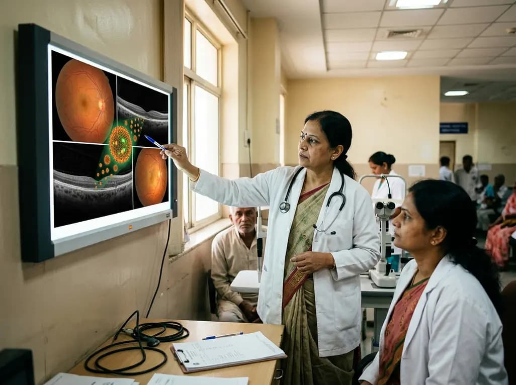
Conventional ophthalmic therapeutics face severe physiological barriers in everyday clinical practice. Tear turnover, corneal epithelial tightness, and nasolacrimal drainage rapidly clear topical drop formulations, resulting in low ocular bioavailability. Consequently, patients require frequent daily dosing, which significantly compromises long-term therapeutic compliance. Furthermore, invasive intraocular injections introduce serious clinical risks such as endophthalmitis, intraocular pressure elevation, and retinal detachment. Recent advances in carbon nanodots ocular disease management offer a highly effective alternative to traditional ocular drug delivery systems. Carbon nanodots are zero-dimensional carbon nanomaterials measuring strictly under ten nanometers in diameter. Their ultrasmall size allows them to navigate complex biological barriers smoothly while causing minimal tissue irritation. Additionally, carbon nanodots exhibit remarkable aqueous solubility, structural stability, and extended retention on ocular surfaces. By bypassing rapid physiological clearance, these nanocarriers maintain sustained therapeutic drug concentrations at targeted tissue sites. As a result, clinicians can lower total drug dosages, minimizing systemic absorption and off-target adverse effects while optimizing overall therapeutic efficacy across diverse clinical settings.
The unique physicochemical profile of carbon nanodots underpins their expanding utility in modern clinical ophthalmology. Synthetic flexibility allows medical researchers to produce these nanostructures from diverse organic precursors via top-down or bottom-up chemistry pathways. Furthermore, facile surface functionalization enables effortless attachment of targeted therapeutic agents, specific cellular ligands, and diagnostic bioimaging tags. Because carbon nanodots possess rich functional surface groups like carboxyl, amino, and hydroxyl species, they interact favorably with hydrophilic drugs and cellular membranes. Moreover, safety remains paramount when developing drug delivery platforms for delicate ocular tissues. Preclinical evaluations consistently indicate that carbon nanodots display minimal cytotoxicity and excellent ocular biocompatibility. Unlike metallic nanoparticles, carbon-based nanostructures do not induce significant local inflammation or oxidative tissue damage at therapeutic concentrations. Additionally, high aqueous dispersibility prevents particle aggregation in tear fluid or vitreous humor. This intrinsic physical stability ensures uniform drug distribution across target ocular tissues, providing a reliable delivery foundation for complex ophthalmic formulations.
Transporting therapeutic agents across corneal and blood-retinal barriers represents a persistent hurdle in modern ophthalmic pharmacology. Carbon nanodots resolve these challenges through enhanced epithelial penetration and sustained retention mechanisms. When loaded with hydrophilic or lipophilic drugs, these nanocarriers shield active pharmaceutical ingredients from rapid enzymatic breakdown in tear film or vitreous humor. Consequently, active therapeutic compounds reach targeted ocular tissues in an intact state. Furthermore, modified surface chemistries allow carbon dots to adhere effectively to mucosal layers on the ocular surface. This mucoadhesive property retards physiological clearance, promoting continuous drug diffusion across corneal tissue layers. In posterior segment applications, surface-functionalized carbon nanodots traverse the vitreous body efficiently, delivering payloads directly to retinal cells. Therefore, researchers observe significantly enhanced drug bioavailability compared to standard drug solutions. Lower dosing requirements enhance patient compliance while minimizing systemic drug exposure, making carbon dots superior delivery vectors for anti-inflammatory, antimicrobial, and anti-VEGF therapies.
Beyond acting as efficient delivery vectors, carbon nanodots serve as dual-action theranostic platforms. Theranostics seamlessly combines diagnostic bioimaging and targeted therapy within a single entity, facilitating real-time monitoring of disease progression and treatment response. Carbon nanodots display robust, tunable photoluminescence and elevated quantum yields. Consequently, clinicians can utilize them for high-resolution ocular fluorescent angiography and live tissue imaging without resorting to toxic synthetic dyes. Additionally, near-infrared emission properties permit deeper tissue penetration with minimal background autofluorescence from surrounding ocular structures. Physicians can non-invasively track nanodot distribution, cellular internalisation, and drug release kinetics across treatment courses. This capability proves vital when managing chronic posterior segment disorders that require individualized treatment adjustments. Furthermore, combining real-time fluorescent tracking with controlled drug delivery empowers clinicians to verify therapeutic delivery immediately after administration. Therefore, carbon nanodots bridge diagnostic precision with therapeutic intervention, driving the evolution of personalized ophthalmic care.
Carbon nanodots show therapeutic promise across both anterior and posterior segment ocular diseases. In anterior segment conditions like bacterial keratitis, carbon dots conjugated with antibacterial agents penetrate dense corneal stroma to eradicate pathogens and reduce corneal inflammation. In glaucoma models, carbon dot formulations enhance intraocular pressure control by promoting sustained aqueous outflow. Conversely, posterior segment conditions benefit significantly from targeted retinal delivery. In neovascular age-related macular degeneration and diabetic retinopathy, carbon nanodots deliver anti-VEGF molecules directly to choroidal vascular beds, suppressing pathological neovascularization and preserving vision. However, translational challenges remain before clinical adoption. Large-scale manufacturing, batch-to-batch consistency, and long-term biosafety assessments must be rigorously established. Establishing standardized regulatory guidelines for theranostic nanomedicines will be crucial. Collaborative interdisciplinary efforts between nanotechnologists and ophthalmologists will accelerate clinical trial evaluation, ultimately providing safer and more effective therapies for vision-threatening ocular conditions.
Carbon nanodots are zero-dimensional carbon-based nanomaterials measuring under ten nanometers in size. In ocular medicine, they serve as advanced drug delivery vehicles and diagnostic imaging agents. Their ultrasmall dimensions allow them to cross tough anatomical eye barriers easily. Furthermore, their surface chemistry enables precise drug conjugation, while their natural fluorescent properties allow clinicians to track drug distribution and monitor disease progression in real time without causing significant tissue toxicity.
Traditional topical eye drops suffer from rapid clearance by tear turnover and low corneal permeability. Carbon nanodots overcome these obstacles through enhanced bioadhesion and superior permeation capabilities. They adhere to mucin layers on the ocular surface, extending retention time and facilitating sustained drug diffusion. Additionally, their stability protects encapsulated therapeutics from degradation, ensuring that higher active drug concentrations reach targeted anterior and posterior ocular tissues with reduced dosing frequency.
Carbon nanodots show therapeutic efficacy across both anterior and posterior ocular diseases. In anterior segment conditions, they effectively deliver antibiotics for bacterial keratitis and pressure-reducing agents for glaucoma. In posterior segment disorders, carbon nanodots deliver anti-VEGF therapies and antioxidants for neovascular age-related macular degeneration and diabetic retinopathy. Furthermore, their bioimaging capabilities assist in diagnosing vitreous opacification, making them versatile platforms for diverse blinding eye conditions.
Disclaimer: This content is for informational and educational purposes only and does not constitute medical advice. Healthcare professionals should rely on their clinical judgment and refer to the latest local and national guidelines for clinical practice.
References

Read summarized clinical updates, watch expert medical content, and earn CME certifications right from your smartphone.
Open your smartphone camera to scan and install.





A comprehensive review highlights how carbon nanodots overcome ocular barriers, offering advanced theranostic and drug delivery solutions for corneal, macular, and retinal diseases.
last month

A randomized trial shows oral DL-3-n-butylphthalide (200 mg thrice daily) significantly slows functional decline in spinocerebellar ataxia type 3 over 12 months, improving SARA and SCAFI scores with favorable safety and zero serious adverse events.
5 days back

Recent research reveals that downregulating YAP1/TAZ-TEAD via Hippo signaling triggers spontaneous human trophoblast syncytialization in 3D cultures, illuminating placental biology and preeclampsia.
Last week

A landmark comparative study evaluates atrial electrophysiology across humans, swine, and rodents. By linking cellular recordings with transcriptomics and in silico modeling, researchers resolve cross-species gaps, explaining translational failures to advance targeted therapies for atrial arrhythmias.
Last week

A novel pathology-adaptive surface engineering strategy uses functionalized plasma polymer coatings to selectively modulate AGE adsorption, reducing oxidative stress and restoring bone formation in diabetic and aging microenvironments.
last month

Researchers have developed an innovative ultrasound-responsive nano-contrast agent that co-delivers CIITA-siRNA and rapamycin directly to inflamed thyroid tissue, significantly reducing autoimmune injury and thyroid autoantibodies in preclinical models of Hashimoto thyroiditis.
Last week
