.webp&w=384&q=75)

Loading, please wait...
Loading, please wait...
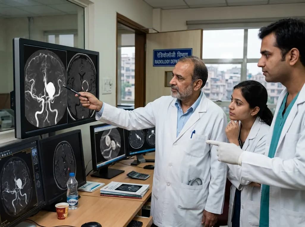
Managing intracranial aneurysms (IAs) remains a significant challenge for neurosurgeons and neurologists worldwide. Historically, clinical decisions relied heavily on luminal imaging techniques such as Digital Subtraction Angiography (DSA) or Computed Tomography Angiography (CTA). While these modalities provide excellent detail regarding the size and shape of the aneurysm, they offer limited information about the vessel wall itself. Recently, Black blood MRI sequences have emerged as a powerful diagnostic adjunct. By suppressing signal from both the blood and cerebrospinal fluid, these sequences allow for direct visualization of the arterial wall. This technological advancement provides clinicians with deep insights into the inflammatory state and stability of the aneurysm. Consequently, this imaging technique is transforming how we manage both ruptured and unruptured intracranial aneurysms in acute settings.
The primary objective of Black blood MRI sequences is to eliminate the 'flow-related' signal that often obscures the vessel wall on traditional MRI scans. This is typically achieved using advanced pulse sequences, such as 3D T1-weighted SPACE (Sampling Perfection with Application-optimized Contrasts using different Flip Angle Evolution). Specifically, these sequences employ variable flip angles to suppress the signal from moving blood, creating a 'black blood' effect. When performed on high-field systems like 3.0 Tesla MRI, the spatial resolution is sufficient to distinguish the thin layers of the intracranial vessel wall. Furthermore, the use of pre- and post-contrast T1-weighted imaging is essential. Contrast enhancement within the aneurysm wall often indicates a breakdown of the internal elastic lamina or active inflammation. Therefore, the technical superiority of these sequences allows for a more granular assessment than traditional time-of-flight MRA. Moreover, clinicians can now identify subtle intramural hematomas or atherosclerotic plaques that were previously invisible. As a result, the diagnostic confidence in neurovascular assessment has increased substantially.
A frequent clinical dilemma occurs when a patient presents with subarachnoid hemorrhage (SAH) and multiple intracranial aneurysms. Determining which of the multiple lesions actually ruptured is critical for planning surgical clipping or endovascular coiling. Traditionally, clinicians looked for markers like size, shape, or 'tit' formation on conventional angiography. However, these morphological features can be misleading. Notably, Black blood MRI sequences offer a more physiological approach by identifying aneurysm wall enhancement (AWE). Evidence suggests that the ruptured aneurysm usually exhibits significantly stronger circumferential enhancement compared to its stable counterparts. This enhancement acts as a surrogate marker for the acute inflammatory response associated with a recent rupture event. Subsequently, identifying the 'culprit' lesion becomes much more reliable, reducing the risk of treating the wrong aneurysm. Additionally, this technique is particularly valuable when the hemorrhage pattern on CT is diffuse or non-localizing. By focusing intervention on the specific enhancing lesion, surgeons can minimize operative time and improve patient outcomes.
The discovery of incidental unruptured intracranial aneurysms (UIAs) often creates immense anxiety for patients and clinicians. While clinical scores like the PHASES score provide a population-based risk estimate, they lack precision for individual cases. This is where Black blood MRI sequences provide an objective biomarker for instability. Specifically, the presence of strong wall enhancement in an unruptured aneurysm is strongly associated with histologic signs of inflammation, such as macrophage infiltration and neovascularization. Furthermore, research indicates that enhancing aneurysms are more likely to grow or rupture during follow-up compared to non-enhancing ones. Therefore, VWI (Vessel Wall Imaging) acts as a functional assessment that complements anatomical measurements. For instance, a small 3mm aneurysm that shows intense wall enhancement may be considered more dangerous than a larger, non-enhancing one. Consequently, radiologists can better stratify which patients require immediate intervention and which can safely undergo conservative surveillance. This individualized approach significantly refines the cost-benefit analysis of invasive treatments.
Following endovascular coiling or flow diversion, surveillance is vital to detect recurrence or incomplete occlusion. Conventional MRA often suffers from metallic susceptibility artifacts, making the vessel wall difficult to evaluate. However, 3D Black blood MRI sequences are remarkably resilient to these artifacts, particularly when using specific sequences designed to minimize signal dropout. Interestingly, the pattern of enhancement after treatment can also provide clues regarding the healing process. Some studies suggest that transient intra-aneurysmal enhancement following coiling represents the organization of thrombus and the formation of a neo-intima. Conversely, persistent or increasing wall enhancement might signal ongoing instability or the failure of the healing process. Moreover, this technique allows for the non-invasive monitoring of the parent artery to ensure there is no significant intimal hyperplasia or stented-segment stenosis. Thus, incorporating VWI into routine follow-up protocols provides a more comprehensive picture of the biological status of the treated aneurysm. Resultantly, clinicians can tailor follow-up intervals based on the patient's unique imaging signature.
Despite its significant advantages, the clinical implementation of Black blood MRI sequences requires careful interpretation. Radiologists must be aware of 'pseudo-enhancement' caused by slow or stagnant blood flow, which can mimic wall enhancement. Specifically, if the blood signal is not fully suppressed, it may appear as a bright layer adjacent to the wall. Therefore, using 3D sequences with high spatial resolution and robust flow suppression is mandatory to ensure diagnostic accuracy. Furthermore, while AWE is a strong marker of inflammation, it is not perfectly sensitive for all rupture events. Some acutely ruptured aneurysms may show minimal enhancement if the inflammatory response is delayed. Additionally, other vasculopathies like CNS vasculitis or intracranial atherosclerosis can also show vessel wall enhancement, potentially confounding the diagnosis. Consequently, the findings from these MRI sequences must always be correlated with the patient’s clinical presentation and other imaging findings. Nevertheless, as the technology matures and protocols become standardized, VWI is set to become an indispensable tool in the neurovascular armamentarium.
These sequences detect aneurysm wall enhancement (AWE), which is a physiological marker of active inflammation or recent rupture. In cases of multiple aneurysms, the lesion that has actually ruptured typically shows significantly stronger and more circumferential enhancement, allowing clinicians to distinguish the culprit from other stable, incidental aneurysms.
Wall enhancement on black blood MRI often correlates with histologic markers of instability, such as macrophage infiltration and neovascularization. Studies indicate that unruptured aneurysms with visible enhancement are more likely to grow or rupture over time, making enhancement a critical biomarker for personalizing treatment decisions beyond simple size measurements.
Optimal imaging usually requires a 3.0 Tesla MRI system and specialized 3D T1-weighted sequences like SPACE or CUBE. These protocols use variable flip angles to suppress luminal blood flow. Comparing pre-contrast and post-contrast images is essential to differentiate between true wall enhancement and baseline mural thickening or artifacts.
Disclaimer: This content is for informational and educational purposes only and does not constitute medical advice, diagnosis, or treatment. Always seek the advice of a physician or other qualified health provider with any questions you may have regarding a medical condition. Refer to the latest local and national guidelines for clinical practice.
References
El Sheikh M et al. Black blood MRI sequences in the acute management of ruptured and unruptured intracranial aneurysms. Br J Neurosurg. 2025 Aug. doi: 10.1080/02688697.2023.2290668. PMID: 38042989.
Mossa-Basha M, et al. (2016). Multicontrast high-resolution vessel wall magnetic resonance imaging and its value in differentiating intracranial vasculopathic processes. Cardiovascular Diagnosis and Therapy. doi: 10.21037/cdt.2016.08.05.
Larsen N, et al. (2018). Vessel Wall Enhancement in Unruptured Intracranial Aneurysms: An Indicator for Higher Risk of Rupture? High-Resolution MR Imaging and Correlated Histologic Findings. American Journal of Neuroradiology. doi: 10.3174/ajnr.A5731.

Read summarized clinical updates, watch expert medical content, and earn CME certifications right from your smartphone.
Open your smartphone camera to scan and install.





High-resolution black blood MRI sequences offer a breakthrough in managing intracranial aneurysms. This case series highlights its role in identifying culprit lesions, surveillance of treated aneurysms, and risk-stratifying unruptured cases, providing critical diagnostic clarity for clinicians.
3 months ago

Depemokimab offers twice-yearly dosing for severe eosinophilic asthma and chronic rhinosinusitis with nasal polyps, significantly cutting exacerbations and hospital admissions.
3 days back

A novel copper metal-organic framework hydrogel remodels the uterine microenvironment to stimulate robust endometrial regeneration, reduce intrauterine fibrosis, and restore reproductive fertility, offering a groundbreaking strategy for managing refractory endometrial damage and Asherman syndrome.
3 days back

New research reveals that non-ablative radiotherapy suppresses ventricular tachycardia by increasing conduction velocity via NaV1.5 upregulation and action potential prolongation. However, sustained sodium current elevation and depressed calcium handling highlight the need for careful clinical translation.
5 days back

A single-cell transcriptomic atlas reveals how peripheral nerve injuries cause dorsal root ganglion remodeling. By evaluating proximal nerve crush and resection, researchers identify distinct glial and immune programs that resolve neuropathic pain and promote regeneration.
3 days back

A breakthrough study demonstrates that fullerol-primed extracellular vesicles generated in a 3D SECCS bioreactor attenuate ferroptosis and restore myogenic regeneration in diabetes via the FOXO3 pathway, offering a cell-free therapeutic strategy for diabetic musculoskeletal degeneration.
3 days back
