.webp&w=384&q=75)

Loading, please wait...
Loading, please wait...
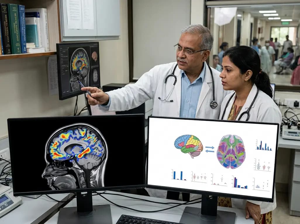
Neurodegenerative conditions such as Alzheimer's disease and Parkinson's disease present immense global healthcare challenges. Understanding their underlying molecular drivers is essential for developing effective therapies. Recent advances in spatial biology have established the Allen Human Brain Atlas as a cornerstone resource for mapping human gene expression across healthy neural structures. By capturing high-resolution transcriptomics across 102 brain regions, this repository allows researchers to cross-reference gene signatures with clinical neuroimaging phenotypes. Consequently, scientists can map transcriptomic correlates against pathological features like amyloid-beta deposition, tau neurofibrillary tangles, and alpha-synuclein propagation. This integrative approach reveals why specific brain regions demonstrate heightened vulnerability to neurodegeneration.
Alzheimer's disease and Parkinson's disease represent two of the most prevalent age-related neurodegenerative disorders globally. Both conditions display progressive neurological decline, yet their topographical patterns of neuronal loss differ significantly across distinct brain structures. To explain these regional disparities, researchers utilize gene expression datasets provided by the Allen Human Brain Atlas. This resource captures baseline transcriptomic profiles across 102 anatomically defined brain regions using multi-site sampling from postmortem controls. Consequently, it offers a standardized reference template for mapping human gene expression against disease-specific neuroimaging patterns.
By linking neuroimaging modalities like positron emission tomography and magnetic resonance imaging with transcriptomic data, investigators identify molecular pathways that align with regional disease severity. Furthermore, these analyses demonstrate that baseline gene expression directly influences local tissue vulnerability. Therefore, mapping spatial gene expression across healthy brain regions allows scientists to uncover biological pathways that render neural circuits susceptible or resistant to degeneration. Ultimately, leveraging comprehensive transcriptomic repositories accelerates the discovery of early diagnostic biomarkers and targeted therapeutic strategies for neurodegenerative disorders.
Alzheimer's disease features abnormal accumulation of amyloid-beta plaques and tau neurofibrillary tangles, whereas Parkinson's disease involves alpha-synuclein aggregation within Lewy bodies. Understanding why these toxic proteins deposit preferentially in specific brain regions remains a major goal in neuroscience. Leveraging data from the Allen Human Brain Atlas, researchers have mapped transcriptomic correlations associated with regional susceptibility to protein aggregation and anatomical propagation.
These transcriptomic investigations reveal that localized susceptibility correlates strongly with gene networks involved in metabolic regulation, immunity, synaptic integrity, and neurotransmission. For instance, brain regions exhibiting high metabolic demands and elevated expression of synaptic transmission genes frequently demonstrate increased vulnerability to tau deposition and alpha-synuclein spread. Additionally, regional differences in immune receptor expression and microglial activation pathways modulate how pathological aggregates propagate across connected networks. Integrating spatial transcriptomic profiles with advanced neuroimaging allows researchers to trace how baseline biological variations create permissive microenvironments for pathology. Consequently, these findings validate that regional transcriptomic heterogeneity forms the molecular basis for distinct clinical phenotypes in neurodegenerative diseases.
Imaging transcriptomics bridges the gap between macroscale clinical imaging phenotypes and microscale molecular processes. Traditional neuroimaging identifies structural atrophy, metabolic dysfunction, and protein deposition, but it cannot reveal the underlying cellular mechanisms driving these changes. By combining neuroimaging datasets with brain-wide transcriptomic atlases, investigators perform cross-modal modeling to correlate radiological features with region-specific gene expression patterns.
Multimodal data integration strategies utilize computational algorithms, including linear mixed models and network diffusion models. These strategies align high-dimensional gene expression matrixes with regional neuroimaging metrics derived from patient cohorts. Consequently, researchers identify gene co-expression modules that account for variation in cortical thinning, functional connectivity disruption, and tracer uptake. Furthermore, this integrative methodology allows clinicians and scientists to construct predictive models that forecast disease progression based on transcriptomic architecture. By providing a systems-level view of brain disease, imaging transcriptomics transforms standard imaging data into a mechanistic discovery tool. As computational methods evolve, multimodal data integration will play a central role in refining diagnostic subtyping and personalizing clinical management.
A persistent mystery in clinical neurology is why certain neurodegenerative conditions target specific neural structures while sparing adjacent tissue. For example, Parkinson's disease primarily affects dopaminergic neurons in the substantia nigra, whereas Alzheimer's disease pathobiology concentrates within the entorhinal cortex and hippocampus. Studies utilizing transcriptomic modeling confirm that local variations in gene expression provide the underlying molecular template for this differential susceptibility across brain regions.
Bioinformatic analyses indicate that molecular pathways involved in energy production, oxidative stress responses, lysosomal degradation, and synaptic vesicle recycling are non-uniformly expressed throughout the brain. Regions demonstrating lower baseline expression of protective chaperone proteins or elevated expression of pro-inflammatory factors exhibit heightened sensitivity to pathological insults. Moreover, transcriptomic modeling reveals that neuroanatomical connectivity interacts with cellular vulnerability, guiding the trans-synaptic spread of toxic protein seeds along specific pathways. Understanding these intrinsic molecular vulnerabilities enables researchers to identify therapeutic targets that fortify resistant microenvironments or protect vulnerable neuronal populations. Therefore, deciphering region-specific transcriptomic architecture offers promising avenues for designing targeted neuroprotective interventions.
The successful application of brain-wide transcriptomic mapping in Alzheimer's disease and Parkinson's disease provides a paradigm that can be extended across other neurodegenerative disorders. Conditions such as amyotrophic lateral sclerosis, frontotemporal dementia, and Huntington's disease exhibit distinct anatomical patterns of neurodegeneration that stem from localized transcriptomic susceptibility. Applying imaging transcriptomics to these broader conditions will accelerate the discovery of shared pathological mechanisms and disease-specific therapeutic targets.
Future clinical research will utilize expanding spatial transcriptomic datasets to uncover the precise gene expression mechanisms driving disease phenotypes. Furthermore, as single-cell and spatial transcriptomic technologies advance, researchers will refine atlas templates with cellular-level resolution. This increased precision will enhance cross-modal predictive models, helping clinicians identify pre-symptomatic stages of neurodegeneration before irreversible structural damage occurs. In addition, identifying specific metabolic, immune, and synaptic gene pathways associated with disease resistance will facilitate the development of novel gene therapies and disease-modifying pharmaceuticals. Ultimately, integrating high-dimensional transcriptomic mapping into clinical translation holds tremendous promise for transforming neurodegenerative disease management globally.
The Allen Human Brain Atlas provides baseline transcriptomic profiles across 102 healthy brain regions. Researchers integrate this spatial gene expression data with neuroimaging scans from Alzheimer's patients. This process helps identify localized molecular pathways—such as those governing metabolism, synaptic function, and neuroinflammation—that correlate with regional amyloid-beta accumulation, tau tangle spreading, and progressive structural atrophy across distinct anatomical brain networks.
Imaging transcriptomics combines macroscale radiological data, such as positron emission tomography or magnetic resonance imaging, with microscale brain-wide gene expression profiles. In Parkinson's disease research, this cross-modal methodology allows investigators to correlate regional alpha-synuclein propagation and dopaminergic degeneration with specific underlying transcriptomic networks. Consequently, it helps explain why certain brain structures demonstrate heightened vulnerability while providing novel molecular targets for neuroprotective therapies.
Yes, analytical frameworks and computational models developed for Alzheimer's and Parkinson's diseases can be extended to various other neurodegenerative disorders. Conditions like frontotemporal dementia, amyotrophic lateral sclerosis, and Huntington's disease also feature selective regional vulnerability. Utilizing brain-wide transcriptomic datasets enables researchers to uncover common transcriptomic pathways driving neurodegeneration, ultimately accelerating biomarker discovery and drug development across the broader field of clinical neurology.
Disclaimer: This content is for informational and educational purposes only. It is not intended as formal medical advice, diagnosis, or treatment. Healthcare professionals should evaluate clinical decisions independently based on patient context. Refer to the latest local and national guidelines for clinical practice.
References

Read summarized clinical updates, watch expert medical content, and earn CME certifications right from your smartphone.
Open your smartphone camera to scan and install.





The Allen Human Brain Atlas provides high-resolution transcriptomics across 102 brain regions, advancing imaging transcriptomics in Alzheimer's and Parkinson's disease. By mapping baseline gene expression against neuroimaging phenotypes, researchers uncover molecular mechanisms driving regional disease vulnerability.
2 months ago

Depemokimab offers twice-yearly dosing for severe eosinophilic asthma and chronic rhinosinusitis with nasal polyps, significantly cutting exacerbations and hospital admissions.
3 days back

A novel copper metal-organic framework hydrogel remodels the uterine microenvironment to stimulate robust endometrial regeneration, reduce intrauterine fibrosis, and restore reproductive fertility, offering a groundbreaking strategy for managing refractory endometrial damage and Asherman syndrome.
3 days back

New research reveals that non-ablative radiotherapy suppresses ventricular tachycardia by increasing conduction velocity via NaV1.5 upregulation and action potential prolongation. However, sustained sodium current elevation and depressed calcium handling highlight the need for careful clinical translation.
5 days back

A single-cell transcriptomic atlas reveals how peripheral nerve injuries cause dorsal root ganglion remodeling. By evaluating proximal nerve crush and resection, researchers identify distinct glial and immune programs that resolve neuropathic pain and promote regeneration.
3 days back

A breakthrough study demonstrates that fullerol-primed extracellular vesicles generated in a 3D SECCS bioreactor attenuate ferroptosis and restore myogenic regeneration in diabetes via the FOXO3 pathway, offering a cell-free therapeutic strategy for diabetic musculoskeletal degeneration.
3 days back
