.webp&w=384&q=75)

Loading, please wait...
Loading, please wait...
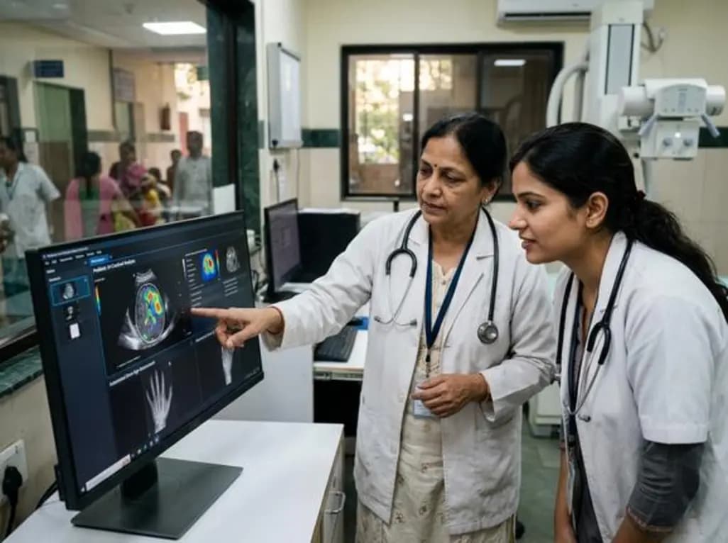
Artificial intelligence (AI) is reshaping healthcare globally, with radiology leading the charge in adoption. In the context of AI in pediatric imaging, the technology addresses critical challenges such as rising imaging demand, complex protocols, and workforce shortages. While adult radiology has seen rapid integration, pediatric translation has lagged due to the unique physiological and developmental characteristics of children. However, recent advancements are narrowing this gap, offering specialized tools for pediatric clinicians.
Deep learning models now support various clinical tasks, from emergency triage to chronic disease management. In chest imaging, AI assists in detecting pneumonia and ensuring the correct placement of tubes and lines. Automated bone age assessment remains one of the most successful examples of AI in pediatric imaging, streamlining a traditionally time-consuming task. Neuroimaging benefits from significantly reduced acquisition times and faster triage of critical findings, which is vital in neonatal and emergency settings.
Cardiovascular applications are also advancing, particularly in detecting congenital heart disease and assessing functional metrics. In pediatric oncology, researchers are exploring AI for precise tumor segmentation and characterization to better plan therapies. These tools do not just interpret images; they enhance the entire workflow by improving structured reporting and patient communication, ultimately allowing clinicians to focus more on direct patient care.
Despite the potential, several hurdles remain for widespread clinical use. The scarcity of high-quality pediatric datasets is a primary concern, as models trained on adults often fail when applied to children. Furthermore, integration into existing hospital systems remains uneven across many regions. Ethical and legal considerations unique to pediatric populations also require careful navigation. To ensure safe adoption, pediatricians and surgeons must collaborate with radiologists to set robust ethical guardrails and validate performance in local clinical settings.
AI improves safety by reducing radiation exposure through optimized imaging protocols and decreasing the need for repeat scans. It also acts as a second pair of eyes, helping to catch subtle findings that might be missed during high-volume shifts.
Children are not just small adults; their anatomy and pathologies evolve rapidly. Adult-trained models may misinterpret normal growth plates as fractures or fail to recognize pediatric-specific disease manifestations, making pediatric-specific training essential.
Pediatricians are essential for defining relevant use cases, curating high-quality datasets, and ensuring that AI tools align with child-centered care goals and ethical standards.
Disclaimer: This content is for informational and educational purposes only and does not constitute medical advice. It is not intended to be a substitute for professional medical judgment, diagnosis, or treatment. Always seek the advice of your physician or other qualified health provider with any questions you may have regarding a medical condition. Refer to the latest local and national guidelines for clinical practice.
References
Gupta A et al. Artificial Intelligence in Pediatric Imaging: A Primer for Pediatric Clinicians. Indian J Pediatr. 2026 Jun 19. doi: 10.1007/s12098-026-06289-4. PMID: 42319740.
Shelmerdine SC et al. Artificial Intelligence Implementation in Pediatric Radiology for Patient Safety: A Multisociety Statement. J Am Coll Radiol. 2026 Jan;23(1):89-101. doi: 10.1016/j.jacr.2025.08.019.
Agrawal A, Agrawal R. Artificial Intelligence in Pediatric Healthcare Part I: Foundations and Basic Concepts. Indian Journal of Child Health. 2026 Apr;13(4).
"
Read summarized clinical updates, watch expert medical content, and earn CME certifications right from your smartphone.
Open your smartphone camera to scan and install.





AI is transforming pediatric radiology by enhancing diagnostic accuracy in neuroimaging, cardiovascular care, and oncology. This primer explores the current landscape of pediatric-specific AI tools, highlighting benefits like automated bone age assessment and the critical need for child-centered validation.
4 months ago

Depemokimab offers twice-yearly dosing for severe eosinophilic asthma and chronic rhinosinusitis with nasal polyps, significantly cutting exacerbations and hospital admissions.
4 days back

A novel copper metal-organic framework hydrogel remodels the uterine microenvironment to stimulate robust endometrial regeneration, reduce intrauterine fibrosis, and restore reproductive fertility, offering a groundbreaking strategy for managing refractory endometrial damage and Asherman syndrome.
4 days back

New research reveals that non-ablative radiotherapy suppresses ventricular tachycardia by increasing conduction velocity via NaV1.5 upregulation and action potential prolongation. However, sustained sodium current elevation and depressed calcium handling highlight the need for careful clinical translation.
6 days back

A single-cell transcriptomic atlas reveals how peripheral nerve injuries cause dorsal root ganglion remodeling. By evaluating proximal nerve crush and resection, researchers identify distinct glial and immune programs that resolve neuropathic pain and promote regeneration.
4 days back

A breakthrough study demonstrates that fullerol-primed extracellular vesicles generated in a 3D SECCS bioreactor attenuate ferroptosis and restore myogenic regeneration in diabetes via the FOXO3 pathway, offering a cell-free therapeutic strategy for diabetic musculoskeletal degeneration.
4 days back
