.webp&w=384&q=75)

Loading, please wait...
Loading, please wait...
High-grade gliomas, encompassing World Health Organization grade III and IV astrocytomas and glioblastomas, represent exceptionally aggressive central nervous system malignancies. Despite aggressive surgical resection, adjuvant radiation, and systemic temozolomide chemotherapy, disease recurrence remains nearly universal. Managing these aggressive neoplasms requires rigorous radiological surveillance and rapid therapeutic adaptations. Clinicians frequently encounter diagnostic dilemmas when interpreting post-treatment brain scans, particularly when distinguishing true tumor progression from radiation necrosis or pseudoprogression. Consequently, neuro-oncologists, neurosurgeons, and neuroradiologists increasingly turn toward advanced artificial intelligence and machine learning methodologies to bolster diagnostic certainty. By analyzing complex neuroimaging datasets, computational algorithms identify subtle patterns that precede visible macrostructural changes. Incorporating these novel tools into routine surveillance offers significant potential for predicting glioma recurrence at earlier, actionable time points. Early identification allows clinical teams to modify treatment regimens, consider secondary surgical resections, or enroll eligible patients into clinical trials for novel therapeutics. Furthermore, reliable risk stratification helps clinical teams formulate tailored management plans while providing clearer prognostic expectations for patients and their caregivers. As computational capabilities expand, artificial intelligence stands poised to refine standard neuro-oncological monitoring.
A recently conducted systematic review evaluated the performance of artificial intelligence and machine learning algorithms in forecasting disease recurrence among high-grade glioma patients. Researchers performed a pooled meta-analysis across fourteen manuscripts encompassing 1,540 patients. Over ninety-nine percent of the included patient cohort had histologically confirmed World Health Organization grade IV glioblastoma, underscoring the clinical focus on highly aggressive malignant tumors. Nine of the included studies examined patients who underwent gross total resection followed by adjuvant radiotherapy, whereas five studies focused on patients receiving subtotal or near-total resections. Across all included studies, baseline pooled diagnostic metrics demonstrated notable performance. The initial random-effects model yielded a pooled sensitivity of eighty-one percent, a specificity of seventy-five percent, and an overall accuracy of seventy-nine percent. However, the initial primary pooled metrics exhibited high statistical heterogeneity across individual study outcomes. To address this variability, investigators performed comprehensive sensitivity analyses. Following sensitivity adjustments, diagnostic performance metrics improved significantly, yielding a sensitivity of eighty-one percent, a specificity of eighty-four percent, and an overall accuracy of eighty-nine percent, alongside minimal residual heterogeneity across the analyzed datasets.
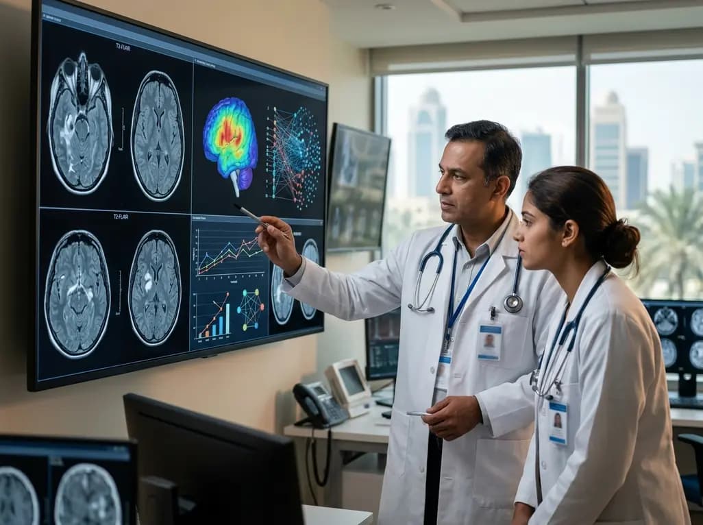
Among the various computational architectures analyzed in the systematic review, the Random Forest model emerged as the most widely applied machine learning algorithm. Random Forest classifiers excel in medical image processing because they construct multiple decision trees, effectively managing high-dimensional radiomic datasets without suffering from severe over-fitting. Regarding imaging modalities, Magnetic Resonance Imaging sequences served as the primary input for model development, with T2-weighted Fluid-Attenuated Inversion Recovery sequences incorporated most frequently. T2-FLAIR imaging provides exceptional sensitivity for hyperintense signal alterations, highlighting peri-tumoral edema, microscopic infiltrating tumor cells, and localized tissue disruptions before contrast enhancement becomes evident. By extracting quantitative radiomic features from T2-FLAIR sequences, machine learning models detect microstructural changes that remain invisible during standard visual inspection. Consequently, combining Random Forest models with multiparametric imaging enables non-invasive, continuous surveillance of high-risk brain regions. Clinicians can leverage these algorithmic insights to detect subtle spatial alterations, thereby facilitating timely therapeutic interventions before extensive clinical deterioration occurs in patients with high-grade gliomas. Furthermore, integrating these radiomic features with multi-sequence imaging enhances overall diagnostic stability across heterogeneous neuro-oncology patient cohorts.
While the synthesized data demonstrates convincing predictive capability, critical examination of included study designs highlights several methodological limitations. Notably, nearly ninety-three percent of the included studies relied on retrospective data collection, with only one study utilizing a prospective design. Retrospective designs inherently introduce potential selection bias, institutional variations in scan acquisition parameters, and risk of model over-fitting to historical training data. Furthermore, heterogeneous imaging protocols across institutions can limit the generalizability of machine learning models to broader patient populations. To overcome these obstacles, future research must prioritize multi-center prospective validation trials across diverse healthcare settings. Standardizing image preprocessing protocols, radiomic feature selection, and algorithmic reporting guidelines will ensure consistent reliability in real-world clinical environments. Additionally, combining radiomic features with molecular biomarkers, such as Isocitrate Dehydrogenase mutation status and MGMT promoter methylation, could significantly enhance model stability and precision. Addressing these critical methodological gaps will help transition artificial intelligence algorithms from academic exploratory models into standardized, point-of-care clinical tools. Moreover, open-source data sharing initiatives can facilitate larger training sets, accelerating the development of universally applicable diagnostic algorithms for complex neuro-oncology applications.
Implementing artificial intelligence models into neuro-oncology practice holds substantial clinical utility for optimizing patient care. Early prediction of tumor recurrence allows multidisciplinary care teams to refine surveillance schedules and initiate personalized treatment strategies proactively. For instance, patients identified as high risk for early relapse can receive tailored follow-up imaging, timely secondary resections, targeted stereotactic radiosurgery, or early enrollment in clinical trial protocols. Conversely, low-risk stratification can prevent unnecessary invasive procedures, decrease frequent clinic visits, and avoid premature discontinuation of effective maintenance therapies. Furthermore, machine learning models assist clinicians in overcoming difficult diagnostic challenges, such as differentiating true tumor progression from treatment-induced radionecrosis or pseudoprogression. Misinterpreting these radiological changes can lead to inappropriate treatment cessation or unnecessary toxicity. By providing high-precision quantitative evaluation, machine learning models reduce diagnostic uncertainty, streamline clinical decision-making, and minimize emotional stress for patients and families. Consequently, integrating artificial intelligence into clinical workflows empowers neuro-oncology teams to deliver timely, individualized, and effective care. As clinical adoption increases, these intelligent tools will serve as valuable diagnostic adjuncts, supporting multidisciplinary decision-making and enhancing overall clinical confidence.
The ongoing evolution of artificial intelligence in neuro-oncology promises to transform how clinicians monitor and manage aggressive brain malignancies. Future developments will focus on integrating longitudinal radiomic analysis with multi-omic data, combining imaging features with genomic, transcriptomic, and liquid biopsy markers to generate comprehensive risk profiles. Furthermore, novel deep learning architectures, including temporal convolutional networks and vision transformers, offer advanced capabilities for analyzing sequential brain scans over extended periods. These temporal learning models track subtle volumetric and microstructural alterations across consecutive scans, dramatically improving predictive accuracy compared to single-timepoint assessments. Establishing prospective multi-institutional registries and standardized validation frameworks will be pivotal for establishing regulatory approval and clinical trust. As artificial intelligence models mature through continuous refinement and prospective validation, they will transition from research tools into essential clinical decision support systems. Ultimately, these advanced predictive tools will enable proactive intervention strategies, optimize resource utilization, and improve long-term survival outcomes for patients facing challenging high-grade glioma diagnoses. Collaborative efforts between data scientists and clinicians will ensure that these innovations remain ethically sound, patient-centered, and seamlessly integrated into global healthcare systems.
In pooled sensitivity analyses, machine learning models demonstrated eighty-nine percent overall accuracy, eighty-one percent sensitivity, and eighty-four percent specificity. These quantitative metrics indicate strong potential for predicting high-grade glioma recurrence. However, performance varies depending on algorithm architecture, imaging sequence quality, and feature selection methods utilized during model training.
The Random Forest classifier was the most frequently utilized model in systematic evaluations, offering high diagnostic stability for complex imaging data. Among Magnetic Resonance Imaging sequences, T2-weighted Fluid-Attenuated Inversion Recovery served as the primary input feature, effectively capturing microstructural peritumoral changes and early tissue infiltration before clinical manifestation.
Most available studies rely on retrospective designs, which introduce potential selection bias and institutional protocol variability. Prospective multi-center validation remains limited. Additionally, standardizing radiomic feature extraction and integrating molecular biomarkers are necessary steps before these computational tools can be broadly implemented into routine clinical workflows.
Disclaimer: This content is for informational and educational purposes only and does not constitute medical advice, diagnosis, or treatment. Always seek the advice of a qualified healthcare provider with any questions regarding a medical condition. Refer to the latest local and national guidelines for clinical practice.
References

Read summarized clinical updates, watch expert medical content, and earn CME certifications right from your smartphone.
Open your smartphone camera to scan and install.





A systematic review demonstrates that machine learning models, particularly Random Forest using T2-FLAIR MRI, predict high-grade glioma recurrence with pooled sensitivity of 81% and accuracy up to 89%, highlighting AI's growing role in neuro-oncology surveillance.
2 months ago

Depemokimab offers twice-yearly dosing for severe eosinophilic asthma and chronic rhinosinusitis with nasal polyps, significantly cutting exacerbations and hospital admissions.
3 days back

A novel copper metal-organic framework hydrogel remodels the uterine microenvironment to stimulate robust endometrial regeneration, reduce intrauterine fibrosis, and restore reproductive fertility, offering a groundbreaking strategy for managing refractory endometrial damage and Asherman syndrome.
4 days back

New research reveals that non-ablative radiotherapy suppresses ventricular tachycardia by increasing conduction velocity via NaV1.5 upregulation and action potential prolongation. However, sustained sodium current elevation and depressed calcium handling highlight the need for careful clinical translation.
6 days back

A single-cell transcriptomic atlas reveals how peripheral nerve injuries cause dorsal root ganglion remodeling. By evaluating proximal nerve crush and resection, researchers identify distinct glial and immune programs that resolve neuropathic pain and promote regeneration.
4 days back

A breakthrough study demonstrates that fullerol-primed extracellular vesicles generated in a 3D SECCS bioreactor attenuate ferroptosis and restore myogenic regeneration in diabetes via the FOXO3 pathway, offering a cell-free therapeutic strategy for diabetic musculoskeletal degeneration.
4 days back
