.webp&w=384&q=75)

Loading, please wait...
Loading, please wait...
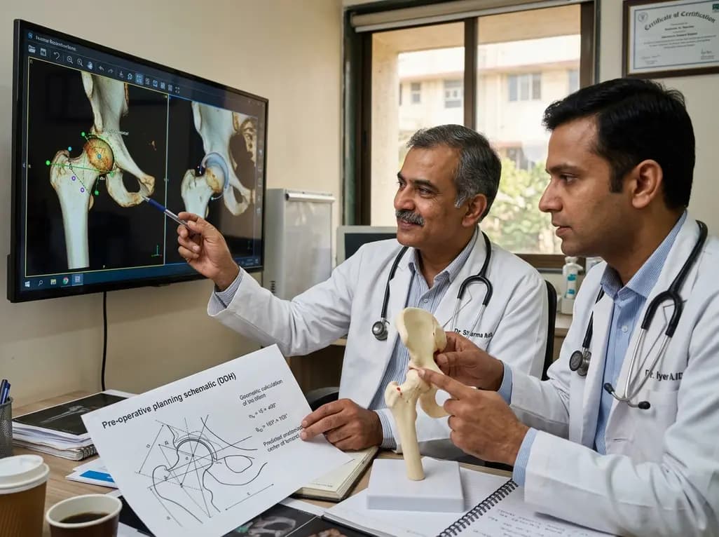
Accurate femoral head center estimation is a cornerstone of successful total hip arthroplasty (THA). However, anatomical deformities in patients with developmental dysplasia of the hip (DDH) often make this task difficult. Therefore, a recent study examined the reliability of using trochanteric landmarks for this purpose. Researchers analyzed 128 femurs to determine if three-dimensional coordinates could provide a more stable reference point than traditional pelvic markers.
The research team retrospectively analyzed female patients who underwent curved periacetabular osteotomy. They utilized the 3D coordinates of the greater and lesser trochanter tips to build multiple regression models. Consequently, they found that these femoral-based landmarks allow for highly precise calculations. Moreover, the study focused on patients with no spinal or knee symptoms to ensure the data specifically reflected hip anatomy.
The study results showed high correlation coefficients, ranging from 0.725 to 0.875. Furthermore, the mean absolute error remained between 2 mm and 3 mm across all three axes. Although the researchers observed slightly higher errors in the anteroposterior direction, the results stayed within clinically acceptable ranges. Specifically, this trochanter-based method outperformed older techniques that relied on pelvic landmarks, which often fail when the pelvis is malformed. Additionally, an error under 3 mm is ideal for restoring femoral offset and maintaining correct leg length.
Ultimately, this 3D approach provides a robust alternative for surgeons when the native femoral head is severely deformed. It offers a reliable adjunct for planning the placement of femoral components. Consequently, this advancement helps mitigate the risks of postoperative instability and limb length discrepancy. Orthopedic surgeons can now integrate these trochanteric landmarks into their digital preoperative templates to improve surgical outcomes in complex DDH cases.
Anatomic deformities in DDH often shift pelvic structures, making them inconsistent reference points for surgery. Therefore, femoral-based landmarks like the trochanters provide a more stable and accurate alternative for preoperative planning.
Most orthopedic studies suggest that an error margin within 3 mm is acceptable for restoring leg length and offset. Consequently, this study's 2-3 mm error range supports the practical applicability of the trochanteric method.
Disclaimer: This content is for informational and educational purposes only. It is not intended to be a substitute for professional medical advice, diagnosis, or treatment. Always seek the advice of your physician or other qualified healthcare provider with any questions you may have regarding a medical condition. Refer to the latest local and national guidelines for clinical practice.
References
Imai N et al. Three-Dimensional Estimation of the Femoral Head Center From Trochanteric Landmarks in Japanese Female Patients With Developmental Dysplasia of the Hip. Orthop Surg. 2026 Jun 22. doi: 10.1111/os.70359. PMID: 42332357.
Imai N et al. Coordinate value of the femoral head center estimated using those of the tip of the greater trochanter and lesser trochanter. Sci Rep. 2023 Feb 16;13(1):2784. doi: 10.1038/s41598-023-29938-1.

Read summarized clinical updates, watch expert medical content, and earn CME certifications right from your smartphone.
Open your smartphone camera to scan and install.





A new study validates a 3D method for estimating the femoral head center using trochanteric landmarks in DDH patients. With an error margin of only 2-3 mm, this technique offers a superior alternative to pelvic-based methods for total hip arthroplasty planning.
4 months ago

Depemokimab offers twice-yearly dosing for severe eosinophilic asthma and chronic rhinosinusitis with nasal polyps, significantly cutting exacerbations and hospital admissions.
4 days back

A novel copper metal-organic framework hydrogel remodels the uterine microenvironment to stimulate robust endometrial regeneration, reduce intrauterine fibrosis, and restore reproductive fertility, offering a groundbreaking strategy for managing refractory endometrial damage and Asherman syndrome.
4 days back

New research reveals that non-ablative radiotherapy suppresses ventricular tachycardia by increasing conduction velocity via NaV1.5 upregulation and action potential prolongation. However, sustained sodium current elevation and depressed calcium handling highlight the need for careful clinical translation.
6 days back

A single-cell transcriptomic atlas reveals how peripheral nerve injuries cause dorsal root ganglion remodeling. By evaluating proximal nerve crush and resection, researchers identify distinct glial and immune programs that resolve neuropathic pain and promote regeneration.
4 days back

A breakthrough study demonstrates that fullerol-primed extracellular vesicles generated in a 3D SECCS bioreactor attenuate ferroptosis and restore myogenic regeneration in diabetes via the FOXO3 pathway, offering a cell-free therapeutic strategy for diabetic musculoskeletal degeneration.
4 days back
