.webp&w=384&q=75)

Loading, please wait...
Loading, please wait...
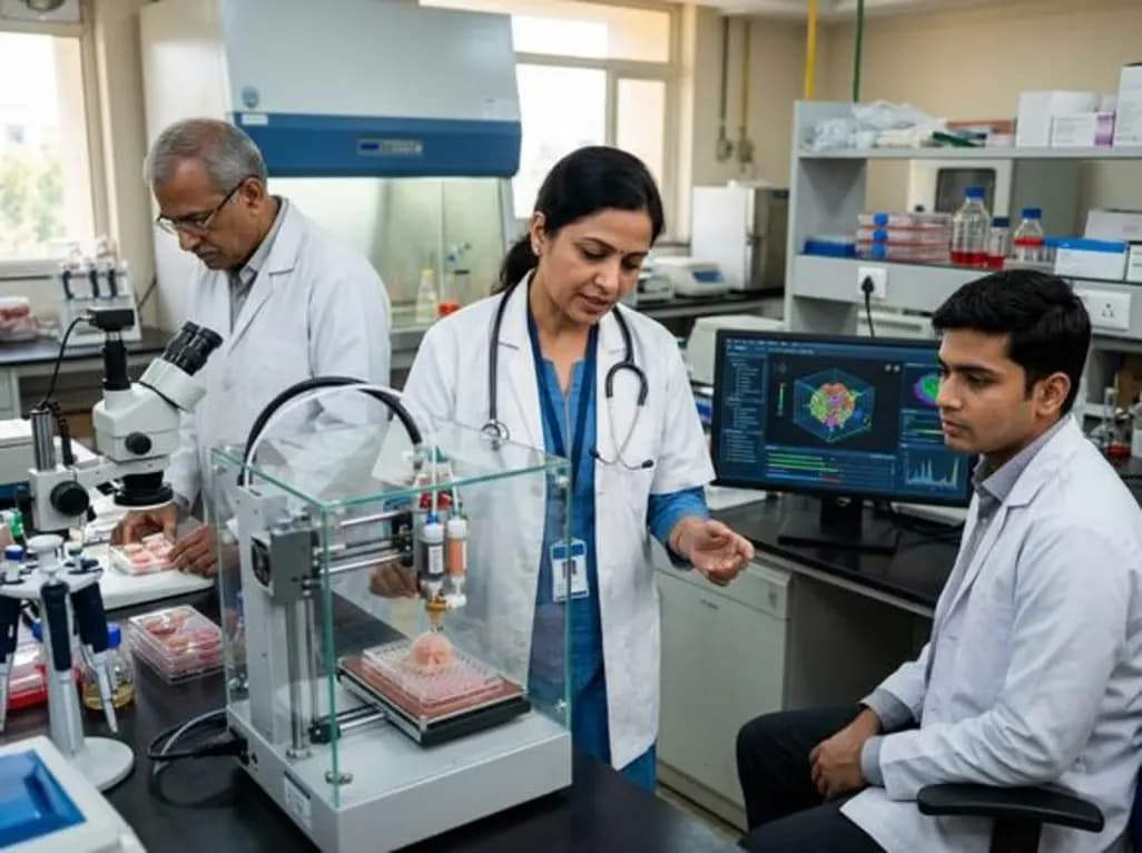
3D bioprinting in oncology has emerged as a promising preclinical technology to explore tumor cell biology, interaction with the microenvironment, and drug sensitivity. Unlike classical 2D systems, these models allow for the inclusion of several cell types and facilitate the migration of immune cells. Consequently, 3D bioprinted tumor models provide more relevant data regarding cell morphology and drug diffusion. This technological shift is crucial for researchers in India looking to enhance the accuracy of preclinical studies.
The application of this technology extends to a large variety of tumor cell lines at a relatively low cost. Researchers can perform these studies over flexible periods, ranging from a few days to several weeks. Furthermore, 3D bioprinting is increasingly applied to primary samples from cancer patients, facilitating personalized medicine. Although technical hurdles remain, the potential for high-throughput drug screening makes it a game-changer in the development of anti-cancer therapies. This innovation addresses the high failure rates often seen in traditional clinical trials.
While the promises of 3D bioprinting in oncology are vast, several challenges must be overcome for full exploitation. These include the optimization of bioinks and the integration of complex vascular networks within the models. Nevertheless, the ability to mimic the human tumor microenvironment accurately offers a significant advantage over animal models. As the technology matures, it will likely become a standard tool in both academic research and pharmaceutical development across the globe.
3D bioprinting creates a multi-layered structure that mimics the actual tumor microenvironment, including cell-cell interactions and drug diffusion, which 2D systems cannot replicate.
Yes, by using primary samples from a patient, 3D bioprinting can help screen specific drugs to determine the most effective treatment for that individual's unique tumor.
The primary benefits include higher physiological relevance, the ability to test multiple cell types simultaneously, and cost-effectiveness compared to some other 3D modeling techniques.
Disclaimer: This content is for informational and educational purposes only. It is not intended to provide medical advice or to be a substitute for professional medical advice, diagnosis, or treatment. Always seek the advice of your physician or other qualified health provider with any questions you may have regarding a medical condition. Refer to the latest local and national guidelines for clinical practice.
References

Read summarized clinical updates, watch expert medical content, and earn CME certifications right from your smartphone.
Open your smartphone camera to scan and install.





3D bioprinting is revolutionizing oncology by creating precise tumor models that mimic the human microenvironment. This technology offers a more accurate alternative to traditional 2D systems for drug sensitivity testing and personalized cancer care, potentially improving clinical outcomes for patients in India.
4 months ago

Depemokimab offers twice-yearly dosing for severe eosinophilic asthma and chronic rhinosinusitis with nasal polyps, significantly cutting exacerbations and hospital admissions.
3 days back

A novel copper metal-organic framework hydrogel remodels the uterine microenvironment to stimulate robust endometrial regeneration, reduce intrauterine fibrosis, and restore reproductive fertility, offering a groundbreaking strategy for managing refractory endometrial damage and Asherman syndrome.
3 days back

New research reveals that non-ablative radiotherapy suppresses ventricular tachycardia by increasing conduction velocity via NaV1.5 upregulation and action potential prolongation. However, sustained sodium current elevation and depressed calcium handling highlight the need for careful clinical translation.
5 days back

A single-cell transcriptomic atlas reveals how peripheral nerve injuries cause dorsal root ganglion remodeling. By evaluating proximal nerve crush and resection, researchers identify distinct glial and immune programs that resolve neuropathic pain and promote regeneration.
3 days back

A breakthrough study demonstrates that fullerol-primed extracellular vesicles generated in a 3D SECCS bioreactor attenuate ferroptosis and restore myogenic regeneration in diabetes via the FOXO3 pathway, offering a cell-free therapeutic strategy for diabetic musculoskeletal degeneration.
3 days back
