.webp&w=384&q=75)

Loading, please wait...
Loading, please wait...
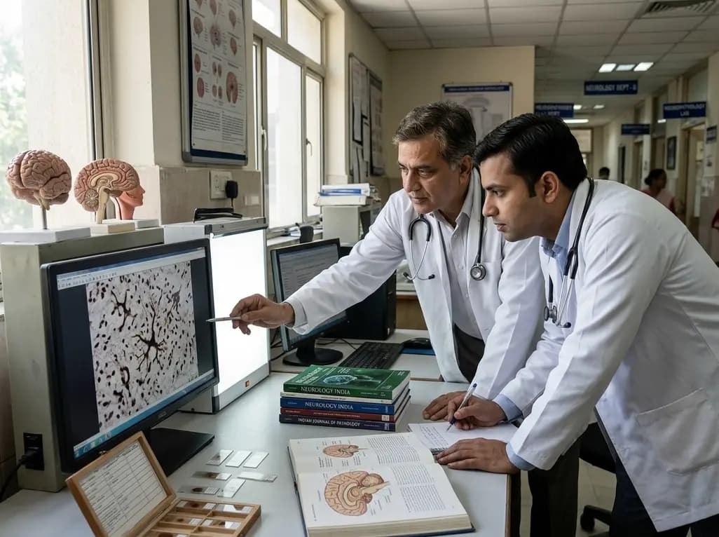
Traumatic brain injury and repetitive head impacts from contact sports trigger complex neurodegenerative cascades in the human brain. Clinical researchers have long recognized neuronal damage after mechanical trauma, yet glial alterations demand equal scientific scrutiny. Specifically, emerging post-mortem evidence confirms that repetitive biomechanical trauma accelerates the development of tau astrogliopathy across multiple cerebral regions. Astrocytes normally preserve the blood-brain barrier and regulate regional extracellular homeostasis. However, severe physical impacts disrupt cytoskeletal architecture within these delicate supporting cells. Consequently, hyperphosphorylated tau accumulates abnormally within astroglial processes, forming distinctive thorn-shaped astrocytes and granular inclusions. While historical neuropathological frameworks linked sports injuries predominantly to chronic traumatic encephalopathy, recent investigations demonstrate far more extensive glial pathology. This widespread astroglial alteration closely resembles patterns previously characterized as aging-related tau astrogliopathy. Furthermore, the persistent presence of these hyperphosphorylated glial lesions highlights ongoing neuroinflammation long after the initial trauma ceases. Clinicians must therefore recognize that biomechanical force affects far more than isolated neuronal networks. Understanding tau astrogliopathy as an enduring footprint of head impacts offers vital insights into late-life neurocognitive decline. Moreover, this perspective expands our diagnostic paradigm toward comprehensive cellular evaluation. Ultimately, evaluating both glial and neuronal changes illuminates the full spectrum of trauma-induced neurodegeneration.
Compelling neuropathological data provides robust confirmation that mechanical impacts accelerate glial tau pathology. In a comprehensive study, investigators examined autopsy-derived brain material from 556 deceased individuals across diverse exposure cohorts. The cohort included 77 individuals with a documented history of moderate-to-severe traumatic brain injury and prolonged post-injury survival. Additionally, researchers examined 45 athletes with confirmed exposure to contact sports. For comparative analysis, the authors included 397 uninjured controls with confirmed neurodegenerative disease and 37 controls without neurodegeneration. Tissue sections from multiple brain regions underwent rigorous immunohistochemical staining for hyperphosphorylated tau using the PHF-1 antibody. Strikingly, contact sports participants demonstrated thorn-shaped astrocytes in 75.6 percent of cases. In contrast, uninjured controls with neurodegenerative diseases displayed these lesions in only 32.5 percent of specimens. Furthermore, individuals surviving moderate-to-severe brain injury exhibited similarly elevated frequencies of astroglial tau deposition. These pathological astrocytes localized prominently at the subpial margin, perivascular spaces, and deep gray nuclei. Statistical analyses confirmed that physical trauma independently elevated the odds of developing tau astrogliopathy. Consequently, these findings verify that repetitive head impacts significantly induce glial pathology beyond normal chronological aging. Therefore, post-mortem tissue provides undeniable evidence linking mechanical trauma directly to premature glial senescence.
The pathophysiological mechanisms connecting physical head impacts to astroglial tau deposition involve complex biomechanical and molecular events. Angular and rotational acceleration forces deliver intense physical shear stress across the brain parenchyma. Because astrocytic end-feet firmly encase the cerebral microvasculature, shear forces directly strain these fragile glial structures. Consequently, mechanical disruption damages the neurovascular unit and compromises blood-brain barrier permeability. This microvascular leakage exposes cerebral tissue to systemic inflammatory mediators and neurotoxic plasma proteins. In response, astrocytes undergo profound phenotypic remodeling, transitioning into reactive phenotypes with altered metabolic capacity. Furthermore, mechanical strain disrupts normal astrocytic cytoskeletal dynamics, impairing molecular transport and physiological tau clearance. When cellular degradation pathways fail, soluble tau monomers misfold and aggregate into hyperphosphorylated oligomers. Over time, these toxic tau proteins assemble into insoluble paired helical filaments within the astrocyte cytoplasm. In addition, activated microglia release inflammatory cytokines, such as interleukin-1 beta and tumor necrosis factor-alpha. These signaling molecules stimulate intracellular protein kinases, driving continuous tau hyperphosphorylation. Therefore, mechanical trauma triggers a persistent feed-forward loop of neuroinflammation and glial proteostatic failure. Ultimately, this chronic biological cascade causes progressive astroglial damage that continues long after sporting careers end.
Distinguishing between overlapping tauopathies presents a critical diagnostic challenge in forensic neuropathology and behavioral neurology. Chronic traumatic encephalopathy neuropathologic change classically features hyperphosphorylated tau deposits within neurons and astrocytic processes around blood vessels. These diagnostic lesions cluster preferentially at the depths of cerebral sulci in a distinct, patchy distribution. In contrast, aging-related tau astrogliopathy presents primarily with thorn-shaped astrocytes and granular fuzzy astrocytes. These astrocytic lesions characteristically occupy subpial, subependymal, and perivascular niches without requiring sulcal depth localization. However, recent evidence confirms that repetitive head trauma frequently incites both pathological processes simultaneously. Thus, clinicians and pathologists must recognize that these conditions frequently coexist rather than represent mutually exclusive diseases. Furthermore, identifying extensive tau astrogliopathy in contact sports athletes challenges the historical notion that trauma damages only neuronal networks. Glial breakdown disrupts potassium buffering and glutamate reuptake, contributing directly to excitotoxicity and cognitive impairment. In addition, widespread glial tau accumulation likely explains why some symptomatic athletes exhibit minimal classical neuronal lesions at autopsy. Consequently, international neuropathology consensus guidelines emphasize detailed morphological evaluation across multiple cerebral regions. Ultimately, recognizing this shared pathological spectrum refines clinical-pathological correlation for patients suffering from persistent traumatic encephalopathy syndrome.
The realization that head trauma accelerates astroglial degeneration demands substantial revisions to athlete safety and clinical care pathways. Because glial tau accumulation develops silently over decades, early clinical identification presents a notable hurdle for practicing physicians. Consequently, aggressive prevention remains our most effective strategy to mitigate lifetime neurodegenerative risk. Governing bodies in contact sports must establish strict contact limits during training, where repetitive subconcussive blows accumulate. Furthermore, sports medicine clinicians must enforce rigorous concussion return-to-play protocols to prevent premature vulnerability to secondary impacts. When treating former athletes or trauma survivors, primary care physicians should routinely document lifetime head trauma exposure. Clinicians must actively monitor for early neuropsychiatric symptoms, including depression, impulsivity, emotional dysregulation, and executive dysfunction. In addition, comprehensive clinical management should address modifiable lifestyle factors to preserve cognitive reserve and cerebrovascular health. Regular aerobic exercise, optimized sleep hygiene, and tight control of hypertension provide substantial neuroprotective benefits. Similarly, specialized cognitive rehabilitation and targeted psychotherapy can help patients adapt to emerging cognitive challenges. Looking ahead, developing blood-based biomarkers of astrocytic injury and refined tau positron emission tomography will revolutionize diagnostic precision. Ultimately, proactive prevention and multidisciplinary clinical vigilance offer the best framework to safeguard long-term brain health.
Alzheimer's disease primarily features extracellular amyloid-beta plaques and intraneuronal neurofibrillary tau tangles. In contrast, tau astrogliopathy involves the abnormal accumulation of hyperphosphorylated tau specifically within astroglial support cells. While Alzheimer's disease damages neuronal transmission pathways directly, astrogliopathy disrupts blood-brain barrier maintenance, metabolic support, and regional neuroinflammatory regulation across brain tissues.
Yes, emerging post-mortem research confirms that repetitive subconcussive head impacts can induce astroglial tau pathology. Even without diagnosed concussions, cumulative shear forces from contact sports damage astrocytic end-feet surrounding cerebral microvessels. Consequently, this recurring mechanical stress triggers chronic neuroinflammation and drives pathological hyperphosphorylated tau aggregation within glial cells over time.
Currently, definitive diagnosis of tau astrogliopathy requires post-mortem histological and immunohistochemical evaluation of brain tissue. However, clinicians can assess clinical risk through detailed head trauma histories and advanced neuroimaging. Ongoing research into novel tau positron emission tomography tracers and blood-based glial biomarkers may soon permit accurate in-vivo clinical detection.
Disclaimer: This content is for informational and educational purposes only... Refer to the latest local and national guidelines for clinical practice.
References

Read summarized clinical updates, watch expert medical content, and earn CME certifications right from your smartphone.
Open your smartphone camera to scan and install.





Autopsy findings reveal that traumatic brain injury and repetitive head impacts from contact sports significantly accelerate tau astrogliopathy. Learn about the cellular mechanisms, histological findings, and clinical strategies essential for protecting athletes and managing post-traumatic neurodegeneration.
Yesterday

India's apex food regulator has launched legal proceedings against top digital retail platforms for distributing hazardous botanical substances and misbranded consumer goods. Discover the regulatory breaches, clinical toxicity risks, and key implications for healthcare providers across the country.
Last week

A retrospective cohort study evaluated 1,000 low-risk singleton births to assess maternal and neonatal outcomes during office-hours versus off-hours. Off-hours deliveries were significantly associated with higher odds of intrapartum cesarean sections and episiotomies, highlighting critical obstetric staffing considerations.
Yesterday

To curb crushing healthcare expenses, Karnataka's Food Safety and Drugs Administration has formally petitioned central authorities to institute a dedicated National List of Essential Medical Devices under the DPCO framework, establishing uniform price ceilings across twenty-four vital clinical categories.
Last week

Nearly 80% of Indians suffer from inadequate Vitamin D, causing silent bone weakening and sudden fractures. This expert guide reviews why mega-dose therapy is critical for high-risk patients, the key factors driving widespread deficiency, and the mandatory medical evaluations needed to protect skeletal health.
3 months ago

A nationwide mixed-methods study evaluated hospital glycemic management systems across 265 hospitals. While real-time alerts and automatic data sync are highly valued, significant disparities in digital maturity and low satisfaction with decision support highlight the need for standardized implementation.
last month
