.webp&w=384&q=75)

Loading, please wait...
Loading, please wait...
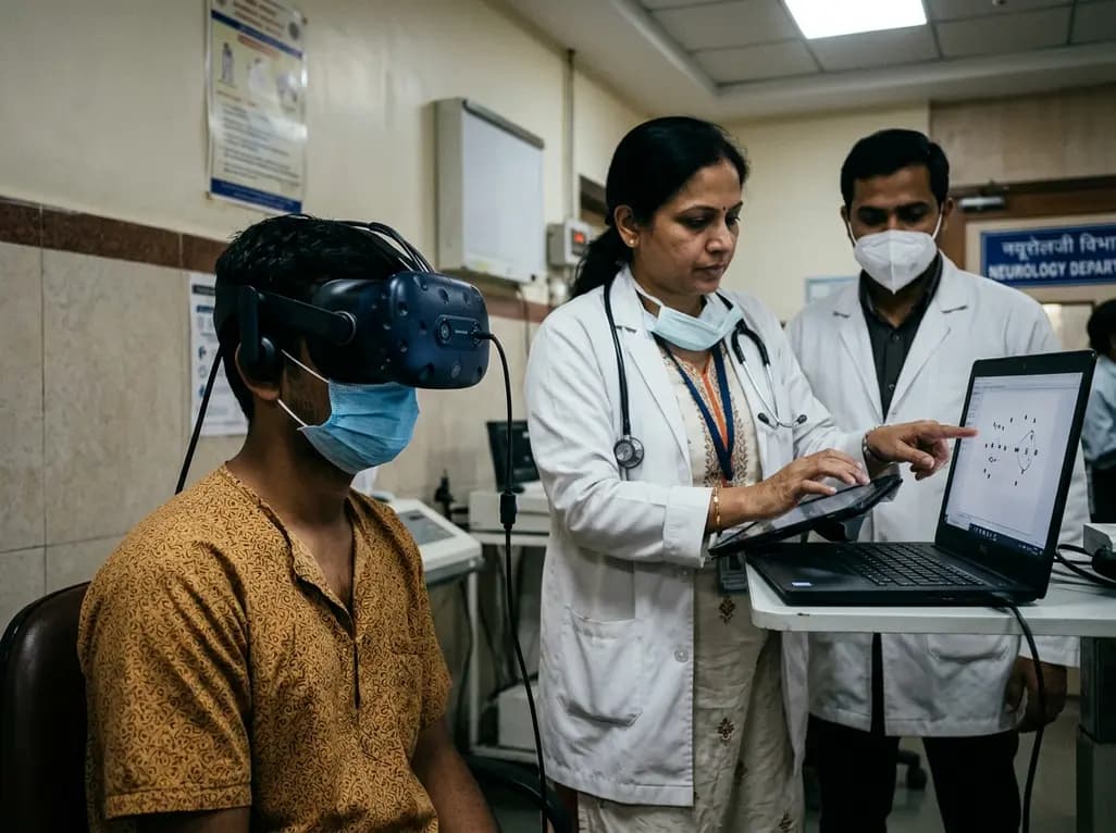
Internuclear ophthalmoplegia represents a distinct ocular motor impairment that occurs due to demyelination or structural disruption within the medial longitudinal fasciculus. This pathway coordinates conjugate horizontal eye movements by connecting the contralateral abducens nucleus to the ipsilateral oculomotor complex. Clinicians frequently encounter this abnormality in patients with multiple sclerosis and diverse brainstem pathologies. However, standard bedside examinations often miss mild or subclinical presentations. Bedside evaluation depends heavily on subjective visual observation to detect subtle slowing of the adducting eye. Consequently, mild adduction lag frequently escapes detection during routine neurological examinations. The introduction of VR-based oculography offers an objective, automated approach to address these diagnostic hurdles. By capturing high-frequency ocular trajectories, digital eye-tracking accurately measures subtle velocity discrepancies between the adducting and abducting eyes. Therefore, quantitative digital assessments provide clinicians with a highly reproducible biomarker that surpasses conventional physical examinations in identifying early brainstem involvement.
Recent technological advances have made immersive virtual reality headsets equipped with infrared eye-tracking cameras accessible for clinical neurophysiology. In a comprehensive prospective monocentric trial, researchers evaluated 471 participants, including 243 individuals with multiple sclerosis, 104 with other neurological disorders, and 124 healthy controls. All participants completed standardized horizontal prosaccade testing at visual angles of ten and twenty degrees. The software algorithms automatically generated quantitative reports comparing peak velocities between adducting and abducting saccades. Masked evaluators reviewed these velocity profiles to establish the presence of internuclear ophthalmoplegia without knowing the clinical diagnosis. Additionally, clinicians performed standard neurological examinations to compare bedside observations directly against digital measurements. The study utilized Cohen's kappa statistics to assess diagnostic concordance between both modalities. Furthermore, multivariable logistic regression evaluated links between automated ocular metrics and established clinical parameters, such as the Expanded Disability Status Scale.
The investigation revealed substantial discrepancies between digital recording capabilities and standard physical examinations. Overall, VR-based assessment identified 43 cases of internuclear ophthalmoplegia across the study cohort, comprising 40 patients with neurological disease and three healthy controls. Concordance between digital eye-tracking and clinical assessment was moderate, demonstrating a Cohen's kappa of 0.42 and an overall agreement rate of 92.5 percent across 414 evaluated participants. Importantly, bedside examination identified only three unique cases that the automated system missed. Conversely, the digital device discovered 28 additional cases that bedside examiners failed to detect. Within the multiple sclerosis subcohort, diagnostic agreement remained consistent with a kappa of 0.45. In this group, the digital platform identified 23 exclusive cases, whereas bedside examination detected only three isolated cases. These results confirm that conventional examination substantially underestimates brainstem oculomotor pathology, while quantitative tracking reliably captures discrete subclinical deficits.
Detecting subclinical oculomotor deficits provides critical insight into underlying neuropathology and structural disease burden. In the multiple sclerosis cohort, the presence of digital internuclear ophthalmoplegia correlated significantly with male sex and advanced age. Furthermore, patients with confirmed digital deficits exhibited higher overall scores on the Expanded Disability Status Scale and elevated brainstem functional system scores. These associations indicate that saccadic adduction deceleration mirrors wider demyelinating injury across central pathways. Brainstem lesions frequently generate cumulative neurological impairment, yet clinicians often struggle to quantify localized brainstem injury using standard physical tests alone. By identifying occult medial longitudinal fasciculus disruption, digital oculography uncovers ongoing structural damage that might otherwise remain hidden. Consequently, objective oculomotor metrics serve as robust indicators of disease progression, helping clinicians stratify patient risk and monitor chronic neurodegenerative trajectories more effectively.
Integrating portable head-mounted eye-tracking devices into outpatient neurology clinics offers practical advantages for patient care. Traditional infrared oculography requires cumbersome tabletop hardware, extensive calibration protocols, and dedicated laboratory environments. In contrast, modern virtual reality headsets provide an immersive, self-contained testing environment that minimizes external visual distractions. The automated testing routine takes only a few minutes to complete, making it feasible for busy clinical workflows. Moreover, standardized stimulus presentation ensures high test-retest reliability across diverse clinical settings. Clinicians can utilize these objective metrics to complement routine physical examinations, especially when patients report ambiguous visual symptoms like intermittent diplopia or reading fatigue. In addition, digital eye-tracking provides clear longitudinal records that help neurologists evaluate treatment responses to disease-modifying therapies or symptomatic interventions such as potassium channel blockers.
Digital ocular motor testing holds immense promise for expanding diagnostic precision across various neuroinflammatory and neurodegenerative conditions. Beyond multiple sclerosis, conditions such as stroke, neuromyelitis optica spectrum disorder, and traumatic brain injury can disrupt brainstem circuits. Digital assessments allow clinicians to detect subtle focal injuries before gross motor deficits manifest clinically. Additionally, software-driven saccadic analysis creates standardized outcome parameters for clinical drug trials targeting remyelination and neuroprotection. However, widespread clinical adoption requires rigorous external multicenter validation and refined normative cutoffs to prevent false-positive classifications in healthy populations. Integrating automated digital analytics into routine neurological evaluations will ultimately refine diagnostic pathways, enabling earlier therapeutic interventions and objective monitoring of complex central nervous system disorders.
Internuclear ophthalmoplegia is an ocular movement deficit caused by lesions in the medial longitudinal fasciculus. It impairs conjugate horizontal gaze, causing adduction slowing. Clinicians frequently miss mild cases during bedside examinations because subtle saccadic decelerations are difficult to detect without high-speed, quantitative tracking tools.
The system uses high-speed infrared cameras embedded within a virtual reality headset to measure horizontal saccadic eye movements. Specialized software calculates and compares the peak velocities of adducting and abducting eyes during gaze shifts, objectively identifying adduction lag indicative of medial longitudinal fasciculus impairment.
Digital oculomotor biomarkers provide objective, reproducible measures of brainstem pathway integrity. Because they correlate with expanded disability status and brainstem functional scores, these digital metrics enable clinicians to detect subclinical disease activity, track neurodegeneration over time, and evaluate therapeutic efficacy accurately.
Disclaimer: This content is for informational and educational purposes only and should not be considered as medical advice. Healthcare professionals should exercise their independent clinical judgment. Refer to the latest local and national guidelines for clinical practice.
References
Reuter E et al. Virtual reality-based oculography detects internuclear ophthalmoplegia in multiple sclerosis and other neurological disorders. J Neurol. 2026 Jun 06. doi: 10.1007/s00415-026-13906-x. PMID: 42250152.
Nij Bijvank JA et al. Diagnosing and quantifying a common deficit in multiple sclerosis: Internuclear ophthalmoplegia. Neurology. 2019;92(20):e2299-e2308. doi: 10.1212/WNL.0000000000007499.
Hof S et al. The prevalence of internuclear ophthalmoparesis in a population-based cohort of individuals with multiple sclerosis. Mult Scler Relat Disord. 2022;63:103824. doi: 10.1016/j.msard.2022.103824.

Read summarized clinical updates, watch expert medical content, and earn CME certifications right from your smartphone.
Open your smartphone camera to scan and install.





A prospective study demonstrates that immersive VR-based oculography significantly improves the detection of internuclear ophthalmoplegia in multiple sclerosis and other neurological conditions compared to standard bedside examination, revealing strong links to overall disease severity and brainstem involvement.
last month

A discrete choice experiment reveals that combining cigarette plain packaging with a 50% price increase reduces smoking uptake by 7.7 percentage points. Integrating fiscal taxation with standardized branding is crucial to overcome brand loyalty and curb tobacco consumption effectively.
Today

A nationwide South Korean study shows underreporting of smoking among women dropped from 57.4% to 36.4% between 2008 and 2021. However, over one-third of female smokers still conceal their habit, highlighting the critical role of cotinine verification in clinical risk assessment.
Today

PepsiCo and Monster Beverage have challenged the FSSAI ban on the energy drink label in Indian courts. While companies highlight supply disruptions, regulatory actions spotlight severe cardiovascular and metabolic risks associated with excessive caffeine, sugar, and taurine consumption, especially in adolescents.
Today
Persistent and recurrent musculoskeletal pain in youth athletes is often dismissed as simple growing pains. A developmentally informed biopsychosocial framework is essential to guide return-to-sport decisions, addressing biological vulnerabilities, psychological appraisals, and sociocultural expectations.
Today

A Bayesian multilevel meta-analysis reveals that aerobic training combined with moderate carbohydrate restriction modestly lowers HbA1c in type 2 diabetes. However, sparse data and very low certainty leave incremental benefits over exercise or diet alone unproven, highlighting the need for individualized care.
Yesterday
