

Loading, please wait...
Loading, please wait...

Dry eye disease presents an escalating clinical challenge as digital screen engagement rises across all age groups. Accurate diagnostic stratification remains crucial for preserving corneal integrity and personalizing therapy. Recent clinical research demonstrates that videographic assessment of spontaneous blinking patterns can uncover subtle kinematic abnormalities that routine slit-lamp examinations frequently miss. Consequently, understanding dynamic eyelid behavior provides vital diagnostic clues. This videographic method bridges patient symptoms with ocular surface integrity, creating an accessible bedside diagnostic tool.
Clinicians recognize that blinking maintains tear film stability and safeguards the ocular surface against desiccating environmental stress. However, routine slit-lamp examinations often fail to capture dynamic eyelid kinematics during unforced natural visual tasks. To overcome this limitation, investigators utilized high-speed smartphone videography to record spontaneous eyelid activity across various patient groups. A masked oculoplastic surgeon evaluated these recordings to identify subtle anatomical parameters, including brow elevation, blepharospasm, lid margin rotation, and delayed lid separation.
Moreover, the mobile recording setup maintained simplicity without intimidating the patient, thereby preserving natural blinking dynamics. Standard clinical measurements, such as the Ocular Surface Disease Index, tear break-up time, Schirmer testing, and corneal fluorescein staining, complemented the video recordings. Consequently, researchers could systematically correlate kinematic eyelid behaviors with objective disease severity metrics. This methodology proves that accessible smartphone hardware can facilitate sophisticated oculoplastic assessments in busy outpatient practices. Therefore, ophthalmologists and optometrists can easily integrate digital video screening into their diagnostic protocols without purchasing costly proprietary equipment. In addition, standardizing smartphone positioning ensures reliable and consistent kinematic evaluations across serial clinical encounters.
The investigation evaluated 54 adult participants, revealing clear links between spontaneous eyelid dynamics and dry eye severity. Specifically, tear break-up time, Schirmer test results, corneal fluorescein staining, and eyelid margin rotation differed significantly across disease cohorts. Patients presenting with moderate-to-severe symptoms demonstrated markedly reduced tear production alongside pronounced ocular surface staining.
Furthermore, eyelid margin rotation emerged as a pivotal clinical finding. Patients suffering from severe ocular surface disease showed abnormal margin twisting during spontaneous blinks far more frequently than individuals with mild symptoms. This mechanical torsion impairs uniform lipid distribution across the ocular surface, accelerating evaporative tear film destabilization. In addition, the persistent shear stress from rotated eyelid margins inflicts direct microtrauma upon the vulnerable corneal epithelium. Thus, eyelid margin rotation serves as both an indicator of severe disease and an active mechanical driver of epithelial breakdown. Recognizing this kinematic anomaly allows clinicians to pinpoint patients at high risk of rapid disease worsening who require targeted anatomical management.
To understand the clinical importance of these observations, physicians must review normal eyelid biomechanics. During an effective blink, the orbicularis oculi and the muscle of Riolan gently compress the tarsal meibomian glands. This dynamic contraction expels protective lipids directly onto the tear film surface. Concurrently, the upper eyelid glides downward across the globe, redistributing mucin and aqueous components into a smooth optical barrier.
However, when eyelid margin rotation or incomplete closure disrupts this motion, tear film homeostasis collapses. The misdirected lid border fails to maintain flush apposition with the bulbar conjunctiva and cornea. Consequently, localized areas of tear thinning emerge swiftly, precipitating rapid tear break-up and painful focal desiccation. Moreover, inadequate margin apposition prevents effective lipid replenishment, fueling an escalating cycle of evaporative tear loss and glandular stagnation. Over time, persistent frictional irritation triggers subclinical inflammatory cascades and tissue remodeling along the eyelid margin. Therefore, ocular surface disease often stems directly from dynamic kinematic failure rather than pure aqueous deficiency.
Incorporating high-speed smartphone videography offers an affordable, practical advantage for daily eye clinics and tertiary centers alike. Traditional interferometry and specialized optical topography systems demand substantial capital investment and extensive clinical space. In contrast, standard modern smartphones possess high-frame-rate video capabilities that capture rapid eyelid closures with exceptional clarity.
Therefore, practitioners can effortlessly record patients during brief conversations to screen for subclinical lagophthalmos, margin rotation, or incomplete blinks. Furthermore, showing these slow-motion recordings to patients provides impactful visual education that reinforces therapeutic compliance. When patients visually recognize how their abnormal blinking habits compromise tear stability, they adhere far better to prescribed warm compresses, blinking routines, and topical regimens. Additionally, this mobile recording modality enables reliable longitudinal monitoring of therapeutic outcomes. Clinicians can accurately verify whether eyelid treatments restore physiological apposition over time. Consequently, smartphone video screening converts subjective clinical observation into an objective, reproducible assessment without inflating institutional healthcare costs. Similarly, clinicians can easily archive these recordings in electronic records to document treatment efficacy across follow-up consultations.
Recognizing kinematic blinking abnormalities fundamentally reshapes clinical management strategies for refractory ocular surface disease. When eye care specialists detect prominent eyelid margin rotation, relying solely on artificial tear substitutes yields disappointing outcomes. Instead, practitioners should implement targeted interventions that address structural eyelid malposition and dynamic muscular imbalances.
For instance, conservative measures like conscious blinking exercises, thermal meibomian therapies, and judicious botulinum toxin application for focal spasms can restore ocular surface equilibrium. Furthermore, when marked eyelid laxity or cicatricial changes predominate, surgical procedures such as lateral tarsal strip stabilization or margin tightening offer definitive structural correction. In addition, future clinical trials should validate these videographic parameters across diverse populations while exploring automated artificial intelligence algorithms for instant motion analysis. Such digital tools could automatically quantify blink completeness and rotational severity directly from brief smartphone recordings. Ultimately, merging accessible mobile imaging with personalized therapeutic interventions represents an exciting advancement in modern ocular surface medicine. Consequently, high-speed videography empowers clinicians to treat both functional kinematics and structural disease components.
Eyelid margin rotation disrupts the precise mechanical contact required between the eyelids and the cornea during a blink. Consequently, this rotational distortion impedes the uniform expression and distribution of meibomian lipids across the ocular surface. As a result, the preocular tear film evaporates prematurely, creating localized dry spots and mechanical friction. Over time, persistent microtrauma from misaligned lid margins damages the corneal epithelium, exacerbating subjective discomfort and accelerating disease progression.
Yes, contemporary smartphones equipped with high-speed video recording provide sufficient frame rates to capture rapid eyelid kinematics accurately. Clinicians can record high-definition footage of spontaneous blinking under ambient lighting without cumbersome diagnostic equipment. Furthermore, slow-motion playback allows practitioners to detect subtle abnormalities, including micro-lagophthalmos, lid margin rotation, and delayed lid separation. Therefore, high-speed mobile videography represents a cost-effective, accessible, and reliable diagnostic adjunct in daily ophthalmology and optometry practice.
Clinical management depends on the underlying etiology of the eyelid dysfunction. Initial non-surgical interventions include deliberate blink training exercises, intense pulsed light, and thermal pulsation to evacuate obstructed meibomian glands. Additionally, clinicians may prescribe preservative-free artificial tears and anti-inflammatory topical therapies to soothe mucosal irritation. However, when severe eyelid margin rotation or significant lid laxity persists, surgical consultation for horizontal lid tightening or anatomical repositioning may be necessary to restore adequate ocular surface protection.
Disclaimer: This content is for informational and educational purposes only and is not intended to replace professional medical advice, diagnosis, or treatment. Healthcare professionals should make decisions based on their independent clinical evaluation and current standard practices. Refer to the latest local and national guidelines for clinical practice.
References

Read summarized clinical updates, watch expert medical content, and earn CME certifications right from your smartphone.
Open your smartphone camera to scan and install.





A clinical study highlights the utility of high-speed smartphone videography in evaluating spontaneous blinking patterns in dry eye disease. Eyelid margin rotation emerged as a significant marker of disease severity, demonstrating how accessible mobile imaging can enhance diagnostic accuracy in ophthalmic practice.
Today

A scoping review of 130 studies reveals that chronic preoperative benzodiazepine use is linked to increased postoperative opioid needs and delirium. Despite widespread use, inconsistent definitions highlight the need for standardized perioperative medication optimization protocols.
Today
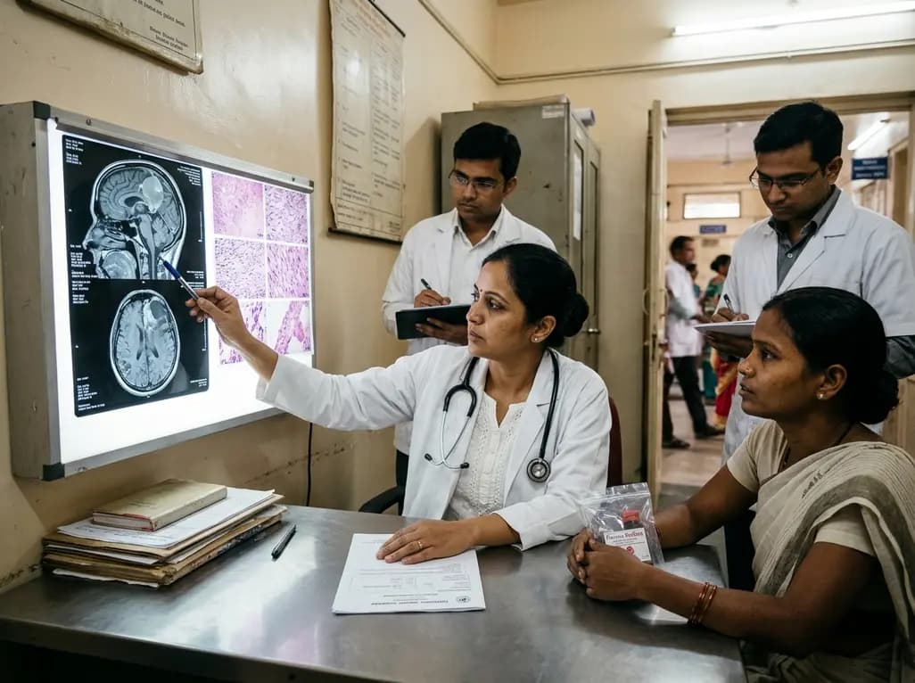
An intracranial EWSR1::CREM fusion sarcoma in a 31-year-old female manifested as a paraneoplastic inflammatory syndrome with anemia and secondary infertility. The dural mass mimicked meningioma, but gross-total resection resolved her systemic inflammation and restored natural fertility within one month.
Today
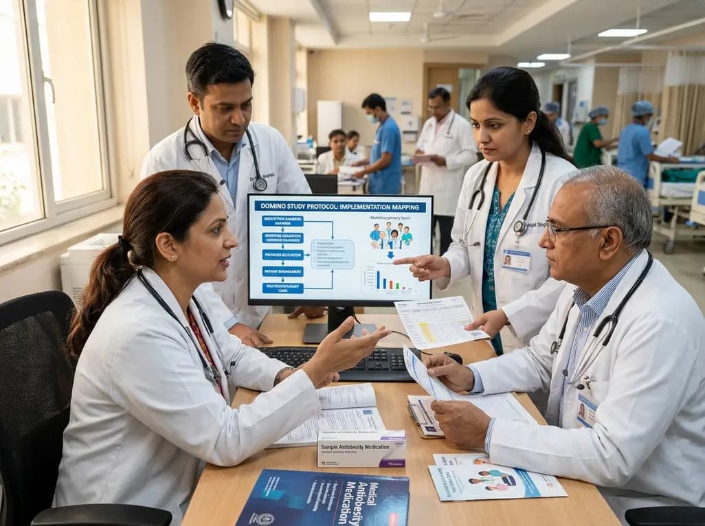
The DOMINO study protocol presents a multidisciplinary implementation mapping approach to overcome provider, patient, and payer barriers to antiobesity medications, offering vital insights for Indian clinicians managing the growing diabesity burden.
Today
An age-matched cross-sectional study reveals that lower muscle quality and impaired lower-extremity function strongly correlate with an increased fear of falling in older women, highlighting the necessity of targeted resistance training and balance interventions in geriatric care.
Today
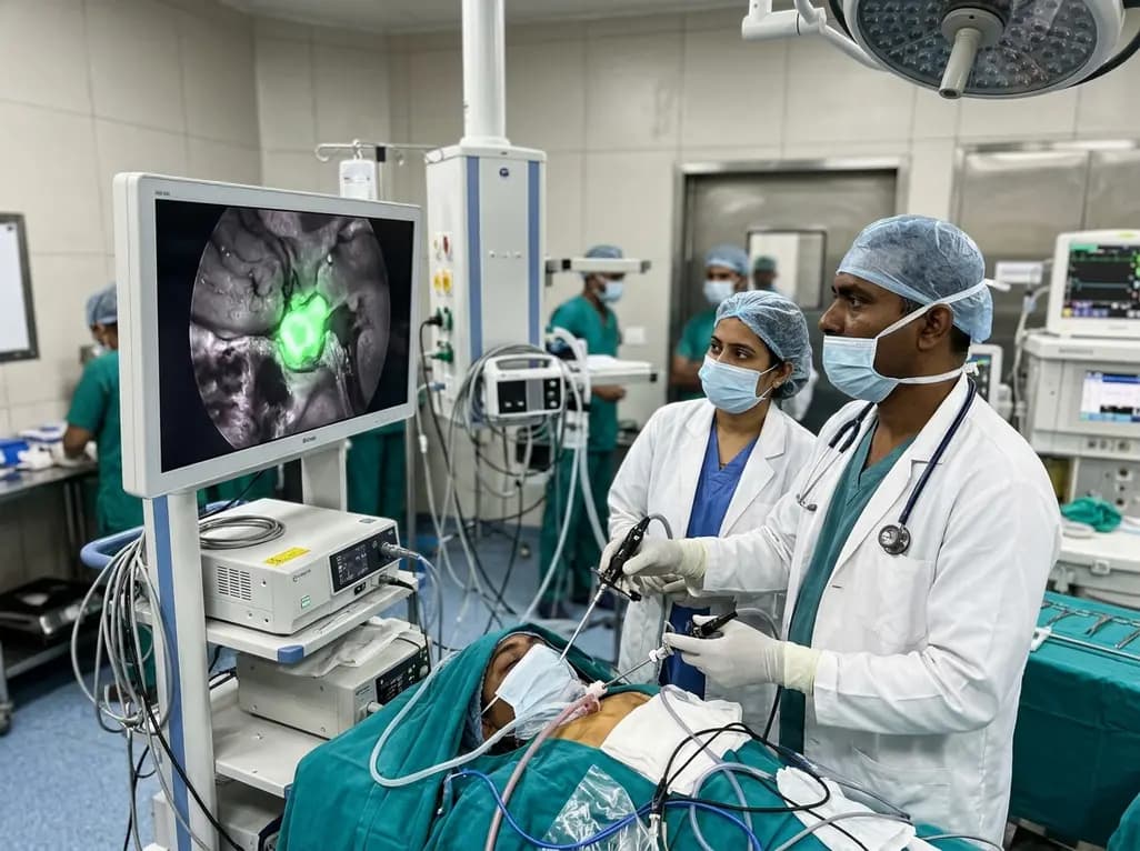
Near-infrared autofluorescence provides real-time label-free optical guidance during endoscopic transsphenoidal pituitary surgery. By highlighting stark signal contrasts between normal pituitary gland and adenomas, this technique aids gross-total tumor resection while safeguarding critical endocrine function.
Today
