.webp&w=384&q=75)

Loading, please wait...
Loading, please wait...

Induction of labor represents one of the most frequent obstetric interventions worldwide. In modern clinical practice, maternal obesity poses distinct physiologic challenges that alter delivery trajectories. Specifically, healthcare providers frequently rely on vaginal misoprostol induction to achieve cervical ripening and initiate uterine contractions. Misoprostol, a synthetic prostaglandin E1 analogue, offers significant advantages due to its cost-effectiveness, thermal stability, and robust uterotonic potency. However, pregnant individuals with severe obesity often demonstrate altered pharmacokinetics and blunted myometrial responsiveness. Consequently, understanding how body mass index influences induction characteristics remains critical for practicing obstetricians. Recent clinical evidence provides valuable clarity regarding dose requirements, labor duration, and maternal-neonatal outcomes in this vulnerable demographic.
Maternal obesity rates continue to rise globally, significantly impacting labor room management across tertiary healthcare centers. Obstetricians increasingly manage pregnancies complicated by elevated maternal weight, where spontaneous labor often fails to occur. Consequently, clinical teams schedule labor induction to prevent stillbirth, macrosomia, and post-term complications. Vaginal misoprostol induction remains a preferred method because clinicians can administer it easily without specialized storage equipment. Nevertheless, severe obesity alters physiological baselines and prolongs labor latency. Research indicates that excess adiposity modifies uterine vascularity and decreases myometrial oxytocin receptor sensitivity. As a result, clinicians frequently observe protracted latent phases during standard induction protocols. Furthermore, excessive maternal soft tissue complicates routine abdominal palpation and digital examinations, which adds technical difficulty to cervical assessment. Therefore, obstetric teams must understand these unique anatomical and physiological variables. By tailoring monitoring strategies to patient body habitus, clinicians can anticipate potential delays and optimize delivery timing effectively.
Misoprostol undergoes rapid de-esterification after administration, converting into its active metabolite, misoprostol acid. In normal-weight parturients, vaginal absorption delivers steady plasma concentrations with sustained local myometrial stimulation. In contrast, individuals with severe obesity exhibit substantial alterations in drug distribution volume and tissue perfusion. Although the vaginal mucosa absorbs the medication locally, increased peripheral vascular resistance and expanded fat mass modify drug disposition. Moreover, excess maternal adipose tissue releases pro-inflammatory cytokines and leptin, which directly interfere with normal uterine contractility patterns. Consequently, myometrial gap junction formation occurs at a slower rate in patients with class III obesity. This fundamental difference explains why ripening agents often take longer to produce coordinated, regular contractions in heavier individuals. Obstetricians must recognize that biological resistance, rather than true induction failure, frequently underlies slower cervical effacement. Understanding these mechanisms allows providers to exercise appropriate patience before classifying an induction as unsuccessful.
Comparative clinical investigations demonstrate significant disparities in labor progression when comparing patients with severe obesity to normal-weight peers. Parturients with a body mass index exceeding 40 kg/m² typically require higher cumulative doses of prostaglandins. In addition, the duration from the initial misoprostol dose to active labor onset is substantially longer in this cohort. Specifically, clinical data show that these patients spend considerably more hours in the latent phase of labor. However, once parturients achieve active cervical dilation beyond six centimeters, the subsequent rate of dilation tends to parallel that of non-obese individuals. Furthermore, secondary oxytocin augmentation is required far more often in obese cohorts to sustain adequate contraction frequency. Therefore, providers should not prematurely intervene with operative delivery during an extended latent phase if maternal and fetal parameters remain reassuring. Recognizing that prolonged latency is an expected physiological characteristic helps prevent unnecessary primary cesarean births.
Evaluating safety outcomes remains crucial when managing high-risk deliveries in individuals with severe adiposity. Fortunately, current evidence indicates that vaginal misoprostol induction does not elevate rates of uterine tachysystole or uterine rupture in unscarred uteri. Nevertheless, prolonged induction processes inherently raise other obstetric risks. In particular, extended duration of ruptured membranes and prolonged labor increase the cumulative risk of intraamniotic infection and postpartum endometritis. Similarly, severe maternal obesity independently elevates the risk of uterine atony, leading to primary postpartum hemorrhage. Neonatal outcomes, including umbilical cord blood gases and neonatal intensive care unit admission rates, generally remain comparable across body mass index categories. However, secondary maternal complications, such as soft-tissue trauma and venous thromboembolism, demand heightened vigilance. Thus, clinical protocols must prioritize proactive third-stage active management and rigorous postpartum surveillance to mitigate hemorrhage risk in these patients.
In India, the dual burden of maternal obesity and gestational diabetes creates complex intrapartum challenges for obstetric clinicians. Most labor wards utilize misoprostol because it provides an economical and thermally stable cervical ripening option. To optimize clinical outcomes, obstetric teams must adopt structured labor induction protocols that accommodate longer latent phases. First, clinicians should avoid escalating misoprostol doses beyond standard recommendations, as aggressive dosing increases hyperstimulation without accelerating delivery. Second, continuous or frequent intermittent fetal monitoring is vital, although thick abdominal panniculi may necessitate internal fetal scalp electrodes. Third, healthcare facilities must ensure immediate availability of multidisciplinary support, including experienced obstetricians, anesthetists, and neonatologists. Additionally, maternity units should prepare early for postpartum hemorrhage by securing adequate intravenous access and active uterotonic agents. Implementing these targeted intrapartum practices empowers clinicians to achieve successful vaginal deliveries while safeguarding maternal and neonatal well-being.
Maternal obesity alters drug pharmacokinetics by increasing the volume of distribution and modifying local tissue perfusion. In addition, elevated adipose tissue concentrations release inflammatory mediators that impair myometrial contractility and gap junction formation. Consequently, individuals with severe obesity often exhibit a delayed clinical response to vaginal misoprostol, leading to an extended latent phase of labor. Obstetricians must expect this slower initial progression rather than presuming medication failure.
Current obstetric evidence does not recommend increasing the starting dose of vaginal misoprostol for patients with severe obesity. Standard doses of 25 micrograms every four to six hours remain appropriate and safe. Increasing individual doses raises the risk of uterine tachysystole and fetal heart rate decelerations without substantially shortening overall labor duration. Instead, clinicians should allow adequate time for cervical ripening while maintaining close cardiotocographic surveillance.
Continuous fetal heart rate monitoring and uterine contraction assessment are essential during labor induction in severely obese parturients. Because excessive maternal abdominal adipose tissue frequently diminishes external ultrasound and tocodynamometer accuracy, clinicians should maintain a low threshold for utilizing internal monitoring devices. Furthermore, providers must secure wide-bore intravenous access early and implement aggressive active management of the third stage to prevent postpartum hemorrhage.
Disclaimer: This content is for informational and educational purposes only and does not constitute medical advice, diagnosis, or treatment. Healthcare professionals should rely on their clinical judgment and verify details independently. Refer to the latest local and national guidelines for clinical practice.
References

Read summarized clinical updates, watch expert medical content, and earn CME certifications right from your smartphone.
Open your smartphone camera to scan and install.





A comparative analysis of labor induction with vaginal misoprostol in individuals with and without severe obesity, highlighting pharmacokinetic differences, labor duration, dosing, and intrapartum safety.
Today

Real-world evidence demonstrates that anti-IL-5 monoclonal antibodies mepolizumab and benralizumab significantly reduce oral glucocorticoid dependence and peripheral eosinophil counts in eosinophilic granulomatosis with polyangiitis (EGPA), enabling complete steroid discontinuation in nearly half of patients.
Today

ATP-citrate lyase (ACLY) bridges energy metabolism and epigenetic regulation by generating acetyl-CoA. This review explores ACLY biology, its disease relevance across cardiometabolic disorders and oncology, and current clinical developments surrounding ACLY inhibitors like bempedoic acid.
Today

A comprehensive umbrella review of 22 meta-analyses and 286,584 adults reveals that calcium supplementation modestly increases bone mineral density and reduces vertebral fractures, but does not decrease hip or wrist fracture risks.
Today
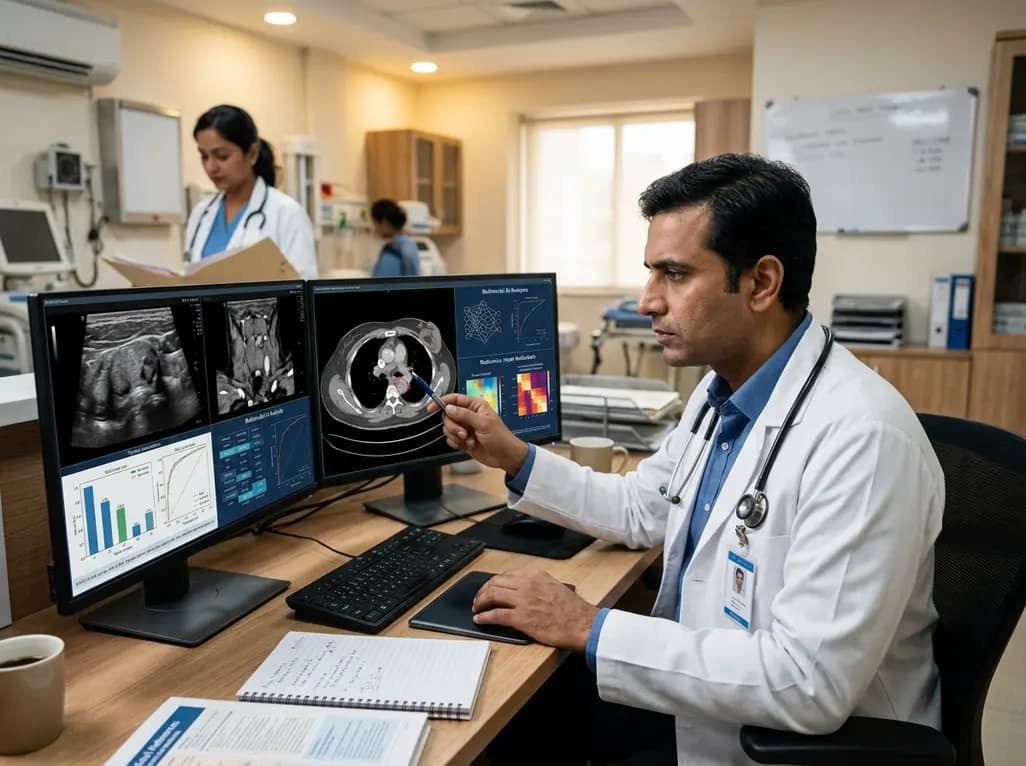
A multimodal AI model integrating ultrasound deep learning, CT perivascular adipose tissue radiomics, and clinical risk factors accurately predicts central lymph node metastasis in papillary thyroid carcinoma, providing clinicians with a non-invasive tool to optimize pre-operative surgical decisions.
Today
