.webp&w=384&q=75)

Loading, please wait...
Loading, please wait...
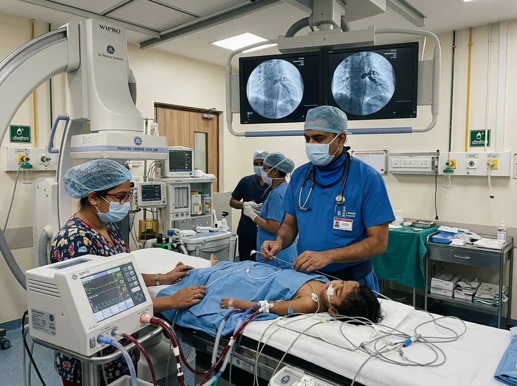
Pediatric pulmonary vein stenosis presents an exceptionally severe challenge in modern pediatric cardiology. This rare and progressive vascular disorder involves fibrointimal hyperplasia that obstructs blood flow from the lungs into the left atrium. Consequently, affected infants and young children rapidly develop post-capillary pulmonary arterial hypertension and progressive right ventricular failure. Without aggressive intervention, multivessel disease carries an extremely high rate of morbidity and mortality. Therefore, interventional pediatric cardiologists frequently perform transcatheter interventions to alleviate vessel narrowing and maintain vessel patency.
However, performing transcatheter pulmonary vein angioplasty or stent implantation in critically ill infants carries profound procedural risks. Balloon inflation across critical pulmonary venous ostia can suddenly occlude all residual pulmonary venous return. As a result, catastrophic hemodynamic collapse, severe hypoxemia, acute pulmonary edema, or sudden cardiac arrest can occur during catheter manipulation. Furthermore, deeply sedated or mechanically ventilated infants with suprasystemic right ventricular pressures have virtually no physiological reserve. Because conventional resuscitation during an active catheter procedure remains extremely difficult, clinical teams need proactive stabilization strategies. Veno-arterial extracorporeal life support provides a reliable mechanical safety net during these precarious transcatheter interventions.
Mechanical circulatory support fundamentally alters the procedural safety profile for high-risk cardiac catheterization. Specifically, veno-arterial extracorporeal life support (VA-ECLS) diverts deoxygenated venous blood away from the overloaded right heart. The extracorporeal circuit oxygenates the blood and returns it directly into the arterial circulation. Consequently, VA-ECLS simultaneously offloads the struggling right ventricle and maintains systemic end-organ perfusion regardless of pulmonary blood flow.
Moreover, this continuous hemodynamic stability allows the interventional team to perform technically demanding vessel recanalizations without the looming threat of sudden cardiovascular collapse. When an operator inflates a high-pressure balloon or deploys a large vascular stent across an obstructed pulmonary vein, transient blood flow cessation does not destabilize the patient. In addition, the circuit ensures optimal cerebral and coronary oxygen delivery even if profound transient hypoxemia develops in the native lungs. Therefore, elective or preemptive VA-ECLS cannulation converts an otherwise life-threatening emergency procedure into a controlled, stepwise intervention. By maintaining gas exchange and cardiac output, clinicians can achieve optimal angiographic results while protecting vital organs from severe ischemic injury.
Careful patient selection remains paramount when considering extracorporeal support for transcatheter interventions. Not every child with pulmonary vascular obstruction requires mechanical circulatory support. Instead, multidisciplinary teams reserve VA-ECLS for patients exhibiting distinct clinical markers of high procedural fragility. Specifically, candidates often have advanced multivessel pediatric pulmonary vein stenosis with bilateral involvement or near-total vessel atresia. Additionally, patients who demonstrate suprasystemic pulmonary artery pressures or severe right ventricular dysfunction face extreme risks during standard sedation.
Furthermore, clinicians evaluate prior procedural histories and anatomical complexity before recommending cannulation. For instance, children who have experienced prior intra-procedural cardiac arrests or severe decompensation during previous catheterizations clearly benefit from planned support. Similarly, infants with single-lung physiology or lone remaining pulmonary veins represent high-risk candidates because any brief vessel occlusion abolishes cardiac preload. Nevertheless, clinicians must carefully weigh these benefits against established ECMO complications. Cannulation carries inherent risks of vascular injury, intracranial hemorrhage, thrombosis, and systemic inflammation. Therefore, comprehensive pre-procedural imaging, echocardiographic evaluation, and multidisciplinary case conferences are mandatory to identify those patients whose survival depends upon extracorporeal protection.
Executing a transcatheter procedure under extracorporeal support requires seamless interdisciplinary collaboration between interventional cardiologists, cardiac surgeons, anesthesiologists, and perfusionists. Before initiating vessel angioplasty, surgical or percutaneous cannulation secures the extracorporeal circuit. In small infants, clinicians frequently utilize neck vessels, such as the right common carotid artery and internal jugular vein, or femoral vessels. Once systemic anticoagulation is established and adequate ECMO flow rates are confirmed, the catheterization procedure begins under full physiological stability.
Subsequently, the interventionalist advances catheters and guidewires across tightly stenotic pulmonary venous orifices. Because the extracorporeal circuit maintains stable hemodynamics, operators can carefully deploy cutting balloons, high-pressure dilation balloons, or customized vascular stents. Furthermore, interventionalists can utilize intravascular ultrasound and detailed rotational angiography without rushing through critical procedural steps. If a vascular rupture, acute pulmonary edema, or transient refractory arrhythmia arises during intervention, the team manages the complication without interrupting life-sustaining perfusion. Consequently, operators can address multiple diseased veins in a single comprehensive session, which significantly reduces the need for repeated urgent procedures.
Following successful transcatheter dilation or stenting, the primary clinical objective shifts toward decannulation and intensive care recovery. Clinicians thoroughly assess pulmonary venous hemodynamics in the catheterization laboratory using direct pressure measurements and angiography. If the reconstructed pulmonary veins demonstrate adequate luminal diameter with low trans-stenotic pressure gradients, right ventricular strain typically decreases immediately. Consequently, the clinical team initiates an active weaning protocol from extracorporeal support.
However, decannulation requires close monitoring of pulmonary compliance, arterial blood gases, and echocardiographic function. Many pediatric patients can successfully separate from VA-ECLS in the catheterization laboratory immediately after the procedure. In contrast, patients with profound pre-existing ventricular dysfunction or severe reperfusion pulmonary edema may require a short period of post-procedural ECMO in the intensive care unit. In addition, meticulous anticoagulation management remains vital during the post-procedural phase to prevent both in-stent thrombosis and surgical site bleeding. By maintaining optimal ventilatory management, inotropic support, and anti-inflammatory therapies, the multidisciplinary intensive care team safely bridges the child through the critical post-catheterization recovery window.
The integration of VA-ECLS into the interventional paradigm marks a significant advance in managing complex pediatric vascular disease. Historically, patients with advanced multivessel stenosis were frequently deemed non-intervenable due to the imminent risk of peri-procedural mortality. Fortunately, using planned mechanical circulatory support offers a viable therapeutic pathway for these critically ill children. This innovative approach allows interventional cardiologists to perform aggressive lumen restoration, effectively prolonging survival and improving overall functional capacity.
Moreover, ongoing research continues to refine therapeutic protocols, incorporating bioabsorbable stents, antiproliferative drug-eluting platforms, and novel anti-remodeling medications. Targeted biologic agents, such as tyrosine kinase inhibitors and mammalian target of rapamycin (mTOR) inhibitors, complement transcatheter interventions by slowing intimal cellular proliferation. Furthermore, advances in pediatric-sized cannulas and low-prime extracorporeal circuits continue to reduce procedural morbidity. As clinical experience grows across specialized centers worldwide, standardized risk-stratification algorithms will help clinicians precisely determine which pediatric patients benefit most from prophylactic extracorporeal support during transcatheter procedures.
Clinicians primarily consider VA-ECLS for children with advanced multivessel pediatric pulmonary vein stenosis who demonstrate severe hemodynamic instability. Specific indications include suprasystemic right ventricular pressures, poor single-lung physiology, lone pulmonary venous drainage, and a documented history of peri-procedural cardiac arrest. In these fragile patients, extracorporeal support prevents catastrophic cardiovascular collapse during balloon dilation or stent deployment, thereby ensuring continuous systemic perfusion and optimal patient safety throughout the entire transcatheter procedure.
VA-ECLS improves procedural outcomes by completely uncoupling systemic oxygenation and blood pressure from native cardiorespiratory function. When interventionalists occlude critically stenosed pulmonary veins during high-pressure balloon inflation or stent placement, mechanical circulatory support maintains uninterrupted end-organ perfusion. Consequently, operators can perform meticulous vessel imaging, precise wire positioning, and accurate stent deployment without the urgency of impending hemodynamic collapse, which substantially enhances procedural safety and anatomical success.
Although VA-ECLS provides crucial hemodynamic stability, it carries distinct clinical risks. Potential complications include vascular injury at the cannulation site, stroke, intracranial hemorrhage, systemic thromboembolism, and significant surgical bleeding related to intra-procedural anticoagulation. Additionally, patients face risks of circuit-induced inflammation and infection. Therefore, clinical teams must carefully balance these invasive procedural hazards against the severe risk of peri-catheterization mortality through rigorous multidisciplinary assessment and patient selection.
Disclaimer: This content is for informational and educational purposes only and should not be considered medical advice. Clinical decisions must always be guided by qualified healthcare professionals based on individual patient circumstances. While the information is grounded in contemporary clinical literature, medical knowledge is constantly evolving. Refer to the latest local and national guidelines for clinical practice.
References

Read summarized clinical updates, watch expert medical content, and earn CME certifications right from your smartphone.
Open your smartphone camera to scan and install.





This clinical review examines the role of veno-arterial extracorporeal life support (VA-ECLS) in facilitating safe, high-risk transcatheter interventions for pediatric pulmonary vein stenosis, detailing patient selection, procedural stabilization, and intensive care management.
last month

Targeting ferroptosis provides a compelling strategy to dismantle hypoxia- and acidosis-driven immunosuppression in solid tumors, enhancing response to modern immunotherapy.
Today

Findings from Project Viva indicate that sustained postpartum weight retention, rather than excessive gestational weight gain alone, directly worsens women's mid-life cardiovascular health, degrading blood pressure and glycemic parameters decades after delivery.
Today

A large UK Biobank study reveals that plasma proteomic signatures capture preclinical organ damage and improve multiorgan risk prediction across early CKM syndrome stages 0 to 2 beyond traditional PREVENT clinical models.
Today

A retrospective CBCT study reveals that superior TMJ space asymmetry correlates moderately with occipital condyle-atlas-axis asymmetry in unilateral TMJ osteoarthritis, highlighting an imaging correlate between jaw biomechanics and upper cervical alignment rather than a standalone diagnostic marker.
Today

A Bayesian multilevel meta-analysis reveals that aerobic training combined with moderate carbohydrate restriction modestly lowers HbA1c in type 2 diabetes. However, sparse data and very low certainty leave incremental benefits over exercise or diet alone unproven, highlighting the need for individualized care.
Today
