.webp&w=384&q=75)

Loading, please wait...
Loading, please wait...
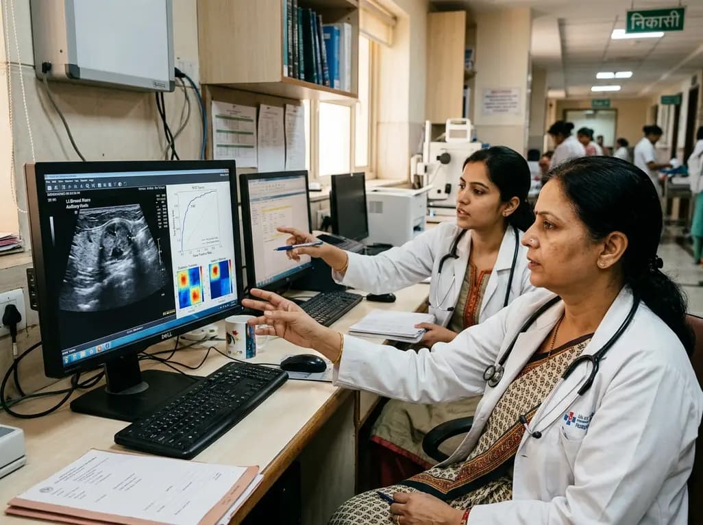
Neoadjuvant chemotherapy has become a cornerstone in managing aggressive human epidermal growth factor receptor 2 (HER2)-positive breast malignancy. Achieving a pathological complete response following systemic treatment strongly correlates with superior event-free survival and overall survival outcomes. However, individual patient responses vary significantly across clinical cohorts. Clinicians currently lack non-invasive, objective biomarkers to identify which patients will achieve pathological clearance prior to initiating systemic regimens. Recent advancements in ultrasound radiomics breast cancer applications offer an innovative solution to this diagnostic dilemma. By extracting high-throughput quantitative features from routine pre-treatment sonograms, artificial intelligence models decode microscopic phenotypic variations within the tumor microenvironment. A groundbreaking dual-center retrospective study demonstrates that combining sonographic radiomics signatures with independent clinical risk factors substantially enhances early response prediction, providing an accessible tool to guide personalized therapeutic strategies.
HER2-positive breast cancer represents an aggressive biological subtype characterized by rapid cell proliferation and heightened metastatic potential. Consequently, standard guidelines recommend neoadjuvant systemic therapy combined with targeted anti-HER2 agents before definitive surgical resection. This therapeutic approach aims to downstage primary breast lesions, eradicate micro-metastatic dissemination, and enable breast-conserving surgery. More importantly, achieving a pathological complete response provides vital prognostic information that directly guides postoperative treatment decisions.
Patients who achieve complete tumor eradication experience markedly reduced recurrence rates. Conversely, individuals with residual invasive disease require intensified secondary regimens, such as antibody-drug conjugates, to mitigate recurrence risks. Therefore, predicting treatment response before administering toxic cytotoxic drugs carries profound clinical implications. Accurate early prediction helps multidisciplinary teams tailor neoadjuvant protocols, avoid ineffective toxicities, and prevent surgical delays. Although baseline magnetic resonance imaging and biopsy profiles offer valuable insights, they possess inherent limitations regarding accessibility, cost, and tissue sampling error.
Conventional diagnostic ultrasound serves as an indispensable, safe, and cost-effective modality in everyday breast oncology practice. However, qualitative ultrasound interpretations depend heavily on operator experience and cannot capture sub-visual heterogeneity across entire tumor volumes. In contrast, ultrasound radiomics breast cancer analytics convert standard medical images into minable digital data pipelines. These mathematical algorithms extract hundreds of texture, shape, intensity, and wavelet descriptors reflecting underlying cellularity, necrosis, and stromal architecture.
To leverage these capabilities, researchers collected imaging and clinical records from 659 HER2-positive breast cancer patients across two major institutions. Center 1 contributed a training cohort of 283 patients alongside an internal validation cohort of 121 patients treated between January 2015 and December 2023. Meanwhile, Center 2 provided an independent external validation cohort of 255 patients treated between January 2018 and December 2024. Surgical pathology specimens served as the definitive reference standard for pathological complete response. By applying advanced feature selection algorithms, investigators established robust predictive signatures that overcome classical single-center overfitting challenges.
Developing dependable radiomics algorithms requires rigorous statistical filtering to eliminate noise and redundant variables. In this study, the investigative team first refined the radiomics signature through systematic redundancy reduction and dimensionality mitigation techniques. Subsequently, they constructed a Least Absolute Shrinkage and Selection Operator regression model. This sophisticated machine learning method isolated the most discriminative textural features associated with systemic treatment efficacy, yielding an optimized radiomics signature score.
Next, the researchers identified independent clinical and pathological risk factors using multivariable logistic regression analysis. They combined these clinical determinants with the extracted radiomics signature to establish a comprehensive integrated nomogram. To evaluate diagnostic accuracy rigorously, investigators compared receiver operating characteristic curves across the standalone clinical model, the radiomics signature model, and the combined predictive tool. Finally, they employed the DeLong test to verify the statistical significance of differences observed between individual area under the curve metrics across all testing datasets.
The statistical findings demonstrated the marked superiority of the combined clinical-radiomics nomogram over traditional single-domain assessments. Within the training cohort, the clinical-only predictive model yielded an acceptable area under the curve of 0.699. Meanwhile, the standalone radiomics signature model achieved an improved area under the curve of 0.817, highlighting the diagnostic power of quantitative image processing. Most impressively, the integrated model demonstrated superior accuracy, achieving an overall area under the curve of 0.861.
Furthermore, DeLong test analysis confirmed that this diagnostic improvement was statistically significant across both internal and external validation cohorts. The integrated model maintained excellent calibration and discriminative stability when evaluated on the independent multi-center dataset of 255 external patients. This consistent external performance proves that combining clinical metrics with high-dimensional sonographic features minimizes institutional scanner variations. Thus, the integrated framework provides an accurate, reproducible, and objective methodology for assessing pre-treatment tumor responsiveness before initiating neoadjuvant cycles.
In developing healthcare settings like India, access to advanced dynamic contrast-enhanced magnetic resonance imaging remains constrained by financial costs, geographic availability, and infrastructure limits. Consequently, ultrasound remains the most ubiquitous, economical, and patient-friendly imaging modality across both rural and urban oncology facilities. Implementing an artificial intelligence-driven sonographic radiomics workflow could democratize precision medicine across diverse healthcare tiers.
By incorporating standardized ultrasound radiomics software into routine pre-chemotherapy workups, Indian oncologists can identify non-responders before initiating standard regimens. Clinicians can subsequently consider adaptive clinical trials, novel drug combinations, or upfront surgical resection for predicted non-responders. In addition, identifying probable responders helps surgical oncologists plan safe breast-conserving surgery or de-escalated axillary interventions well in advance. However, successful clinical translation will require seamless Picture Archiving and Communication System integration, automated tumor segmentation tools, and prospective clinical validation in local demographic cohorts.
The success of pre-treatment ultrasound radiomics opens exciting avenues for longitudinal tumor surveillance throughout systemic therapy. Future investigations must explore delta-radiomics, which evaluates quantitative feature changes between successive chemotherapy cycles. Combining baseline radiomics with mid-treatment ultrasound metrics may offer even higher predictive accuracy. Furthermore, merging sonographic radiomics with serum biomarkers, such as circulating tumor DNA, could establish powerful multi-modal predictive ecosystems.
Nevertheless, clinicians must recognize current operational challenges before widespread deployment occurs. Variations in transducer frequencies, acoustic settings, and operator-dependent scanning angles can introduce feature instability. Therefore, academic medical societies must prioritize the development of standardized ultrasound acquisition protocols and vendor-neutral radiomic harmonization tools. As machine learning algorithms become more interpretable and accessible, quantitative sonography will likely evolve from an experimental research tool into an indispensable daily clinical ally.
Conventional ultrasound relies exclusively on visual qualitative interpretations by radiologists, which inherently subject the evaluation to subjective inter-observer variability. In contrast, ultrasound radiomics uses automated mathematical algorithms to extract hundreds of invisible quantitative data points from digital image pixels. These computational features capture fine tumor heterogeneity, microstructural density, and stromal patterns that human vision cannot detect, enabling objective, reproducible predictive modeling of systemic therapeutic efficacy.
Achieving a pathological complete response indicates total eradication of invasive cancer cells from the resected breast and axillary lymph nodes. This outcome serves as a robust surrogate marker for prolonged relapse-free survival and reduced mortality. Accurately predicting this response allows oncologists to personalize systemic regimens, de-escalate toxic therapies for likely responders, and promptly explore alternative novel targeted combinations for predicted non-responders.
Yes, the clinical-radiomics model utilizes standard digital sonograms that hospitals routinely acquire during baseline breast cancer evaluations without requiring additional invasive procedures or expensive imaging agents. By integrating automated radiomics feature-extraction algorithms directly into hospital Picture Archiving and Communication Systems, multidisciplinary teams can generate immediate, objective predictive scores alongside standard clinical reports to guide neoadjuvant treatment planning efficiently.
Disclaimer: This content is for informational and educational purposes only and does not constitute medical advice, diagnosis, or treatment. Healthcare professionals should exercise independent clinical judgment and verify all information before making clinical decisions. Refer to the latest local and national guidelines for clinical practice.
References
Du LW et al. Early Prediction Of Treatment Response To Neoadjuvant Chemotherapy Based On Pre-treatment Ultrasound Radiomics of HER2-positive Breast Cancer Patients by Radiomics-based Model: A Dual-center Retrospective Study. Acad Radiol. 2026 Aug 18. doi: undefined. PMID: 42613278.
Jiang M, et al. Pretreatment ultrasound-based deep learning radiomics model for the early prediction of pathologic response to neoadjuvant chemotherapy in breast cancer. Eur Radiol. 2023;33(9):6420-6431.
Guo C, et al. Radiomics of Multimodal Ultrasound for Early Prediction of Pathologic Complete Response to Neoadjuvant Chemotherapy in Breast Cancer. Ultrasound Med Biol. 2024;50(12):1812-1821.

Read summarized clinical updates, watch expert medical content, and earn CME certifications right from your smartphone.
Open your smartphone camera to scan and install.





A dual-center study reveals that an integrated model combining pre-treatment ultrasound radiomics and clinical risk factors accurately predicts pathological complete response to neoadjuvant chemotherapy in HER2-positive breast cancer, achieving an AUC of 0.861 and paving the way for personalized oncology care.
last month

A nationwide multicenter study identified three COPD clinical phenotypes, finding that mild smoking males experience the highest rate of rapid lung function decline. A CatBoost model using six routine clinical and socioeconomic variables accurately stratifies high-risk patients for early therapeutic intervention.
Today
Discover the crucial distinctions between mesonephric and mesonephric-like proliferations of the female genital tract. Learn the key histomorphologic, immunohistochemical, and molecular differences essential for accurate diagnosis and clinical management.
Today

A large UK Biobank study reveals that plasma proteomic signatures capture preclinical organ damage and improve multiorgan risk prediction across early CKM syndrome stages 0 to 2 beyond traditional PREVENT clinical models.
Today
A narrative review investigates whether the articularis genus muscle functions as an independent anatomical entity or blends with the vastus intermedius. We evaluate its morphology, role in retracting the suprapatellar bursa, and direct clinical significance in anterior knee pain and arthroplasty.
Today

A Bayesian multilevel meta-analysis reveals that aerobic training combined with moderate carbohydrate restriction modestly lowers HbA1c in type 2 diabetes. However, sparse data and very low certainty leave incremental benefits over exercise or diet alone unproven, highlighting the need for individualized care.
Today
