.webp&w=384&q=75)

Loading, please wait...
Loading, please wait...
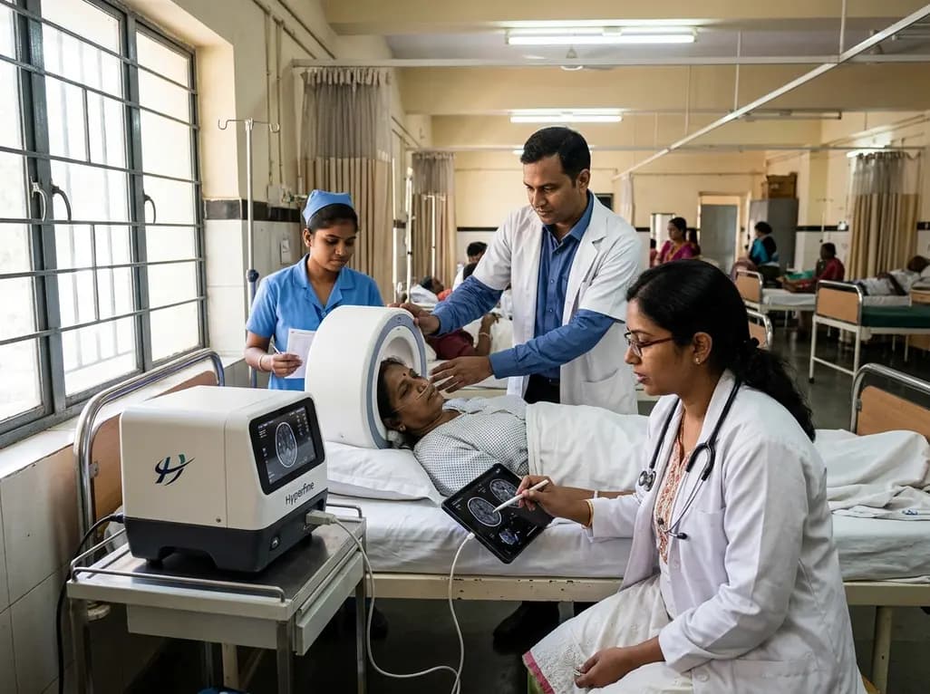
Clinicians managing advanced multiple sclerosis often face severe logistical hurdles during routine disease monitoring. Traditional high-field neuroimaging requires patient transfer to specialized radiology suites. However, individuals suffering from severe physical disability frequently cannot tolerate conventional scanners due to posture difficulties, spasticity, and transport burdens. Consequently, clinical research often excludes severely disabled populations, leaving a critical knowledge gap regarding end-stage neurodegeneration. Portable ultra-low-field MRI provides a transformative solution to this diagnostic dilemma. Operating at 0.064 Tesla, this innovative technology enables direct bedside scanning in routine wards or skilled nursing facilities. Clinicians can now evaluate intracranial structures without moving fragile individuals from their hospital beds. Furthermore, recent technological advances allow automated brain tissue volumetry from these low-field acquisitions. By mitigating immobility barriers, point-of-care imaging opens new avenues for inclusive clinical care. Therefore, researchers sought to determine whether low-field imaging could reliably quantify tissue atrophy in severe disease. Understanding these technical capabilities is crucial for modern neurotherapeutics and bedside management.
To establish clinical validity, investigators designed a rigorous prospective trial evaluating two distinct participant cohorts. Cohort 1 enrolled 14 healthy controls and 33 individuals with multiple sclerosis who underwent both portable ultra-low-field MRI and standard 3 Tesla examinations. This paired design allowed direct cross-platform comparison of brain volumetry across differing magnetic field strengths. Cohort 2 enrolled 40 individuals with progressive disease, categorizing 24 participants as severely disabled and 16 as less severe. Remarkably, the portable scanner achieved high scan completion rates. Only two individuals in the progressive cohort failed quality control due to technical artifacts. In Cohort 1, ultra-low-field measurements revealed robust disease differences that mirrored high-field metrics. Specifically, artificial intelligence segmentation using WMH-SynthSeg demonstrated substantial effect sizes for whole brain volume and cortical gray matter. Thalamic volumetric estimations also yielded excellent concordance between modalities. Consequently, these findings validate portable scanners as dependable alternatives for volumetric assessments when standard 3 Tesla machines remain inaccessible.
Quantitative analysis at ultra-low magnetic field strengths introduces distinct computational challenges because of reduced signal-to-noise ratios. Therefore, investigators systematically compared conventional segmentation software, such as SIENAX, against modern deep learning algorithms like WMH-SynthSeg. Each processing pipeline demonstrated unique technical advantages depending on the specific brain region examined. Conventional SIENAX processing excelled at capturing global tissue boundaries and cortical loss. For instance, SIENAX produced the largest effect sizes when differentiating severe from less severe disease in whole brain volume and cortical gray matter. Conversely, deep learning tools handled deep white matter structures with higher statistical power. WMH-SynthSeg yielded the highest effect size for total white matter volume estimation. However, AI-derived cortical gray matter segmentations showed greater variability based on input pulse sequences. Thus, clinicians and neuroscientists must carefully select their analytic pipelines when interpreting bedside data. Applying standardized, validated post-processing algorithms remains essential for meaningful quantitative monitoring.
Characterizing neurodegenerative progression in late-stage disease requires accurate differentiation between gray matter and white matter atrophy patterns. Historically, inflammatory demyelination dominates early relapse-remitting phases, whereas progressive disease manifests pervasive, relentless neuroaxonal loss. The study highlighted that gray matter atrophy represents the primary radiological differentiator in advanced clinical stages. Specifically, cortical gray matter volume exhibited the strongest statistical divergence between severely disabled individuals and less impaired cohorts. Patients with severe progressive disease demonstrated profound cortical thinning that aligned with clinical immobility. In contrast, white matter volumetric differences presented smaller effect sizes between clinical severities. These structural results suggest that late-stage functional decline stems primarily from widespread neocortical neurodegeneration rather than isolated white matter lesion accumulation. Furthermore, portable imaging effectively captured these microstructural shifts right at the bedside. Consequently, monitoring cortical preservation could serve as a valuable metric during future palliative and restorative therapeutic trials.
Establishing clinical relevance requires imaging metrics to correlate meaningfully with physical disability and cognitive function. During the trial, clinicians assessed participant mobility alongside validated neuropsychological batteries. Interestingly, the strength of clinicoradiological associations depended heavily on the volumetric processing algorithm employed. In Cohort 2, conventional SIENAX segmentation produced the most consistent correlations with clinical disability and functional scores. Reduced whole brain and gray matter volumes tracked closely with deteriorating ambulatory function. On the other hand, AI-based metrics displayed divergent correlations depending on the image contrast utilized during deep learning segmentation. While artificial intelligence provided swift automated segmentations, its clinical correlations exhibited noticeable instability across cortical regions. Therefore, conventional edge-based registration pipelines currently provide superior clinical interpretability for bedside monitoring. Clinicians must recognize these methodological nuances when evaluating real-world bedside data. Combining robust automated pipelines with clinical vigilance guarantees reliable tracking of ongoing neurodegeneration.
The successful deployment of portable scanning addresses major systemic inequities in neurologic care. Historically, severely disabled patients residing in chronic care facilities could not participate in therapeutic trials due to scanning logistics. Now, portable devices permit longitudinal evaluations in non-traditional healthcare environments, including nursing homes and intensive care units. In resource-constrained settings, such as regional Indian hospitals, low-field systems offer substantial infrastructure savings. They require standard electrical wall outlets and completely eliminate expensive cryogen cooling or magnetic shielding rooms. In addition, point-of-care neuroimaging facilitates prompt detection of opportunistic central nervous system complications in immunocompromised individuals. Clinicians can safely scan patients without removing supportive life-support apparatus or dealing with ferromagnetic equipment hazards. As analytical software advances, ultra-low-field imaging will increasingly bridge hospital and community neurology. Ultimately, this technology democratizes neuroimaging, ensuring that fragile, bedbound individuals receive optimal structural monitoring throughout their disease course.
Ultra-low-field MRI operates at a magnetic strength of 0.064 Tesla, whereas conventional clinical scanners utilize 1.5 Tesla or 3 Tesla systems. Consequently, ultra-low-field systems sacrifice high spatial resolution to gain mobility, low power consumption, and direct bedside point-of-care accessibility for severely disabled or immobile patients.
Conventional segmentation pipelines like SIENAX proved more reliable and robust for analyzing cortical gray matter atrophy and whole brain loss at ultra-low field strengths. Meanwhile, artificial intelligence-based algorithms excelled in quantifying white matter loss and subcortical structures, demonstrating that analytical pipeline choice directly dictates volumetric precision.
Bedside MRI eliminates complex transport logistics, physical transfer risks, and specialized monitoring requirements for severely disabled patients. Furthermore, this portable technology facilitates longitudinal neuroimaging in long-term care facilities, expanding clinical trial enrollment to historically excluded populations suffering from advanced progressive forms of multiple sclerosis.
Disclaimer: This content is for informational and educational purposes only and does not substitute professional medical advice, diagnosis, or treatment. Refer to the latest local and national guidelines for clinical practice.
References

Read summarized clinical updates, watch expert medical content, and earn CME certifications right from your smartphone.
Open your smartphone camera to scan and install.





A prospective study demonstrates that portable 0.064 T ultra-low-field MRI enables reliable bedside neuroimaging and brain tissue volumetry in severely disabled multiple sclerosis patients, capturing key gray and white matter atrophy patterns without the logistical hazards of conventional MRI transfers.
4 weeks back

A meta-analysis of 14 RCTs reveals that dietary fiber significantly increases bowel movements and stool consistency in functional constipation while modulating gut-brain axis biomarkers, including short-chain fatty acids, serotonin, and inflammation, highlighting benefits beyond simple stool bulking.
Today

A retrospective study of 824 patients undergoing over 2,200 intradetrusor onabotulinumtoxinA injections identifies sex- and etiology-specific predictors of 2-week post-void residual, highlighting the influence of baseline residual volumes, toxin dosage, and distinct urodynamic parameters on urinary retention risk.
Today
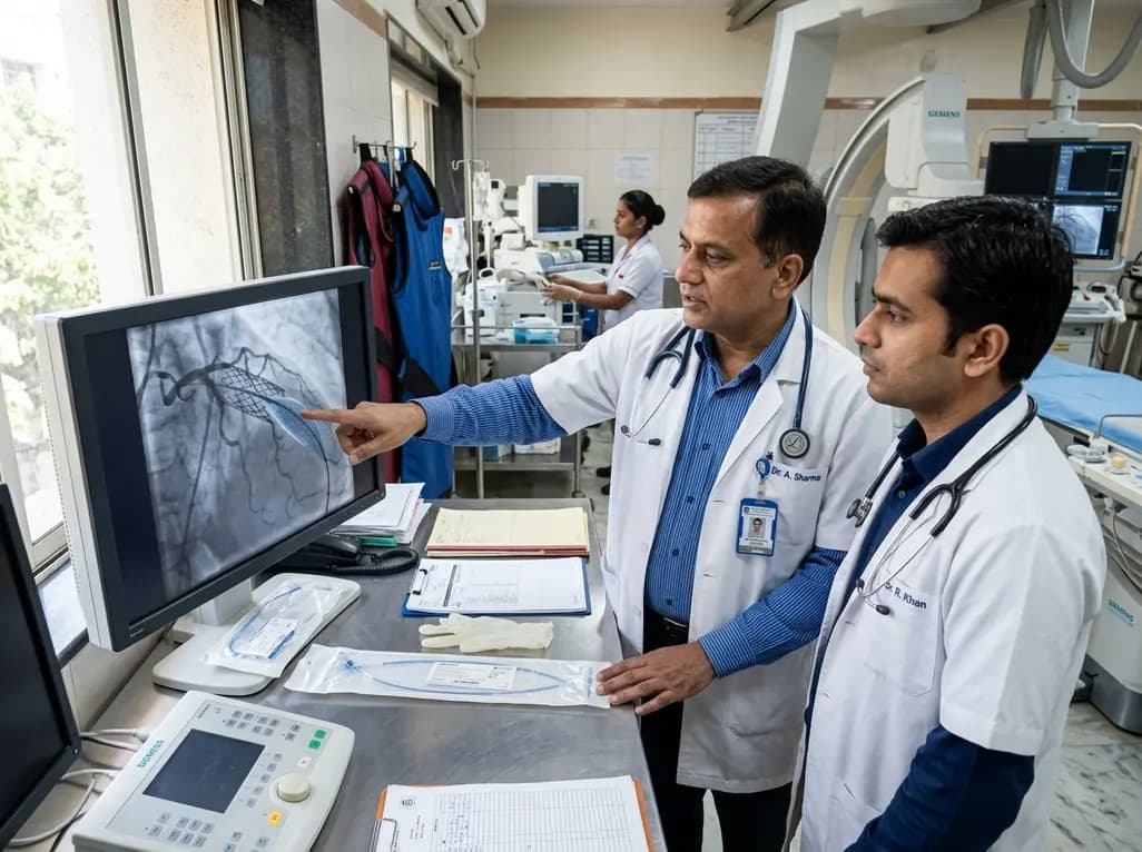
The US FDA has granted approval to the SELUTION SLR drug-eluting balloon for treating coronary in-stent restenosis. This milestone introduces a sustained-release sirolimus platform that eliminates the need for additional permanent metallic stents while maintaining robust vascular healing and luminal patency.
Today

The Thumbs-Out Classification (TOC) is a reliable screening tool for detecting thumb-in-palm deformity in children with cerebral palsy. Utilizing traffic-light tiers for radial abduction, TOC enables clinicians to monitor upper limb function and initiate timely therapeutic or surgical interventions.
Today

A comprehensive review analyzes periconceptional GLP-1 receptor agonist exposure, revealing reassuring data on congenital anomalies and adverse perinatal outcomes while emphasizing the need for cautious patient counseling.
Today
