.webp&w=384&q=75)

Loading, please wait...
Loading, please wait...
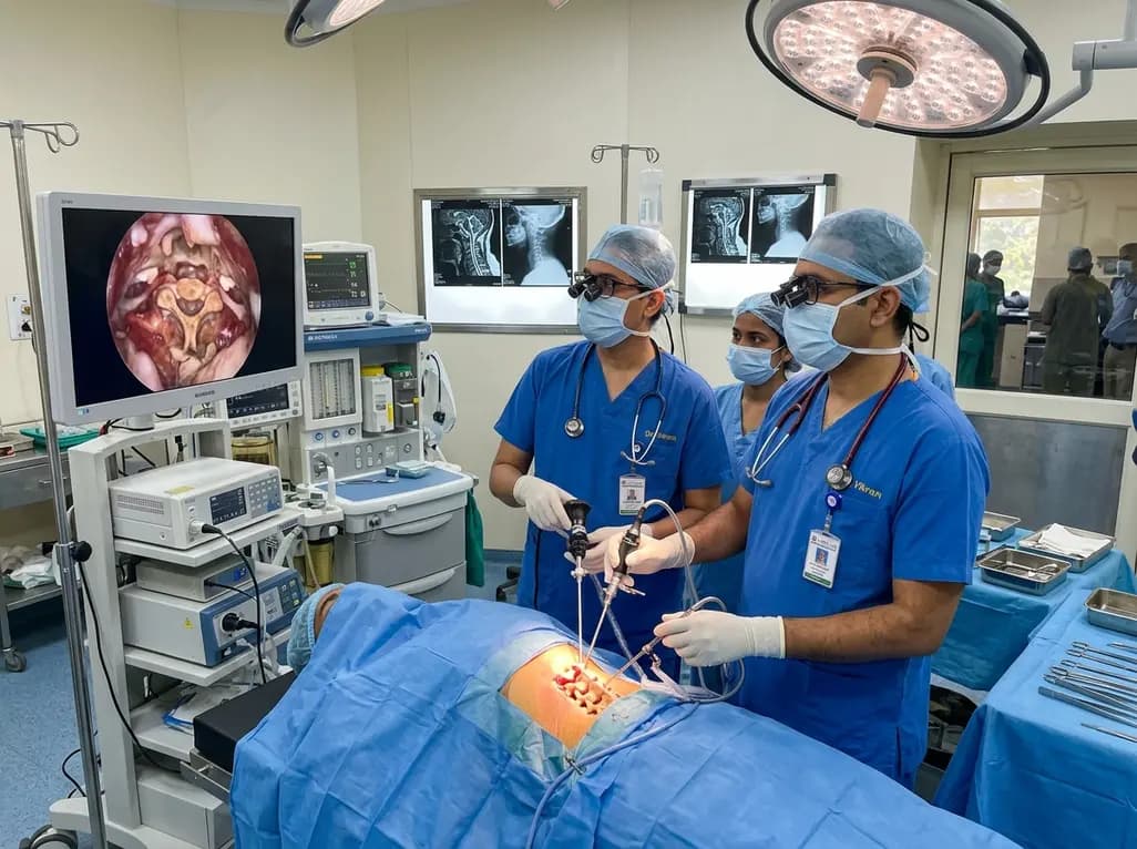
Surgical management of complex cervical spine disease has evolved toward minimally invasive techniques that decompress neural structures while sparing biomechanical integrity. In patients presenting with combined spinal cord and nerve root compression, UBE cervical laminectomy has emerged as an innovative motion-preserving intervention. This procedure addresses both central canal stenosis and bilateral neuroforaminal narrowing through a minimally disruptive posterior portal approach. Consequently, surgeons can achieve extensive neural decompression without the morbidity of extensive open exposure or the mandatory instrumentation associated with fusion.
The operative method utilizes two independent percutaneous portals to achieve expansive visualization and unconstrained instrument triangulation. Initially, the surgeon establishes viewing and working portals on the symptomatic or more accessible side. Through continuous saline irrigation, the visual field remains clear and epidural bleeding diminishes significantly. The surgical team performs an ipsilateral posterior cervical foraminotomy by resecting the medial facet joint and unroofing the exiting nerve root. Subsequently, the surgeon floats the base of the spinous process to facilitate access across the midline.
Furthermore, contralateral supralaminar drilling allows direct visualization of the contralateral lamina and neuroforamen without excessive muscle detachment. The operative team then performs contralateral decompression beneath the over-the-top trajectory. Therefore, bilateral nerve roots and the central spinal cord undergo thorough decompression from a single unilateral approach. This dual-portal mobility provides critical angulation advantages that standard tubular systems cannot replicate.
Clinical investigations demonstrate substantial functional gains following biportal decompression for cervical myeloradiculopathy. Patients experience rapid relief of both axial neck pain and radiating brachialgia within the immediate postoperative period. Moreover, long-term follow-up at one year confirms sustained improvements across standard validated indices. Neck Visual Analog Scale and arm Visual Analog Scale scores decline markedly after surgery, reflecting adequate relief of central and foraminal neural compression.
Additionally, patient functional capacity improves significantly as quantified by the Neck Disability Index and the modified Japanese Orthopaedic Association score. Patients suffering from myelopathic motor deficits, clumsy hands, and gait instability demonstrate meaningful neurological recovery. Importantly, the minimally invasive nature of the biportal portals decreases postoperative wound pain and reduces narcotic reliance during early recovery. Consequently, patients achieve accelerated rehabilitation and an earlier return to regular daily activities compared to historical open cohorts.
Quantitative radiological assessments provide objective evidence of anatomical decompression across the treated segments. High-resolution postoperative magnetic resonance imaging demonstrates significant enlargement of the dural sac cross-sectional area. Central stenotic narrowing resolves completely, allowing circumferential cerebrospinal fluid restoration around the cervical spinal cord. Furthermore, axial computed tomography scans confirm successful enlargement of the neuroforaminal dimensions bilaterally.
Importantly, the surgical technique preserves the critical biomechanical structures required for posterior column stability. Although facet length and cross-sectional area decrease following targeted foraminotomy, bone resection remains controlled and within safe physiological limits. Because the contralateral approach utilizes an oblique sub-laminar corridor, facet joint preservation on the contralateral side is maximized. Consequently, the structural integrity of the zygapophyseal joints remains robust enough to resist pathological translation or hypermobility under physiological loads.
Preserving physiological alignment represents a primary objective when performing non-instrumented posterior cervical decompression. Conventional open laminectomy frequently carries a risk of progressive postoperative kyphosis due to extensive detachment of posterior muscular attachments and interspinous ligaments. In contrast, unilateral biportal endoscopic procedures spare the contralateral paraspinal musculature and preserve crucial midline tension band structures. Therefore, the physiological load-bearing mechanism of the cervical spine remains intact.
Radiological evaluations at one-year follow-up confirm that global cervical lordosis from C2 to C7 is maintained without kyphotic deterioration. Segmental lordotic angles and intervertebral disc heights also show stable measurements over time. Crucially, flexion-extension dynamic radiographs reveal no evidence of delayed segmental instability, spondylolisthesis, or hypermobility. Consequently, these findings validate the procedure as a viable motion-preserving alternative that avoids unnecessary spinal arthrodesis in carefully selected patients.
Achieving optimal surgical outcomes with biportal endoscopy requires meticulous attention to surgical indications and fluid dynamics. Surgeons must maintain controlled irrigation pressure throughout the procedure to prevent elevated epidural hydrostatic pressures or fluid extravasation into paraspinal tissue planes. Furthermore, careful haemostasis using radiofrequency ablation prevents visualization loss in tight posterior cervical spaces.
Additionally, accurate patient selection remains critical for long-term clinical success. The technique is ideally suited for patients with multi-level or single-level myeloradiculopathy without severe baseline kyphotic deformity or gross preoperative instability. Although the technical learning curve is steep, master surgeons benefit from panoramic visualization and independent instrument handling. Therefore, this motion-sparing endoscopic decompression represents an increasingly valuable addition to modern minimally invasive spine surgery.
UBE cervical laminectomy minimizes soft tissue trauma and preserves posterior cervical musculature. The technique provides magnified visualization through continuous irrigation, enabling precise central and bilateral foraminal decompression without extensive muscle detachment, significant blood loss, or prolonged postoperative hospital stays.
Clinical studies show no significant postoperative cervical instability at one year. Because the procedure preserves key posterior ligamentous attachments and limits facet joint resection to safe physiological thresholds, patients maintain baseline lordosis without developing progressive kyphosis or segmental hypermobility.
Ideal candidates are individuals diagnosed with cervical myeloradiculopathy caused by canal stenosis and bilateral foraminal narrowing. Candidates should possess preserved baseline cervical lordosis without gross spinal instability, significant spondylolisthesis, or severe rigid kyphotic deformities requiring corrective instrumentation.
Disclaimer: This content is for informational and educational purposes only and does not constitute medical advice, diagnosis, or treatment. Healthcare professionals should exercise their independent clinical judgment when evaluating medical information. The views expressed herein do not necessarily represent the official policy or position of any medical institution or regulatory body. Refer to the latest local and national guidelines for clinical practice.
References
Jung J et al. Unilateral biportal endoscopic cervical laminectomy with bilateral foraminotomy for cervical myeloradiculopathy: clinical and radiological outcomes at 1 year. Neurosurg Rev. 2026 Aug 03. doi: 10.1007/s10143-026-04417-4. PMID: 42545519.
Kim JY, Kim HS, Jeon I. Biportal Endoscopic Spine Surgery: Technical Nuances and Application in Posterior Cervical Decompression. Neurospine. 2023;20(1):45-56.
Park MK, Son SK, Park W. Biportal Endoscopic Posterior Cervical Foraminotomy: Indications, Surgical Techniques, and Clinical Outcomes. J Minim Invasive Spine Surg Tech. 2022;7(2):189-198.

Read summarized clinical updates, watch expert medical content, and earn CME certifications right from your smartphone.
Open your smartphone camera to scan and install.





A 1-year study evaluated unilateral biportal endoscopic (UBE) cervical laminectomy with bilateral posterior foraminotomy for cervical myeloradiculopathy. The procedure significantly improved pain scores and functional outcomes while expanding the canal and preserving cervical stability without requiring spinal fusion.
last month

This clinical review examines the dangerous intersection of climate change and antimicrobial resistance (AMR). It highlights thermal selection, altered transmission pathways, and the imperative for One Health and Planetary Health adaptation frameworks to preserve therapeutic efficacy.
Today
Discover the crucial distinctions between mesonephric and mesonephric-like proliferations of the female genital tract. Learn the key histomorphologic, immunohistochemical, and molecular differences essential for accurate diagnosis and clinical management.
Today

A large UK Biobank study reveals that plasma proteomic signatures capture preclinical organ damage and improve multiorgan risk prediction across early CKM syndrome stages 0 to 2 beyond traditional PREVENT clinical models.
Today

A narrative review investigates whether the articularis genus muscle functions as an independent anatomical entity or blends with the vastus intermedius. We evaluate its morphology, role in retracting the suprapatellar bursa, and direct clinical significance in anterior knee pain and arthroplasty.
Today

A Bayesian multilevel meta-analysis reveals that aerobic training combined with moderate carbohydrate restriction modestly lowers HbA1c in type 2 diabetes. However, sparse data and very low certainty leave incremental benefits over exercise or diet alone unproven, highlighting the need for individualized care.
Today
