.webp&w=384&q=75)

Loading, please wait...
Loading, please wait...

Risk assessment prior to invasive sampling represents a crucial step in contemporary urological diagnostics. In this setting, evaluating tumor density in prostate cancer has emerged as a valuable strategy to guide clinical decisions. Traditional prostate-specific antigen tests and multiparametric imaging identify suspicious lesions with notable sensitivity. However, these conventional modalities occasionally prompt redundant tissue sampling in equivocal cases. Therefore, integrating precise lesion volume relative to overall gland size offers essential diagnostic clarity.
Clinicians historically relied on two-dimensional diameter measurements to quantify suspected prostatic tumors. However, simple linear dimensions cannot adequately capture the irregular geometric shapes of invasive lesions. Furthermore, prostate gland volume varies considerably across individual patients. A fifteen-millimeter lesion occupies substantial relative volume within a small, twenty-gram prostate. In contrast, that identical lesion represents only a minor fraction of an enlarged eighty-gram gland. Consequently, absolute millimeter measurements generate diagnostic ambiguity when stratifying oncological risk. Additionally, standard systematic biopsies often miss anterior lesions or produce indeterminate sampling. Multiparametric magnetic resonance imaging significantly improved target visualization over traditional methods. Nevertheless, conventional imaging categories still rely partly on linear thresholds. Therefore, urologists require normalized volumetric parameters that integrate individual gland architecture. This proportional evaluation allows clinicians to differentiate indolent nodules from aggressive malignancies more effectively. Moreover, volumetric normalization accounts for the confounding influence of benign hyperplasia. As a result, surgical teams gain clearer diagnostic confidence before recommending invasive interventions. Consequently, standardized volumetric density measurements resolve these persistent imaging challenges effectively.
Recent evidence confirms that tumor density markedly enhances lesion-level cancer detection. Specifically, physicians calculate this metric by dividing software-derived lesion volume by total magnetic resonance prostate volume. This simple calculation normalizes tumor volume against the specific organ dimensions of the patient. Consequently, clinicians eliminate the confounding distortion caused by widespread adenomatous background tissue. Recent clinical investigations demonstrated that lesions harboring clinically significant disease exhibit substantially higher median density values. In fact, significant tumors demonstrated more than double the median density of non-significant lesions. Furthermore, multivariable regression models confirm that tumor density serves as an independent predictor of significant malignancy. This lesion-level metric also delivers superior discriminatory performance compared to isolated diameter thresholds. Therefore, urologists can utilize this ratio to prioritize urgent biopsy procedures for high-density lesions. Additionally, calculating this parameter requires no supplemental blood tests or proprietary tissue profiling assays. Thus, busy clinical centers can immediately implement this metric within existing pre-biopsy workflows. Ultimately, this practical approach optimizes diagnostic stratification without adding fiscal strain.
The route of tissue sampling also plays a fundamental role in optimizing patient outcomes. Historically, urologists favored transrectal needle biopsies due to widespread outpatient familiarity. However, the transrectal approach carries substantial risks of severe rectal bleeding and urinary tract sepsis. In contrast, transperineal access routes pass through clean perineal skin rather than rectal mucosa. Consequently, transperineal biopsies virtually eliminate hospital readmissions for life-threatening bacteremia. Furthermore, the transperineal trajectory permits superior mechanical access to complex anatomical locations. Anterior stromal tumors and apical lesions remain notoriously difficult to sample via transrectal angles. Transperineal needles enter parallel to the prostatic urethra, facilitating comprehensive sampling along the longest tumor axis. Additionally, modern software-assisted fusion systems overlay pre-acquired magnetic resonance imaging onto live ultrasound. This spatial synchronization ensures that the needle precisely penetrates the exact volume of interest. Therefore, coupling transperineal fusion biopsy with tumor density creates an exceptionally accurate diagnostic pathway. As a result, surgical teams obtain representative tissue cores while preserving patient safety.
Clinical decision-making presents particular difficulty when diagnostic imaging reports indeterminate abnormalities. Specifically, lesions categorized as PI-RADS 3 or smaller PI-RADS 4 exhibit highly variable malignancy rates. While obvious PI-RADS 5 lesions clearly necessitate immediate biopsy, equivocal findings frequently perplex urologists. Consequently, clinicians risk either over-biopsying benign nodules or delaying necessary oncological interventions. Fortunately, tumor density provides vital discriminatory power in these ambiguous clinical situations. For example, an indeterminate target showing elevated tumor density indicates a high probability of aggressive disease. Therefore, clinicians should promptly recommend targeted needle sampling for such high-risk indeterminate lesions. Conversely, a very low tumor density within a large prostate suggests a favorable benign etiology. In such instances, physicians can safely counsel patients toward structured monitoring rather than immediate intervention. Moreover, decision-curve analyses demonstrate that incorporating tumor density enhances net clinical benefit. This refined stratification protects patients from procedural discomfort while ensuring timely detection of aggressive malignancies. Thus, volumetric ratios provide objective criteria that support nuanced shared clinical decision-making.
Implementing volumetric metrics into routine urological practice does not require costly capital investments. Most modern commercial fusion systems already calculate three-dimensional target volumes during routine segmentation. Therefore, urologists can determine tumor density within seconds by executing a simple mathematical division. Furthermore, integrating this parameter into multidisciplinary tumor boards improves diagnostic concordance between radiologists and urologists. This standardized communication ensures consistent evaluation across diverse institutional settings. In Indian healthcare environments, optimizing biopsy indications holds exceptional clinical and economic significance. Healthcare facilities frequently operate under heavy patient volumes and constrained procedural theatre access. Consequently, prioritizing biopsies based on tumor density helps conserve valuable healthcare resources for urgent cases. Additionally, preventing unnecessary biopsies spares patients avoidable financial expenditures and psychological distress. Clinicians can confidently direct men with low-density equivocal lesions toward safe active surveillance pathways. Meanwhile, surgical teams can promptly biopsy individuals with elevated density scores. Furthermore, this streamlined strategy enhances overall clinical efficiency across busy outpatient departments. Thus, tumor density represents an accessible, reproducible tool that elevates the quality of personalized prostate care.
Clinicians calculate tumor density by dividing the exact three-dimensional lesion volume by the total prostate volume measured on magnetic resonance imaging. Advanced fusion biopsy software automatically generates both volumetric measurements during pre-procedural image segmentation. This standardized ratio normalizes the tumor burden relative to overall gland size, which provides a far more precise assessment of malignant potential than traditional two-dimensional diameter measurements alone.
The transperineal approach significantly improves patient safety by eliminating needle traversal through the rectal mucosa, which drastically reduces the incidence of severe infectious complications and sepsis. Furthermore, transperineal needle trajectories provide superior anatomical access to anterior and apical prostatic zones, regions frequently missed by traditional transrectal biopsies. When combined with magnetic resonance imaging fusion guidance, transperineal sampling delivers outstanding diagnostic accuracy with minimal procedural morbidity.
Indeterminate PI-RADS 3 lesions often present diagnostic uncertainty because their cancer rates vary widely across patient populations. Evaluating tumor density helps stratify this risk effectively. Lesions exhibiting elevated tumor density correlate with a substantially higher risk of clinically significant disease, justifying prompt targeted biopsy. Conversely, low tumor density in an enlarged benign gland reassures clinicians, allowing them to recommend safe active surveillance and avoid unnecessary invasive procedures.
Disclaimer: This content is for informational and educational purposes only and does not constitute medical advice, diagnosis, or treatment. Always consult a qualified healthcare provider with any questions regarding a medical condition. Refer to the latest local and national guidelines for clinical practice.
References

Read summarized clinical updates, watch expert medical content, and earn CME certifications right from your smartphone.
Open your smartphone camera to scan and install.





Evaluating tumor density in prostate cancer by dividing exact MRI lesion volume by total prostate volume significantly improves the detection of clinically significant disease. This volumetric approach enhances transperineal fusion biopsy accuracy while avoiding unnecessary procedures.
last month
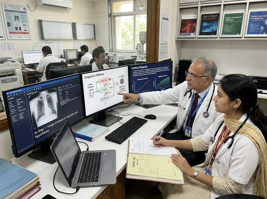
A breakthrough peer-reviewed study demonstrates that a sequential AI diagnosis system can mirror real-world clinical reasoning. By ordering stepwise tests and updating hypotheses dynamically, this multi-agent orchestrator achieved superior diagnostic accuracy while reducing investigative costs.
Today

A prenatal case highlights severe glutaric acidemia type II presenting as bilateral hyperechoic cystic kidneys at 18 weeks. Whole-exome sequencing revealed a novel homozygous ETFDH frameshift variant, demonstrating the power of rapid genetic testing in fetal renal anomalies.
Today

Review how tailored exercise programs benefit frail older patients with heart failure. Explore pathophysiological mechanisms, individualized training prescriptions, and strategies to overcome adherence barriers in clinical practice.
Yesterday

This comparative study evaluates posterior labral tears with and without glenohumeral instability, highlighting distinct demographic patterns, MRI underestimation in stable shoulders, and successful arthroscopic repair outcomes.
Yesterday

A clinical study demonstrates that early intervention with tocilizumab, either as monotherapy or bridged with corticosteroids, achieves significant proptosis reduction and resolves active inflammation in treatment-naive thyroid eye disease.
Yesterday
