.webp&w=384&q=75)

Loading, please wait...
Loading, please wait...
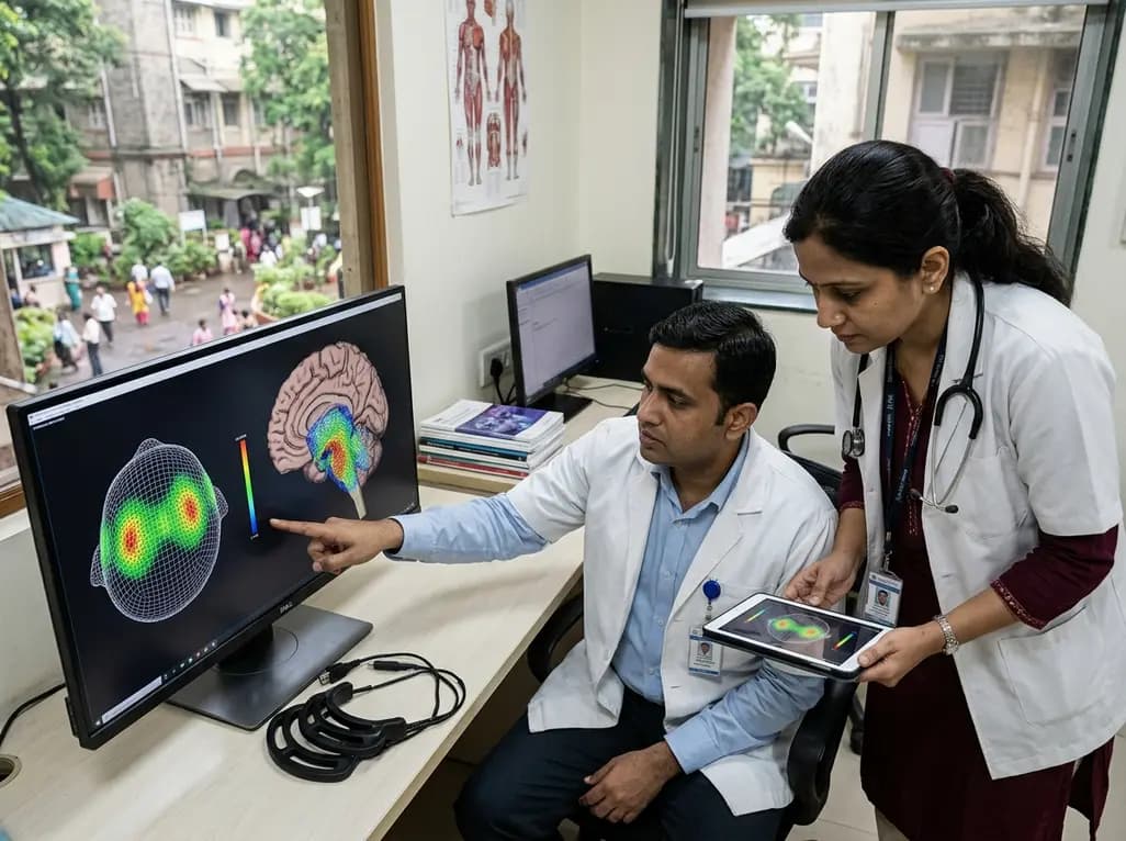
Tumor Treating Fields (TTFields) represent a significant shift in neuro-oncology, providing a non-invasive, regional treatment modality that utilizes low-intensity alternating electric fields. Currently, the FDA has approved this technology primarily for supratentorial glioblastoma, where it disrupts the mitotic spindle of rapidly dividing cancer cells. By interfering with polar microtubules during metaphase and anaphase, these electric fields induce mitotic arrest and subsequent apoptosis. Consequently, patients with supratentorial tumors have seen improved survival outcomes when combining TTFields with standard chemotherapy. However, applying this technology to infratentorial regions, specifically for TTFields brainstem gliomas, remains technically challenging due to the deep anatomical location and complex surrounding structures. Historically, standard transducer array placements focused on the larger cortical surfaces, leaving the brainstem with potentially sub-therapeutic dose distribution. Therefore, clinicians must investigate whether specialized array configurations can reach the required field intensities in the posterior fossa. Understanding the feasibility of these layouts is essential for expanding the therapeutic reach of TTFields to patients who currently have very limited treatment options. Because brainstem gliomas are often surgically inaccessible, non-invasive innovations like TTFields offer a crucial glimmer of hope for future clinical management.
Researchers recently conducted a sophisticated simulation study to address these geographic delivery hurdles. Using high-resolution MRI and CT imaging from seven distinct patients with brainstem gliomas, the team employed the MAXPOINT planning platform. This software utilizes finite element method (FEM) calculations to model the complex dielectric properties of various head tissues, including the scalp, skull, and cerebrospinal fluid. Specifically, the study defined the clinical target volume (CTV) as the enhancing tumor seen on T1 post-contrast imaging plus a three-millimeter peritumoral expansion. By utilizing patient-specific modeling, the platform could optimize the arrangement of scalp-only transducer arrays rather than relying on a one-size-fits-all approach. This level of customization is vital because even minor variations in skull thickness or tumor orientation can significantly alter the path of electric fields. Furthermore, the researchers compared these optimized layouts against standard, unplanned configurations to quantify the potential benefits of digital planning. Such simulations are critical because they allow scientists to predict field intensity without exposing patients to experimental risks. Through this computational approach, the study aimed to prove that scalp-only arrays could indeed project therapeutic energy deep into the brainstem and posterior fossa regions.
The primary metric used to evaluate success in this study was the local minimum field intensity (LMiFI), measured in Volts per centimeter (V/cm). Scientists generally consider an LMiFI of 1.0 V/cm or higher as the therapeutic reference threshold necessary to inhibit glioma cell proliferation effectively. Additionally, the study assessed the local minimum power density (LMiPD), which provides a measure of energy absorption within the tissue. Results indicated that the MAXPOINT-optimized layouts achieved significantly higher field intensities across all targeted regions compared to standard layouts. Specifically, the median LMiFI in the gross tumor volume (GTV) reached 1.1 V/cm in the optimized group, whereas the standard layout barely met the 1.0 V/cm threshold. The difference was even more pronounced in the broader brainstem and posterior fossa areas. In the posterior fossa, for instance, the median LMiFI rose to 1.5 V/cm with optimization, representing a substantial increase over the 1.2 V/cm seen in standard placements. Consequently, these findings suggest that precise array placement can reliably deliver doses that meet or exceed the established therapeutic requirements. This data provides the first robust evidence that TTFields brainstem gliomas treatments are dosimetrically feasible using current wearable technology.
One of the most significant revelations of this simulation was the superiority of optimized scalp-only array layouts over conventional designs. While clinicians previously worried that the dense bone of the skull base might shield the brainstem from electric fields, the optimization process successfully circumvented this issue. By strategically repositioning the four transducer arrays, the MAXPOINT platform maximized the field intersection within the infratentorial space. This optimization led to p-values of 0.019 or lower across all analyzed regions, reinforcing the statistical significance of the improvement. Moreover, the study highlighted that the clinical target volume, which includes the peritumoral expansion, received a median LMiFI of 1.1 V/cm. This ensures that the treatment covers not just the visible mass but also the microscopic infiltrative edges of the tumor. Because brainstem gliomas are notoriously infiltrative, achieving a therapeutic dose in the CTV is just as important as targeting the GTV. The study also found that power density was significantly higher in optimized models, which further supports the biological efficacy of the planned treatment. These advancements in array design demonstrate that we can overcome anatomical barriers through intelligent, patient-specific engineering.
For oncologists and neurologists, these simulation results offer a clear roadmap for future clinical trials involving infratentorial tumors. Currently, the prognosis for brainstem gliomas remains dismal, with limited efficacy from traditional radiotherapy and chemotherapy. Introducing a non-toxic, localized therapy like TTFields could alter the treatment paradigm significantly. However, clinicians must emphasize the importance of personalized treatment planning to ensure that every patient receives the maximum possible dose. Without optimization, some patients might receive field intensities that hover at or below the therapeutic threshold, potentially leading to treatment failure. Furthermore, the use of scalp-only arrays is a major advantage for patient comfort and compliance. Unlike invasive electrodes, these arrays are wearable and allow patients to maintain their daily activities. If subsequent clinical trials confirm these simulation results, TTFields could become a standard adjunct therapy for brainstem malignancies. Similarly, the methodology used in this study could be adapted for other infratentorial cancers, such as medulloblastoma or ependymoma. Therefore, this research serves as a foundational step toward expanding the reach of bioelectric medicine in the most challenging areas of the central nervous system.
Looking ahead, the next logical step is to translate these simulation findings into prospective clinical evaluations. While the computational data is compelling, researchers must now verify these results in real-world settings with diverse patient populations. Future studies should focus on the safety and tolerability of higher field intensities in the brainstem, as this region controls vital functions such as respiration and heart rate. Additionally, combining optimized TTFields with novel systemic agents might provide a synergistic effect that further improves survival rates. Researchers are also exploring the use of advanced imaging to monitor real-time changes in tumor dielectric properties during treatment. This could allow for dynamic array adjustments throughout the course of therapy. Overall, the feasibility evaluation performed by the MAXPOINT platform provides a strong rationale for moving forward with infratentorial TTFields applications. By bridging the gap between computational physics and clinical oncology, we are moving closer to a future where brainstem gliomas are no longer untreatable. The success of this simulation underscores the power of personalized medicine in tackling the most difficult frontiers of cancer care.
TTFields are low-intensity, alternating electric fields delivered via wearable transducer arrays. They specifically target rapidly dividing cancer cells by disrupting the mitotic spindle during cell division. This mechanism leads to cell death or growth arrest without affecting the non-dividing healthy cells, providing a localized and non-toxic treatment option.
The MAXPOINT platform uses patient-specific MRI and CT data to create a 3D model of the head. It then calculates the most efficient transducer array placement to maximize the electric field intensity within the tumor. This optimization ensures that even deep-seated tumors, like brainstem gliomas, receive a therapeutic dose.
While this simulation study confirms that therapeutic doses can be delivered to the brainstem, prospective clinical trials are necessary to fully establish safety in this region. Early data suggest that TTFields are generally well-tolerated, with the most common side effect being mild skin irritation under the transducer arrays.
Disclaimer: This content is for informational and educational purposes only and does not constitute medical advice, diagnosis, or treatment. Always seek the advice of your physician or other qualified health provider with any questions you may have regarding a medical condition. Refer to the latest local and national guidelines for clinical practice.
References
Chen AY et al. Feasibility evaluation of tumor treating fields for brainstem gliomas. J Neurooncol. 2026 Jul 06. doi: 10.1007/s11060-026-05697-y. PMID: 42406140.
Stupp R et al. Effect of Tumor-Treating Fields Plus Maintenance Temozolomide vs Maintenance Temozolomide Alone on Survival in Patients With Glioblastoma: A Randomized Clinical Trial. JAMA. 2017;318(23):2306–2316.
Ballo MT et al. Tumor Treating Fields: A New Frontier in Oncology. Frontiers in Oncology. 2019;9:1158. doi: 10.3389/fonc.2019.01158.
Taphoorn MJB et al. Health-Related Quality of Life in Patients With Newly Diagnosed Glioblastoma Treated With Tumor-Treating Fields Plus Temozolomide: A Secondary Analysis of the EF-14 Phase 3 Randomized Clinical Trial. JAMA Oncol. 2018;4(4):495-504.

Read summarized clinical updates, watch expert medical content, and earn CME certifications right from your smartphone.
Open your smartphone camera to scan and install.





A new simulation study demonstrates that patient-specific modeling using scalp-only transducer arrays can effectively deliver therapeutic-level Tumor Treating Fields (TTFields) to brainstem gliomas, offering a potential non-invasive treatment path for infratentorial tumors.
3 months ago

As monsoon seasons elevate arboviral disease threats, Tamil Nadu has initiated an ICMR-monitored evaluation of the tetravalent dengue vaccine in Tenkasi district. Clinicians emphasize clinical monitoring, public prevention, and high-quality early pediatric care to reduce disease morbidity across endemic regions.
Today

A comparative cohort study evaluates vNOTES vs TLH hysterectomy for benign conditions, demonstrating shorter operative duration, faster return to daily activities, and equivalent female sexual function scores without increased dyspareunia.
Today

Review how tailored exercise programs benefit frail older patients with heart failure. Explore pathophysiological mechanisms, individualized training prescriptions, and strategies to overcome adherence barriers in clinical practice.
Today

This comparative study evaluates posterior labral tears with and without glenohumeral instability, highlighting distinct demographic patterns, MRI underestimation in stable shoulders, and successful arthroscopic repair outcomes.
Today

A clinical study demonstrates that early intervention with tocilizumab, either as monotherapy or bridged with corticosteroids, achieves significant proptosis reduction and resolves active inflammation in treatment-naive thyroid eye disease.
Today
