.webp&w=384&q=75)

Loading, please wait...
Loading, please wait...

Primary trigeminal neuralgia presents as excruciating, paroxysmal facial pain that severely impairs patient quality of life. Traditionally, clinicians view this neuropathic disorder through the lens of peripheral nerve root pathology. However, contemporary neuroimaging reveals that altered central pain processing plays an equally decisive role in symptom perpetuation. Evaluating trigeminal neuralgia functional connectivity through resting-state functional magnetic resonance imaging provides objective insight into these maladaptive neural networks. Although classical trigeminal neuralgia and idiopathic trigeminal neuralgia exhibit overlapping clinical symptoms, their central neural signatures show critical divergences. Resting-state blood oxygen level-dependent signals allow investigators to map spontaneous cerebral activity without active task performance. Consequently, researchers can identify subtle dysfunctions in distributed networks responsible for sensory discrimination, cognitive appraisal, and emotional affective responses. By evaluating spontaneous low-frequency fluctuations, advanced functional imaging captures persistent neuroplastic reorganization triggered by chronic nociceptive volleys. Understanding these central dynamics shifts the clinical paradigm beyond simple nerve root compression toward holistic network medicine.
Neurovascular compression at the root entry zone remains the anatomical hallmark of classical trigeminal neuralgia. Nevertheless, structural compression alone fails to explain why certain patients suffer persistent pain despite surgical decompression. Conversely, many asymptomatic individuals harbor anatomical contact without experiencing any neuropathic symptoms. Therefore, researchers hypothesize that peripheral insult incites widespread cortical and subcortical remodeling. A groundbreaking neuroimaging study examined ninety-three subjects, including fifty surgical patients with verified compression status and forty-three healthy controls. Functional metrics revealed significant topological alterations across frontoparietal, limbic, and default mode networks. Specifically, persistent paroxysmal pain appears to compromise descending inhibitory pathways while hypersensitizing sensory gating structures. Increased spontaneous activity in frontal regions indicates compensatory recruitment of cognitive networks trying to modulate intractable pain. Moreover, these neuroimaging findings explain why chronic pain syndromes frequently co-occur with affective distress, sleep disturbances, and executive dysfunction. Peripheral vascular pulsatility initiates the cascade, but central brain dynamics sustain the clinical syndrome.
Differentiating classical trigeminal neuralgia from idiopathic presentations is essential for guiding surgical decision-making. In classical disease, surgical exploration consistently confirms morphological neurovascular compression. In contrast, idiopathic disease exhibits minimal or no verifiable root distortion. Investigating these distinct cohorts has uncovered unique central reorganization patterns. Specifically, patients with trigeminal neuralgia demonstrated increased functional connectivity between the medial prefrontal cortex and the left planum temporale. Concurrently, they exhibited reduced connectivity between the medial prefrontal cortex and the left superior frontal gyrus. When comparing subtypes, classical cases showed pronounced functional disconnection between the left insula and the left occipital pole. Furthermore, classical patients exhibited markedly reduced neural activity in the right temporal pole compared to idiopathic cohorts. These localized divergences suggest that severe mechanical root distortion drives distinct neuroplastic trajectories. Consequently, idiopathic forms might depend more heavily on central dysregulation or microstructural channelopathies, whereas classical forms reflect severe secondary reorganization from chronic peripheral pulsation.
Translating resting-state connectome alterations into individual clinical utility requires sophisticated computational modeling. Group-level statistical comparisons highlight biological trends, yet individualized diagnostic tools remain paramount for clinical translation. To bridge this divide, researchers applied support vector machine algorithms to functional connectivity metrics and spontaneous activity parameters. The machine learning model successfully discriminated neuralgia patients from healthy controls with seventy-four percent accuracy. Additionally, the classification algorithm achieved an area under the receiver operating characteristic curve of 0.80. These metrics validate the discriminatory power of spontaneous neural fluctuations as objective neuropathic pain biomarkers. Furthermore, feature selection algorithms highlighted connectivity within the medial prefrontal cortex and insular hubs as the most informative predictive weights. Machine learning removes subjective observer bias and captures nonlinear interactions across multidimensional neuroimaging datasets. In the future, automated classifiers may help clinicians objectively confirm borderline diagnoses and predict individualized procedural responses.
For neurologists, neurosurgeons, and pain physicians, these neuroimaging insights offer profound practical implications. Microvascular decompression remains the primary curative surgery for patients with confirmed neurovascular compression. However, central connectivity changes might explain persistent sensory dysesthesias or post-decompression pain recurrence. If preoperative imaging reveals profound disruption of descending modulatory networks, clinicians can anticipate heightened central sensitization. Consequently, multidisciplinary teams can introduce targeted neuromodulation or centrally acting pharmacotherapies earlier in the disease trajectory. In addition, recognizing idiopathic neuralgia as a distinct central pathophysiological entity prevents inappropriate, aggressive surgical interventions. Non-invasive repetitive transcranial magnetic stimulation and functional neurofeedback might selectively target dysregulated nodes like the prefrontal cortex or insula. Integrating functional imaging into routine diagnostic workups could refine surgical selection criteria and reduce treatment failures. Personalized pain management requires clinicians to treat the central pain connectome alongside peripheral nerve pathology.
As functional imaging resolution advances, connectome mapping will unlock personalized neurosurgical planning. Future investigations must track dynamic functional connectivity changes longitudinally across preoperative and postoperative phases. Such prospective datasets will clarify whether successful microvascular decompression normalizes pathological functional connectivity or leaves permanent central scars. In addition, integrating resting-state metrics with diffusion tensor imaging will elucidate the exact coupling between white matter microstructure and cortical function. Combining multi-omics data with artificial intelligence platforms could ultimately create comprehensive predictive algorithms for facial pain. Clinicians could evaluate genetic susceptibility, anatomical nerve morphology, and whole-brain network integrity within a unified framework. Furthermore, multicenter registries across diverse geographic populations will ensure that machine learning diagnostic models remain generalizable and robust. Moving beyond qualitative interpretation toward quantitative neuroimaging biomarkers heralds a transformative era in neuropathic pain management.
Resting-state functional magnetic resonance imaging measures spontaneous low-frequency fluctuations in blood oxygenation levels while the patient rests quietly. By assessing synchronous activity across distant brain areas, clinicians can map functional connectivity, detect circuit disruptions, and identify centralized neuroplastic alterations caused by persistent neuropathic pain.
Differentiating these subtypes is essential because classical neuralgia involves definitive neurovascular compression responsive to surgical microvascular decompression. Conversely, idiopathic cases lack severe mechanical compression. Identifying distinct central neural patterns helps clinicians avoid unnecessary invasive surgeries and prioritize appropriate pharmacotherapy or neuromodulation strategies.
Current support vector machine models trained on resting-state imaging metrics achieve moderate diagnostic accuracy, reaching approximately seventy-four percent with an area under the curve of 0.80. Although not yet ready for standalone clinical diagnosis, these models provide objective biomarkers that assist clinical decision-making.
Disclaimer: This content is for informational and educational purposes only and does not constitute medical advice, diagnosis, or treatment recommendations. Clinical decisions should always be made by qualified healthcare professionals based on individual patient assessments and established medical standards. Refer to the latest local and national guidelines for clinical practice.
References
Wu M et al. Stratifying trigeminal neuralgia and characterizing an abnormal property of brain functional organization: a resting-state fMRI and machine learning study. J Neurosurg. 2025 Jul 01. doi: 10.3171/2024.11.JNS241935. PMID: 40153851.
Bendtsen L et al. Advances in diagnosis, classification, pathophysiology, and management of trigeminal neuralgia. Lancet Neurol. 2020;19(9):784-796. doi: 10.1016/S1474-4422(20)30233-7.
Chen DQ et al. Multivariate pattern analysis of resting-state fMRI in trigeminal neuralgia. Pain. 2019;160(4):805-814. doi: 10.1097/j.pain.0000000000001452.

Read summarized clinical updates, watch expert medical content, and earn CME certifications right from your smartphone.
Open your smartphone camera to scan and install.





A landmark neuroimaging study combines resting-state fMRI and machine learning to map distinct trigeminal neuralgia functional connectivity patterns. The findings reveal critical central biomarkers that differentiate classical from idiopathic subtypes, offering new avenues for targeted therapeutic interventions.
Today
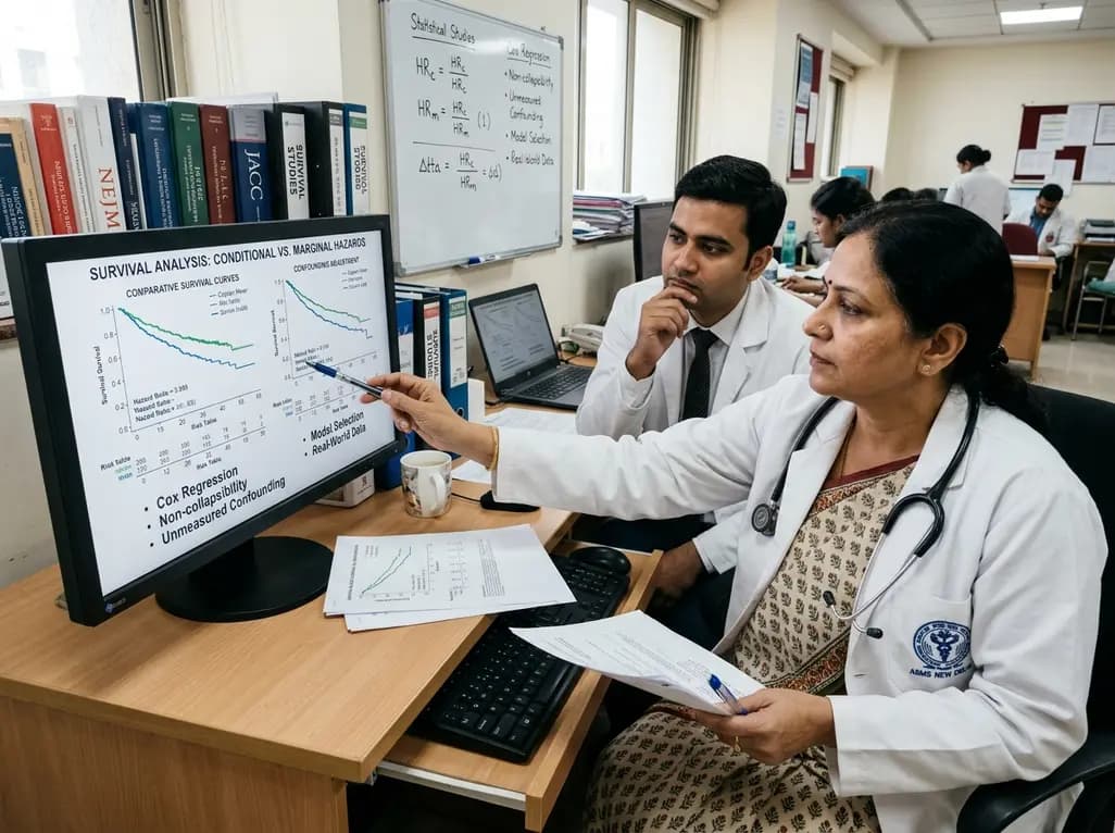
Survival analyses often report hazard ratios, but mathematical non-collapsibility creates crucial differences between conditional and marginal estimates. Clinicians must account for unmeasured confounding and model selection when interpreting trial endpoints and real-world cardiovascular outcomes.
Today

A cross-sectional study of 700 college students highlights significant contraceptive knowledge disparities between sexually active and inactive young adults. The findings demonstrate a critical need for proactive, comprehensive sexual health counseling before sexual debut.
Yesterday
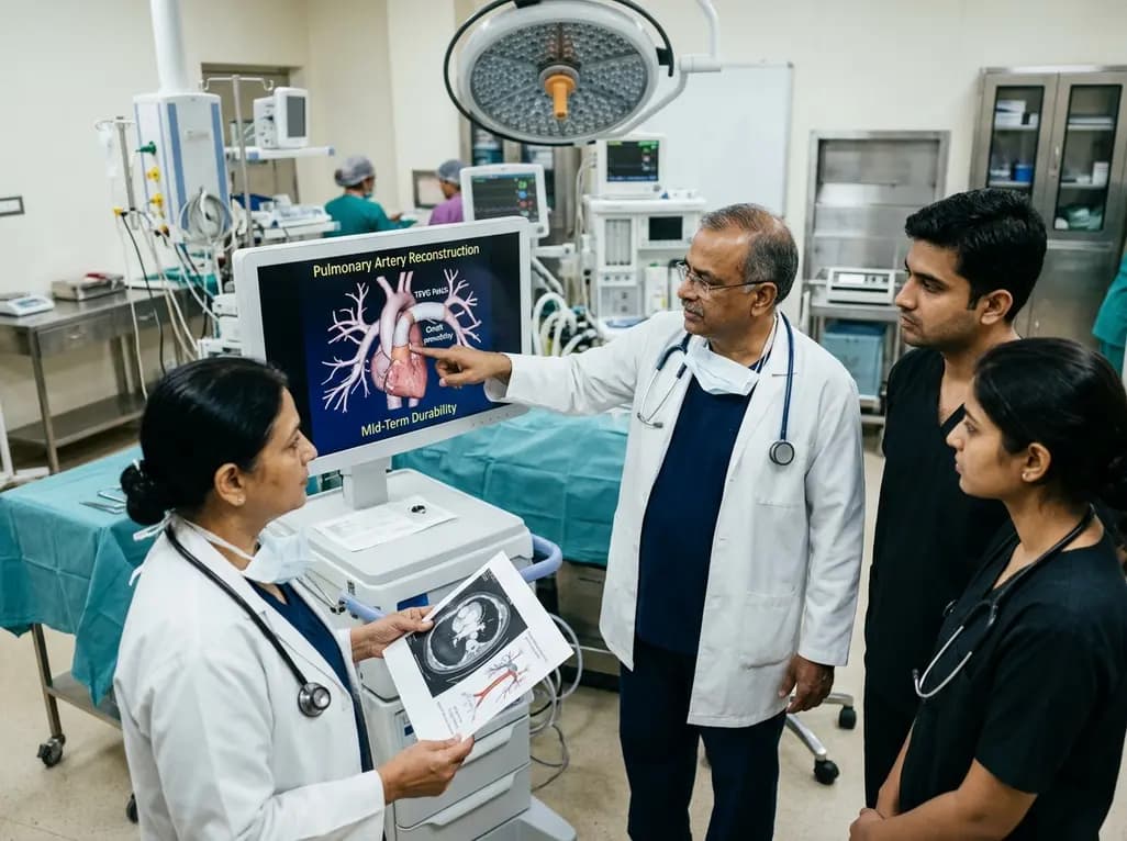
Autologous tissue-engineered vascular graft patches created via in-body tissue architecture show excellent mid-term durability and safety for pulmonary artery reconstruction in congenital heart surgery, particularly for peripheral branch augmentation.
Today

A novel cluster analysis of real-world datasets reveals four distinct CPPD disease phenotypes, validating existing EULAR categories while uncovering previously unrecognized monoarticular and axial presentations to refine patient management.
Today

Recent pharmacokinetic research demonstrates that trace dermal exposure to microgram quantities of trenbolone and metenolone produces detectable urinary metabolites for days to over a week, providing critical objective data for evaluating unintentional contamination claims in sports drug testing.
Today
